ABSTRACT
Objectives: To determine the incidence of crack formation and propagation in apical root dentin after retreatment procedures performed using ProTaper Universal Retreatment (PTR), Mtwo-R, ProTaper Next (PTN), and Twisted File Adaptive (TFA) systems.
Materials and Methods: The study consisted of 120 extracted mandibular premolars. One millimeter from the apex of each tooth was ground perpendicular to the long axis of the tooth, and the apical surface was polished. Twenty teeth served as the negative control group.
One hundred teeth were prepared, obturated, and then divided into 5 retreatment groups.
The retreatment procedures were performed using the following files: PTR, Mtwo-R, PTN, TFA, and hand files. After filling material removal, apical enlargement was done using apical size 0.50 mm ProTaper Universal (PTU), Mtwo, PTN, TFA, and hand files. Digital images of the apical root surfaces were recorded before preparation, after preparation, after obturation, after filling removal, and after apical enlargement using a stereomicroscope. The images were then inspected for the presence of new apical cracks and crack propagation. Data were analyzed with χ2 tests using SPSS 21.0 software.
Results: New cracks and crack propagation occurred in all the experimental groups during the retreatment process. Nickel-titanium rotary file systems caused significantly more apical crack formation and propagation than the hand files. The PTU system caused significantly more apical cracks than the other groups after the apical enlargement stage.
Conclusions: This study showed that retreatment procedures and apical enlargement after the use of retreatment files can cause crack formation and propagation in apical dentin.
Keywords: Apical crack; Endodontics; Nickel-titanium; Retreatment; Root Canal Preparation
INTRODUCTION
The most important purpose of root canal treatment is to prepare the root canals chemo- mechanically and fill them hermetically. However, root canal preparation may result in weakening of the remaining tooth structure and lead to the formation of crack lines and
Research Article
Received: Apr 27, 2017 Accepted: Oct 9, 2017
Özyürek T, Tek V, Yılmaz K, Uslu G
*Correspondence to Gülşah Uslu, DDS, MSc Research Assistant, Department of Endodontics, Ondokuz Mayis University Faculty of Dentistry, Samsun, 55137, Turkey.
Tel: +90-362-312-1919 Fax: +90-362-457-3062
E-mail: gulsah.turkkan@hotmail.com Copyright © 2017. The Korean Academy of Conservative Dentistry
This is an Open Access article distributed under the terms of the Creative Commons Attribution Non-Commercial License (https://
creativecommons.org/licenses/by-nc/4.0/) which permits unrestricted non-commercial use, distribution, and reproduction in any medium, provided the original work is properly cited.
Conflict of Interest
No potential conflict of interest relevant to this article was reported.
Author Contributions
Conceptualization: Özyürek T, Yılmaz K, Uslu G; Data curation: Yılmaz K, Uslu G; Formal analysis: Özyürek T; Funding acquisition:
Özyürek T, Yılmaz K, Uslu G; Investigation:
Özyürek T, Yılmaz K, Uslu G; Methodology:
Özyürek T, Uslu G; Project administration:
Özyürek T; Resources: Özyürek T, Yılmaz K, Uslu G, Tek V; Supervision: Özyürek T;
Taha Özyürek ,1 Vildan Tek ,2 Koray Yılmaz ,3 Gülşah Uslu 1*
1Department of Endodontics, Ondokuz Mayıs University Faculty of Dentistry, Samsun, Turkey
2Department of Endodontics, Bülent Ecevit University Faculty of Dentistry, Zonguldak, Turkey
3Çorum Oral and Dental Center, Çorum, Turkey
Incidence of apical crack formation and propagation during removal of root canal filling materials with
different engine driven nickel-titanium
instruments
Visualization: Özyürek T; Writing - original draft: Özyürek T, Yılmaz K, Uslu G, Tek V;
Writing - review & editing: Özyürek T, Yılmaz K, Uslu G, Tek V.
ORCID iDs Taha Özyürek
https://orcid.org/0000-0003-3299-3361 Vildan Tek
https://orcid.org/0000-0001-7557-279X Koray Yılmaz
https://orcid.org/0000-0001-6096-7385 Gülşah Uslu
https://orcid.org/0000-0003-3176-1251
and restorative or endodontic procedures, these crack lines and microcracks can propagate and produce oblique root fractures, leading to endodontic treatment failure [1,2]. Factors affecting the progress of vertical root fractures include root canal instrumentation, filling techniques [3], retreatment procedures [4], tooth anatomy [5], post replacement [6], and a high concentration of sodium hypochlorite (NaOCl) [7]. Retreatment procedures are performed when the initial root canal treatment fails. These procedures involve removing the root canal filling material, controlling microbial infection, and reshaping the canals, followed by obturation to produce an impermeable seal. The additional mechanical instrumentation involved in these retreatment procedures weakens the structure of the root dentin wall [4].
Nickel-titanium (NiTi) rotary systems feature technologically advanced properties and are used in root canal retreatment because they provide easier and faster preparation than manual instrumentation. Although the canal preparation time is shorter using rotary systems than using hand instrumentation, they generate more stress on the dentin wall, which leads to cracked lines and microcracks [8]. Several techniques can be used to remove gutta-percha, including the use of heat, solvents, mechanical instruments, and various combinations of these methods [9]. NiTi rotary systems with different designs have been developed to improve the efficiency of gutta-percha removal.
ProTaper Universal Retreatment (PTR; Dentsply Maillefer, Ballaigues, Switzerland) and Mtwo-R (VDW, Munich, Germany) are the most commonly used NiTi retreatment systems.
Both these systems work in a continuous rotational motion. PTR is used for removing root filling material and comprises 3 different instruments: D1 (30/0.09), D2 (25/0.08), and D3 (20/0.07). The cross-section areas of the instruments are convex triangular, similar to that of ProTaper Universal (PTU; Dentsply Maillefer) files. The Mtwo-R retreatment system consists of 2 NiTi files: R15/0.05 and R25/0.05. The instruments have an S-shaped cross-section and an active cutting tip.
ProTaper Next (PTN; Dentsply Maillefer) consists of 5 instruments, namely, X1 (17/0.04), X2 (25/0.06), X3 (30/0.07), X4 (40/0.06), and X5 (50/0.06). PTN uses M-Wire technology, which improves instrument strength and flexibility [10]. According to the manufacturer, the asymmetric rotary motion of the instrument reduces screwing by minimizing contact between the instrument and dentin wall [11]. Twisted File Adaptive (TFA; SybronEndo, Orange, CA, USA) is one of the new-generation of NiTi file systems. It employs a combination of 2 different motions (continuous rotation and reciprocating) based on the stress
encountered by the instrument in the canal. The TFA system is produced using R-phase heat treatment and unique surface conditioning [12], which the manufacturer claims increases of strength, flexibility, and resistance to fatigue [13].
A comprehensive literature review revealed no studies of apical crack formation and propagation during removal of root canal filling material with PTN and TFA. Therefore, this study aimed to evaluate the incidence of crack formation and propagation in apical dentin after retreatment procedures using PTR, Mtwo-R, PTN, and TFA NiTi rotary systems and conventional hand files, with additional instrumentation. The null hypothesis was that there would be no difference in apical crack formation among the groups.
MATERIALS AND METHODS
Specimen preparation
After obtaining approval from the Ethics Committee of Ondokuz Mayıs University (No.
2016/713), 120 mandibular premolar teeth with straight and single root canals extracted because of periodontal reasons were included in the study. Soft and hard tissues around the teeth were mechanically removed using a periodontal curette. The crowns of the teeth were removed from the cementoenamel junction under water cooling to allow 14 mm of standard root length. The teeth were radiographically examined in the mesio-distal and bucco-lingual directions. Teeth found to have calcification, a history of previous root canal treatment, internal and/or external resorption, cracks, or fractured and/or immature roots were excluded. Teeth with a deviated apical foramen were excluded from the study for standardization. The selected teeth were kept in distilled water at 4°C until the experiment.
As in previous studies, the roots of the teeth were coated with aluminum foil and then embedded in acrylic resin (Imicryl, Konya, Turkey) [14,15]. After the acrylic had set, the teeth were taken out of the resin, and the foils were removed. To simulate the periodontal ligament, the resin blocks were filled with a silicon impression material (Express XT Light Body Quick, 3M ESPE, Seefeld, Germany), and the specimens were returned to the resin blocks. The apical 4 mm of the root was exposed to capture images during the procedure. In accordance with Adorno et al. [16], 1 mm apical root segments were ground perpendicular to the tooth axis using waterproof 320-grit silicon carbide abrasive paper.
The ground apical surface was polished using waterproof 1,000-grit and 1,200-grit silicon carbide abrasive paper to obtain a finely polished surface to ensure that high-quality images could be obtained. The polished apical segment of the root was dipped in water during the experimental procedure to avoid dehydration [17]. The canal diameter of the specimens 5 mm from the apex was checked using a size 120 K-file (Dentsply Sirona, Ballaigues, Switzerland).
Teeth with canal diameters larger than 1.2 mm at 5 mm from the apex were excluded from the study. In all the specimens, baseline images were captured using a digital camera (Canon EOS 500D, Canon, Inc., Tokyo, Japan) attached to a stereomicroscope (Olympus BX43, Olympus Co., Tokyo, Japan). Twenty teeth were randomly stored as the negative control group.
Root canal preparation
A size 10 K-file (Dentsply Maillefer) was placed into the roots until the tip could be seen from the apex. The working length (WL) was set as 1 mm short of this length. In all the specimens, the root canal was enlarged in accordance with the crown-down method using K-files (Dentsply Maillefer) to ensure an apical diameter of 0.40 mm, with a 2% taper. During the root canal preparation, each of the specimens was irrigated with 20 mL of 1% NaOCl. For the final irrigation, 2 mL of 17% EDTA for 1 minute, followed by 2 mL of 1% NaOCl was used to remove the smear layer in each tooth. Each new set of files was used to prepare 4 canals.
Images of the apical surface of the samples after root canal preparation were captured using a digital camera attached to the stereomicroscope.
Root canal obturation
The canals were dried using paper points (Diadent, Diadent Group International, Cheongju, Korea). The root canals were obturated using gutta-percha and AH Plus root canal sealer (Dentsply DeTrey, Konstanz, Germany). 40/0.02 master cones (Diadent) were coated with sealer and placed into the canal to the WL. The root canals were obturated using
(Dentsply International, Johnson City, TN, USA). The quality of root canal obturation was confirmed by mesio-distal and buccal-lingual radiographs. Samples with inadequate or nonhomogeneous root canal obturation were replaced with new ones. Temporary filling material (Cavit-G, 3M ESPE) was used for sealing the coronal orifice. All procedures were executed by a single operator.
Following obturation, the teeth were stored at 37°C in 100% humidity for 14 days for sealer setting. Images of the apical surfaces of the obturated root canals were captured using a digital camera attached to a stereomicroscope. The specimens were divided into 5 groups (n = 20 in each group). A total of 45 apically cracked specimens were divided equally into 5 groups to ensure standardization (n = 9 in each group) after obturation.
Hand file group
In this group, size 3 Gates Glidden drills (Dentsply Maillefer), followed by size 2 drills operating at 1,500 rpm were used to remove coronal filling material. The canals were re-instrumented with Hedström files (Dentsply Maillefer) of sizes 30, 25, 20, and 15 in a circumferential, quarter-turn, push–pull filing motion to remove the filling material until the WL was achieved. Once the WL was reached with a size 15 file, sizes 20, 25, 30, and 35 files were used at the WL. Additional instrumentation was then performed using a size 50 Hedström file (Dentsply Maillefer) at the WL.
PTR group
In the PTR group, the retreatment procedure was performed using the crown-down technique and PTR D1 (30/0.09), D2 (25/0.08), and D3 (20/0.07) retreatment files. The files were activated with an X-Smart endodontic motor (Dentsply Maillefer). Gutta-percha and sealer in the coronal and middle thirds of the canal were removed using D1 and D2 files at 550 rpm and 200 g/cm−1 torque. A D3 file at 250 rpm and 150 g/cm−1 torque was used for gutta- percha and sealer removal at the apical third.
The files were used with a brushing motion against the lateral walls. The final apical preparation was achieved using a PTU F5 (50/0.05) file (Dentsply Maillefer) at 250 rpm and 200 g/cm−1 torque.
Mtwo-R group
The Mtwo R25/0.05 instruments were used in the crown-down technique. The instruments were activated by the torque control endodontic motor (X-Smart, Dentsply Maillefer) at 280 rpm and 120 g/cm−1 torque according to the manufacturer's instructions. The files were used with a brushing motion against the lateral walls. The final apical preparation was achieved using an Mtwo (50/0.04) file (VDW) at 200 rpm and 200 g/cm−1 torque.
PTN group
The PTN X3 (30/0.07) and X2 (25/0.06) instruments were used in the crown-down technique.
The PTN X3 file was used to remove the gutta-percha and sealer from coronal one-third of the WL, and the X2 file was used in the full WL. The instruments were activated using a torque control endodontic motor (X-Smart, Dentsply Maillefer) at 300 rpm and 200 g/cm−1 torque according to the manufacturer's instructions. The files were used with a brushing motion against the lateral walls. The final apical preparation was achieved with a PTN X5 (50/0.06) file (Dentsply Maillefer) at the same speed and torque values.
TFA group
In this group, the TFA ML2 (35/0.06) and ML1 (25/0.08) instruments were used in the crown- down technique, as described in the PTN group. The gutta-percha and sealer in the coronal third of the canal were removed using the TFA ML2 file. The TFA ML1 file was then used to reach the WL. The instruments were activated with an Elements Motor in the TFA program.
The final apical enlargement was performed with a TFA ML3 (50/0.04) file (SybronEndo), using the same program.
The canals in each group were irrigated with 2 mL of 1% NaOCl solution during each instrument change or pecking motion. A total of 20 mL of 1% NaOCl solution was used during the retreatment process. Four root canals were retreated with a new set or a new file, and the files were then discarded [18]. During the root canal preparation and retreatment process, to avoid an excessive load, the operator's finger was placed axially over the tooth. All the procedures were performed by an experienced endodontist.
Stereomicroscopic examination
Five images were taken of each specimen in the experimental groups: baseline, after
preparation, after obturation, after filling removal, and after apical enlargement (Figures 1-3).
Two calibrated examiners who were blinded to the group assignment evaluated each image.
Each image was matched with a previous image, and any visible crack line on the ground
A B
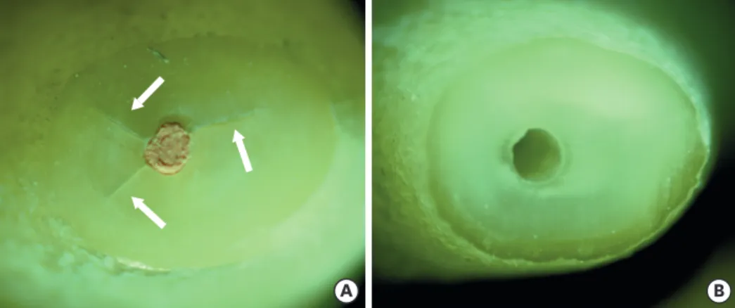
Figure 1. (A) Crack formation after root canal filling removal (white arrows). (B) Propagation of the same cracks after apical enlargement (white arrows).
A B
Figure 2. (A) The specimen showing no crack after root canal obturation. (B) New crack formation in the same
apical surface that was not present in the previous image was defined as a crack [19]. The images were compared with the baseline image, and the presence or propagation of a crack during the procedures was noted.
Statistical analysis
Pearson's χ2 test was employed in the intergroup statistical analysis of the incidence of dentinal defects. Cohen's kappa test was conducted to calculate inter-examiner reliability.
The level of statistical significance was set to 5%. All the statistical analyses were performed using SPSS version 21 (IBM-SPSS Inc., Chicago, IL, USA).
RESULTS
The inter-examiner agreement was 93% for surface deformation of the tested files (kappa test). The incidences of new apical crack formation and propagation using the tested files are shown in Table 1. The baseline images of the specimens showed no apical cracks. After root canal instrumentation, 35 of the 100 specimens showed apical cracks. After root canal obturation, 10 new crack formations were observed after root canal obturation. A total of 45 cracks formed after the root canal instrumentation and obturation. After removal of root canal filling material, 6 new cracks were observed in the PTU group, and 5 new cracks were
A B
Figure 3. (A) New crack formation after root canal obturation (white arrows). (B) The sample without any crack after root canal filling removal and apical enlargement
Table 1. Incidence of new crack formation and crack propagation for tested nickel-titanium (NiTi) files
Group Baseline Root canal preparation Root canal obturation Root canal filling removal Apical enlargement
Total New Total New Total New Propagation Total New Propagation Total
t/N n t/N n t/N n p/P t/N n p/P t/N
Control 0/20 - - - - - - - - - -
Hand file 0/20 7 7/20 2 9/20 1a 0/9a 10/20a 1a 1/10a 11/20a
ProTaper Next 0/20 7 7/20 2 9/20 5b 3/9b 14/20b 1a 5/14b 15/20b
ProTaper Universal
Retreatment 0/20 7 7/20 2 9/20 6b 3/9b 15/20b 3b 7/15b 18/20b
Twisted File Adaptive 0/20 7 7/20 2 9/20 5b 4/9b 14/20b 1a 5/14b 15/20b
Mtwo-R 0/20 7 7/20 2 9/20 5b 4/9b 14/20b 1a 5/14b 15/20b
p value - - - - - < 0.05 < 0.05 < 0.05 < 0.05 < 0.05 < 0.05
ProTaper Next, Dentsply Maillefer, Ballaigues, Switzerland; ProTaper Universal Retreatment, Dentsply Maillefer, Ballaigues, Switzerland; Twisted File Adaptive, SybronEndo, Orange, CA, USA; Mtwo-R, VDW, Munich, Germany. Different superscripts indicate statistical difference at 5%.
t, total number of crack; N, total number of specimens; n, number of new cracks; p, number of propagated crack; P, number of previous cracked teeth.
observed in the PTN, TFA, and Mtwo-R groups. The number of new cracks that formed after root canal filling removal in the NiTi file groups was significantly greater than that in the hand file group (p < 0.05). The NiTi rotary file systems caused significantly more apical crack propagation than the hand file group (p < 0.05). After apical enlargement, the PTU system caused significantly more apical cracks than the other groups (p < 0.05).
DISCUSSION
The primary objective of retreatment is to remove root canal filling materials, disinfect the root canal system, and obturate the root canals [20]. Removal of root canal filling material from the apical third is the most difficult part of retreatment because of the anatomic variability of the apical third [21]. Thus, in most cases, apical enlargement is an important stage of retreatment procedures. In this situation, apical enlargement may also cause apical cracks.
No previous studies compared apical crack formation and propagation during root canal filling removal in PTN and TFA retreatment procedures. Therefore, the present study aimed to evaluate the incidence of new crack formation and propagation in apical dentin after retreatment procedures using PTR, Mtwo-R, PTN, and TFA NiTi rotary systems and conventional hand files, with additional instrumentation. The results showed that the tested NiTi rotary file systems caused more apical cracks than the hand files. This result was similar to that reported in previous studies, which found that NiTi rotary files were associated with apical crack formation [16,19]. However, there was no statistically significant difference in apical crack formation among the tested NiTi rotary systems. Therefore, the null hypothesis of the present study was rejected.
The present study consisted of extracted mandibular premolar teeth. Adorno et al. [16]
reported that a deviated apical foramen could cause crack formation on the apical surface.
Thus, in the present study, teeth with a deviated apical foramen were excluded to ensure standardization. In addition, the present study captured images of all stages of the retreatment procedures, with 1 mm apical root segments ground and polished prior to image capture. Comparing crack formation and propagation in the grounded apical surface eliminated potential damage to dentin caused by the sectioning process. Prior to dividing the teeth into the treatment groups, the teeth were examined under a stereomicroscope to detect the presence of any fractures or cracks, as cracks in some teeth might not be seen externally. The absence of any dentinal defects in the negative control group in the present study indicates that the teeth were free of cracks. The absence of any dentinal defects in the negative control group is consistent with that reported in other studies [22,23].
To prevent file fracture, manufacturers recommend discarding the files after they have been used a few times in root canals. The complexity of canals (calcification and curvature) also plays a role in file fracture. However, there is no consensus in the literature on how many times files can be used [24-26]. The manufacturers of the files used in the present study recommended single patient usage. Therefore, in the preparation of a maxillary first molar with 4 canals, each new file set was used to prepare 4 root canals and then discarded [23,27].
Previous studies reported that a high concentration of NaOCl solution decreased the elastic modulus and hardness level of dentin [7,28]. In the present study, a 1% NaOCl solution was
of dentin. Thus, dentinal defects that occurred were attributed mainly to the mechanical preparation.
The results indicated that 35 of 100 specimens exhibited new apical crack formation after initial root canal preparation with hand files. This finding is in accordance with that of previous studies, which reported that apical cracks formed after root canal preparation using hand files [19,29]. Moreover, in the present study, after root canal obturation, 10 of 65 specimens showed new crack formation. The teeth were obturated in accordance with the vertical compaction method. In contrast to previous studies that using lateral compaction technique [19,29], dentinal defects were observed after root canal obturation using vertical compaction technique.
The results of the present study showed that the NiTi rotary systems caused significantly more new apical crack formation and propagation than the hand files after root canal filling removal. This finding was consistent with that of a previous study, which suggested that fewer apical cracks in the hand file group could be due to less aggressive movement of hand files in the canal as compared to that of NiTi rotary files. Topçuoğlu et al. [19] found no significant difference in apical crack formation and propagation between Mtwo-R and PTR NiTi rotary systems after a retreatment process, in common with the findings of the present study. Previous studies revealed that PTN and TFA were effective in the removal of root canal filling materials [30,31]. However, no previous study compared apical crack formation and propagation during root canal filling removal using PTN and TFA. Thus, the results of the present study cannot be directly compared with those of other studies.
The present study evaluated different NiTi file systems with different motion kinematics (continuous rotation and adaptive motion). The results revealed no significant difference between TFA and the other NiTi rotary systems. Zhou et al. [32] compared apical crack formation of NiTi rotary systems during root canal preparation and concluded that TFA systems caused significantly less apical crack formation than PTU and WaveOne (Dentsply Maillefer) systems. In the present study, adaptive motion did not affect apical crack formation and propagation during root canal retreatment.
Previous studies reported that the design and taper of a file could affect crack formation because of different stress levels caused by the files on the canal walls [8,23]. In the present study, all the NiTi rotary files caused significantly more apical cracks and crack propagation than the hand files. Furthermore, significantly more new crack formation occurred using the PTU system with a 5% taper than the NiTi file systems after the apical enlargement stage.
According to the manufacturer, the offset design of PTN files reduces the contact of files with the canal walls during root canal preparation and allows for more effective removal of debris.
The offset design could be the main reason for the fewer apical cracks in the PTN group as compared with the number of cracks in the PTR group after apical enlargement.
Although all efforts were made to simulate clinical conditions in a laboratory environment, external factors, such as storage of teeth until the slicing phase after extraction, could have affected the mechanical properties of the teeth and the results obtained in the present study [33]. Despite simulating clinical conditions, the dry condition of the teeth during the experimental procedures might also have influenced the results. Another limitation of the present study was the difficulty in the standardization of apical pressure applied by the operator during the root canal shaping procedure, and nonstandardization could have influenced the results.
CONCLUSIONS
Within the limitations of the present study, all the tested files caused apical cracks during the retreatment procedures, with crack formation varying, depending on the files used. In all stages of the retreatment procedures, hand instruments caused significantly fewer apical cracks than rotary NiTi instruments. There was no significant difference in crack formation among the rotary NiTi instruments.
REFERENCES
1. Peters OA. Current challenges and concepts in the preparation of root canal systems: a review. J Endod 2004;30:559-567.
PUBMED | CROSSREF
2. Wilcox LR, Roskelley C, Sutton T. The relationship of root canal enlargement to finger-spreader induced vertical root fracture. J Endod 1997;23:533-534.
PUBMED | CROSSREF
3. Onnink PA, Davis RD, Wayman BE. An in vitro comparison of incomplete root fractures associated with three obturation techniques. J Endod 1994;20:32-37.
PUBMED | CROSSREF
4. Shemesh H, Roeleveld AC, Wesselink PR, Wu MK. Damage to root dentin during retreatment procedures.
J Endod 2011;37:63-66.
PUBMED | CROSSREF
5. Wu MK, van der Sluis LW, Wesselink PR. Comparison of mandibular premolars and canines with respect to their resistance to vertical root fracture. J Dent 2004;32:265-268.
PUBMED | CROSSREF
6. Kishen A. Mechanisms and risk factors for fracture predilection in endodontically treated teeth. Endod Topics 2006;13:57-83.
CROSSREF
7. Sim TP, Knowles JC, Ng YL, Shelton J, Gulabivala K. Effect of sodium hypochlorite on mechanical properties of dentine and tooth surface strain. Int Endod J 2001;34:120-132.
PUBMED | CROSSREF
8. Bier CA, Shemesh H, Tanomaru-Filho M, Wesselink PR, Wu MK. The ability of different nickel-titanium rotary instruments to induce dentinal damage during canal preparation. J Endod 2009;35:236-238.
PUBMED | CROSSREF
9. Friedman S, Moshonov J, Trope M. Efficacy of removing glass ionomer cement, zinc oxide eugenol, and epoxy resin sealers from retreated root canals. Oral Surg Oral Med Oral Pathol 1992;73:609-612.
PUBMED | CROSSREF
10. Alapati SB, Brantley WA, Iijima M, Clark WA, Kovarik L, Buie C, Liu J, Ben Johnson W. Metallurgical characterization of a new nickel-titanium wire for rotary endodontic instruments. J Endod 2009;35:1589-1593.
PUBMED | CROSSREF
11. Ruddle CJ, Machtou P, West JD. The shaping movement: fifth-generation technology. Dent Today 2013;32:94, 96-9.
PUBMED
12. Gambarini G, Gergi R, Naaman A, Osta N, Al Sudani D. Cyclic fatigue analysis of twisted file rotary NiTi instruments used in reciprocating motion. Int Endod J 2012;45:802-806.
PUBMED | CROSSREF
13. Pedullà E, Lo Savio F, Boninelli S, Plotino G, Grande NM, Rapisarda E, La Rosa G. Influence of cyclic torsional preloading on cyclic fatigue resistance of nickel - titanium instruments. Int Endod J 2015;48:1043-1050.
PUBMED | CROSSREF
14. Üstün Y, Topçuoğlu HS, Düzgün S, Kesim B. The effect of reciprocation versus rotational movement on the incidence of root defects during retreatment procedures. Int Endod J 2015;48:952-958.
PUBMED | CROSSREF
15. Capar ID, Arslan H, Akcay M, Uysal B. Effects of ProTaper Universal, ProTaper Next, and HyFlex instruments on crack formation in dentin. J Endod 2014;40:1482-1484.
PUBMED | CROSSREF
16. Adorno CG, Yoshioka T, Jindan P, Kobayashi C, Suda H. The effect of endodontic procedures on apical crack initiation and propagation ex vivo. Int Endod J 2013;46:763-768.
PUBMED | CROSSREF
17. Liu R, Kaiwar A, Shemesh H, Wesselink PR, Hou B, Wu MK. Incidence of apical root cracks and apical dentinal detachments after canal preparation with hand and rotary files at different instrumentation lengths. J Endod 2013;39:129-132.
PUBMED | CROSSREF
18. Zuolo AS, Mello JE Jr, Cunha RS, Zuolo ML, Bueno CE. Efficacy of reciprocating and rotary techniques for removing filling material during root canal retreatment. Int Endod J 2013;46:947-953.
PUBMED | CROSSREF
19. Topçuoğlu HS, Düzgün S, Kesim B, Tuncay O. Incidence of apical crack initiation and propagation during the removal of root canal filling material with ProTaper and Mtwo rotary nickel-titanium retreatment instruments and hand files. J Endod 2014;40:1009-1012.
PUBMED | CROSSREF
20. Kesim B, Üstün Y, Aslan T, Topçuoğlu HS, Şahin S, Ulusan Ö. Efficacy of manual and mechanical instrumentation techniques for removal of overextended root canal filling material. Niger J Clin Pract 2017;20:761-766.
PUBMED
21. Gergi R, Sabbagh C. Effectiveness of two nickel-titanium rotary instruments and a hand file for removing gutta-percha in severely curved root canals during retreatment: an ex vivo study. Int Endod J 2007;40:532-537.
PUBMED | CROSSREF
22. Tamse A. Vertical root fractures in endodontically treated teeth: diagnostic signs and clinical management. Endod Topics 2006;13:84-94.
CROSSREF
23. Yoldas O, Yilmaz S, Atakan G, Kuden C, Kasan Z. Dentinal microcrack formation during root canal preparations by different NiTi rotary instruments and the self-adjusting file. J Endod 2012;38:232-235.
PUBMED | CROSSREF
24. Yared GM, Bou Dagher FE, Machtou P. Cyclic fatigue of ProFile rotary instruments after clinical use. Int Endod J 2000;33:204-207.
PUBMED | CROSSREF
25. Gambarini G. Cyclic fatigue of ProFile rotary instruments after prolonged clinical use. Int Endod J 2001;34:386-389.
PUBMED | CROSSREF
26. Alapati SB, Brantley WA, Svec TA, Powers JM, Mitchell JC. Scanning electron microscope observations of new and used nickel-titanium rotary files. J Endod 2003;29:667-669.
PUBMED | CROSSREF
27. Topçuoğlu HS, Demirbuga S, Tuncay Ö, Pala K, Arslan H, Karataş E. The effects of Mtwo, R-Endo, and D-RaCe retreatment instruments on the incidence of dentinal defects during the removal of root canal filling material. J Endod 2014;40:266-270.
PUBMED | CROSSREF
28. Kuttler S, McLean A, Dorn S, Fischzang A. The impact of post space preparation with Gates-Glidden drills on residual dentin thickness in distal roots of mandibular molars. J Am Dent Assoc 2004;135:903-909.
PUBMED | CROSSREF
29. Topçuoğlu HS, Düzgün S, Akpek F, Topçuoğlu G. Effect of glide path and apical preparation size on the incidence of apical crack during the canal preparation using Reciproc, WaveOne, and ProTaper Next systems in curved root canals: a stereomicroscope study. Scanning 2016;38:585-590.
PUBMED | CROSSREF
30. Nevares G, de Albuquerque DS, Freire LG, Romeiro K, Fogel HM, Dos Santos M, Cunha RS. Efficacy of ProTaper NEXT compared with Reciproc in removing obturation material from severely curved root canals: a micro–computed tomography study. J Endod 2016;42:803-808.
PUBMED | CROSSREF
31. Özyürek T. Cyclic fatigue resistance of reciproc, WaveOne, and WaveOne gold nickel-titanium instruments. J Endod 2016;42:1536-1539.
PUBMED | CROSSREF
32. Zhou X, Jiang S, Wang X, Wang S, Zhu X, Zhang C. Comparison of dentinal and apical crack formation caused by four different nickel-titanium rotary and reciprocating systems in large and small canals. Dent Mater J 2015;34:903-909.
PUBMED | CROSSREF
33. Liu R, Hou BX, Wesselink PR, Wu MK, Shemesh H. The incidence of root microcracks caused by 3 different single-file systems versus the ProTaper system. J Endod 2013;39:1054-1056.
PUBMED | CROSSREF