INTRODUCTION
Gliofibroma is a very rare bimorphic neoplasm composed of both glial and mesenchymal components (1-5). The term, gliofibroma, was first introduced by Friede in 1978 (6). Since this initial report, about 23 cases have been reported (1-17) mostly in the first two decades of life (1). Because of the paucity of the literature with regard to this neoplasm, its exact biologi- cal behavior is not fully known (1-4). In the World Health Organization classification of tumors of the nervous system, the gliofibroma was not included as a distinct entity (18).
We herein present a case of gliofibroma involving the left parietal region in a young adult man, describing the histologic findings and immunohistochemical profile including MIB-1 and p53, with a review of the literature. This is the first case report of gliofibroma in Korea.
CASE REPORT
The patient was a 25-yr-old man who had suffered from gen- eralized tonic clonic seizure for 6 months. The seizure was ac- companied by tingling sense as a prodromal symptom. A ma- gnetic resonance scan of the brain showed an enhancing mass occupying the left parietal lobe with a central area displaying low signal intensity. Since this lesion showed high attenuation on computed tomographic scan, it was considered to be calci- fication. He underwent gross total excision of the mass under the clinical and radiological diagnosis of oligodendroglioma or metastatic tumor. At operation, the mass was firmly palpable
at the cortical surface and located in the deep cortex. Neither chemotherapy nor radiotherapy was given. For 2 months after the operation, the patient showed no symptoms or signs of recurrence of tumor.
On gross examination, the submitted specimen was a hard mass with attached mucoid tissue, measuring 3.2×2.6×2.2 cm. On sections, the hard mass measured about 1.5 cm and exhibited calcified areas.
Under the light microscope, the tumor was a relatively well circumscribed mass without encapsulation but showed an infil- trative growth pattern (Fig. 1). The tumor consisted of round- to-oval and spindle cells and abundant extracellular collagenous stroma. Tumor cells were arranged in small groups or islands separated by bundles of fibrous connective tissue in most areas.
There was extensive calcification in the sclerotic tumor tissue (Fig. 2). The cellularity of tumor varied from area to area and was closely related with the abundance of collagenous stroma.
In areas with abundant hyalinized collagenous stroma, fib- roblast-like spindle cells and tumor cells were sparsely scat- tered (Fig. 3A). The periphery of the tumor showed hypercel- lular aggregates of small round cells with a scanty collagenous stroma (Fig. 3B). The nuclei of tumor cells were round to oval or elongated with a fine chromatin pattern and perinuclear halo (Fig. 3B). The nuclei were not uniform, and angulated nuclei and nuclear grooves were frequently observed. Some round-to- spindle cells showed wavy cellular processes. Neither mitosis nor necrosis was observed throughout the tumor tissue. The tumor was hypervascular with sclerosis of the vascular wall. The collagenous tissue in the tumor was strongly stained with Mas- son-trichrome. The reticulin stain showed abundant reticulin
Yoonjung Kim, Yeon-Lim Suh, Changohk Sung, Seung-Chyul Hong*
Departments of Diagnostic Pathology and Neurosurgery*, Samsung Medical Center, Sungkyunkwan University School of Medicine, Seoul, Korea
Received : 5 June 2002 Accepted : 23 August 2002
Address for correspondence Yeon-Lim Suh, M.D.
Department of Diagnostic Pathology, Samsung Medical Center, 50 Ilwon-dong, Kangnam-gu, Seoul 135-710, Korea
Tel : +82.2-3410-2761, Fax : +82.2-3410-0025 E-mail : ylsuh@smc.samsung.co.kr
625
Gliofibroma: A Case Report and Review of the Literature
Gliofibroma is a rare astrocytic tumor, composed of a glial component ranging from benign to high grade of malignancy and a consistently benign mesenchymal com- ponent. Its exact biological behavior is not fully known. In addition, histogenesis and prognostic factors are also still debatable. We herein present a rare case of gliofibroma in a 25-yr-old male with seizure. A computed tomographic scan of the brain showed a 1.5 cm-sized, enhancing mass with calcification. Histologically, the tumor consisted of glial fibrillary acidic protein (GFAP)-positive glial cells admixed with a mesenchymal component and extensive collagen lay down. The glial cells displayed variable cellularity, but without mitosis or necrosis. Since the MIB-1 label- ing index was up to 35.8% in the cellular areas of the glial component, it could be considered to be a predictor of worse prognosis.
Key Words : Astrocytoma; Fibroma; Glioma; Brain Neoplasms
fibers outlining the islands or nests of tumor cells (Fig. 4). The tumor cells lacked periodic acid-Schiff (PAS)-positive mucin or glycogen in their cytoplasm.
Immunohistochemically, most of the round-to-elongated tumor cells were strongly reactive for glial fibrillary acidic pro- tein (GFAP) (dilution 1:3,000; Biogenex, CA, U.S.A.), vime- ntin (dilution 1:80; DAKO, Glostrup, Denmark), and S-100
protein (dilution 1:2,000; DAKO). Their cellular processes were evident by GFAP immunostain (Fig. 5A, B). None of the tumor cells expressed EMA (dilution 1:50; DAKO) or cytok- eratin AE1/AE3 (dilution 1:80; Zymed, San Francisco, CA, U.S.A.). There were some spindle cells that were GFAP- negative but vimentin-positive (Fig. 5C). However, tumor cells were negative for phosphorylated neurofilament (dilu-
Fig. 1.Histologically, the tumor is relatively well circumscribed (ar- rows) and shows reactive gliosis in the surrounding brain parenchy- ma (Masson-trichrome stain, ×200).
Fig. 2.Tumor cells are arranged in small groups or islands in most areas with abundant collagen deposit and calcification (H&E stain,
×200).
Fig. 3.The tumor contains a sparse population of fibroblast-like spindle cells and glial cells in abundant connective tissue stroma (A, H&E stain, ×200). Hypercellular areas of the tumor with scanty collagenous stroma show round to oval and elongated nuclei with an irregular contour and perinuclear halo (B, H&E stain, ×400).
A B
tion 1:200; Biogenex, CA, U.S.A.) or synaptophysin (dilution 1:40; DAKO), in contrast to the strong expression in the entrapped neurons. p53 (dilution 1:80; Zymed) was not ex- pressed in tumor cells. MIB-1 (dilution 1:50; DAKO) was almost negative in the sclerotic areas of the tumor, but its label- ing index was up to 35.8% in the cellular areas of the tumor periphery. These histologic and immunohistochemical find- ings of the tumor were consistent with a gliofibroma.
DISCUSSION
Since the first description of gliofibroma in 1978, only spo- radic cases of tumors designated as “gliofibroma” have ap- peared in the literature (1-17). Table 1 summarizes the clinical and histologic findings of the cases. The tumor showed no ap- parent gender predilection. It has been described as arising both in the supratentorial and infratentorial regions, including several cases developing in the spinal cord. The age at presen- tation varies, although most patients were in the first two de- cades of life. There are several adult cases including the present patient. Histologically, the majority of these neoplasms have a benign histology and show no recurrence or metastasis after resection. Necrosis and prominent vascular proliferation are not typical features of gliofibroma (2). However, five cases showed anaplastic or malignant features in their glial compo- nent, such as increased mitotic activity and cellularity, abnormal mitosis, and marked nuclear pleomorphism (1, 3, 6, 10, 11).
Among them, four patients died of the disease (1, 4, 6, 10).
Gliofibroma display either close intermingling of mesenchy- mal (fibroblastic) and glial tissues or alternate areas of glial and fibroblastic elements (1, 2). Our case showed the former pat- tern. The glial cells of the present case showed fried-egg appea- rance or artifact of oligodendroglioma, but their nuclear fea- tures with angulated nuclei and nuclear grooves were reminis- cent of astrocytoma. Moreover, astrocytic differentiation of those cells was confirmed by immunohistochemistry for GFAP.
Clear cell changes of the glial element and extensive perivascu- lar sclerosis were considered to be unique features of the present case, as compared with previously reported cases (1-7, 10, 11, 13-15, 17).
Among the rare bimorphic neoplasms of mixed mesenchy-
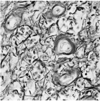
Fig. 4.The tumor contains abundant reticulin fibers outlining the islands or nests of tumor cells (reticulin stain, ×200).
Fig. 5. Results of immunohistochemical stains. (A, B) Most of the tumor cells are strongly reactive for GFAP, and their cellular processes are evident by GFAP immunostains (×400). (C) Some spindle cells are GFAP-negative but vimentin-positive (×400).
A B C
mal and glial elements, gliosarcoma is the most common and well-recognized lesion comprised of a malignant astrocytic component (glioblastoma) and also a malignant mesenchymal component (sarcoma) (1, 2). The sarcomatous element is fre- quently accompanied by an increased deposition of collagen material (2). In gliofibroma with a malignant behavior, the glial component exhibits features of anaplasia, while the his- tology of mesenchymal component is consistently benign (1).
Unfortunately the term “gliofibroma’’ has been used in the lit- erature for both benign and malignant forms. Actually the prognostic factors of the tumor are still a matter of debate (1).
Nevertheless, the MIB-1 or Ki-67 antibody appears to be a marker of cell proliferation (1). The present case showed focal areas with increased celluarity in the glial element of tumor, in which the MIB-1 index was 35.8%. The MIB-1 index of
our case is the highest among the gliofibroma cases reported in the literature (1, 2, 17). With existence of high cellular area, the high Ki-67 positivity may be considered to be a possible predictor of worse prognosis.
Histologically, gliofibromas should be distinguished from other collagen-producing tumors of the central nervous system, including clear cell meningioma, gliosarcoma, and desmoplas- tic infantile astrocytoma and ganglioglioma (DIA/DIG). Clear cell meningioma is easily excluded by the negative EMA stain- ing of the tumor cells. In addition, the cells have no PAS-posi- tive cytoplasmic glycogen. Gliosarcoma can be distinguished by the presence of a malignant mesenchymal element. Gliofi- broma may be included under a broader category of desmoplas- tic astrocytic tumors with DIA/DIG (2, 13, 19). In spite of their striking similarities, there are also several differences,
Authors, Year (Reference)
Case No.
Age (yr),
Sex Location of tumor Pathologic findings Treatment Outcome
(duration of follow-up) Friede 1978 (6) 1 3.7, F Lower medulla Dedifferentiation in the glial RT/chemoT No surgery
component Died 3 mos
after presentation Budka and Sunder-Plassmann 2 45, F Cervical spinal cord Moderately increased Surgery (GTR) Alive (1 yr)
1980 (7) cellularity in the glial component
Iglesias et al. 1984 (5) 3 11 days, M Thoracic spinal cord Benign gliofibroma Surgery (GTR) Alive (4 yr)
Reinhardt et al. 1984 (8) 4 16, F Rt temporal lobe NA Surgery Alive (6 mos)
Bonin et al. 1990 (9) 5 32, F 4th ventricle NA NA NA
Snipes et al. 1991 (10) 6 2 mo, F Thalamus, post. Increased MFs in the Surgery (STR) Died (16 mos)
fossa glial component
Vazquez et al. 1991 (4) 7 9, F spinal cord (C, T) Benign gliofibroma Surgery (STR)/RT Died (1.5 yr) 8 5.5, M spinal cord (T, S) Benign gliofibroma Surgery (GTR)/RT Alive (2.5 yr) 9 11 mo, F Rt temporal lobe Foci of pleomorphism and Surgery (GTR) Alive (2 yr)
numerous MFs in the glial component
Schober et al. 1992 (11) 10 18, M Rt frontal lobe Small foci of anaplasia and Surgery (GTR) Alive (7 days) giant cells in the glial component
Iglesias-Rozas et al. 1992 (12) 11 14 mo, F Lt frontoparietal lobe NA Surgery Alive (18 mos) Rushing et al. 1993 (13) 12 6 mo, F Posterior fossa Benign gliofibroma Surgery (GTR) Alive (2 yrs) Cerda-Nicolas et al. 1993 (14) 13 9, M Lt parietal lobe Benign gliofibroma Surgery (GTR) Alive (5.5 mos)
14 4, F 4th ventricle Foci of increased cellularity & NA NA giant cells in the glial component
Windisch et al. 1995 (15) 15 5 mo, M T10-11 Benign gliofibroma, abundant Surgery (STR) Alive (7 mos) small thick-walled vessels
Caldemeyer et al. 1995 (3) 16 8, M Temporal lobe Numerous MFs and increased chemoT NA cellularity in the glial component
17 6 mo, F Cerebellum Benign gliofibroma Surgery (GTR) NA
Prayson, 1996 (2) 18 3 mo, M Lt frontoparietal lobe Benign gliofibroma 0.9% of PI Surgery (STR) Alive (31 mos)
Molenkamp et al. 1998 (16) 19 NA NA NA NA NA
Sharma et al. 1998 (1) 20 24, F T6-8 Benign gliofibroma 0% of PI Surgery (STR) Alive (2 yr) 21 10, M Temporal lobe Benign gliofibroma 0% of PI Surgery (GTR) Alive (3 mos) 22 54, F Rt parietal lobe Increased cellularity and Surgery (GTR)/RT Died (6 mos)
MFs in the glial component, 10.5% of PI
Matsumura et al. 2002 (17) 23 12, F Cervical spinal cord Increased cellularity 1% of PI Surgery (GTR) Alive (33 mos) Present case, 2003 24 25, M Lt parietal lobe Foci of increased cellularity Surgery (GTR) Alive (2 mos)
up to 35.8% of PI Table 1.Clinical findings of gliofibromas reported in the literature
NA: not available, C: cervical, T: thoracic, S: sacral, Rt: right, Lt: left, RT: radiation therapy, chemoT: chemotherapy, GTR: gross total resection, STR:
subtotal resection, yr: year, mo: month, MFs: mitotic figures, PI: proliferation index.
DIA/DIG are presented as a large cystic lesion in the superfi- cial cortex of the brain and usually affect infancy or early child- hood. DIA/DIG is considered to be a benign neoplasm (WHO grade I) with a low MIB-1 labeling index less than 5% (20).
In DIA/DIG, the mesenchymal fibroblastic element is absent.
In the present case, clear cell features of the glial component resembled those of glioneuronal tumors such as neurocytoma, which prompted us to do immunostain using the neuronal markers and electron microscopic study. However, tumor cells showed no evidence of neuronal differentiation.
The most salient feature of these desmoplastic neoplasms may be their ability to generate connective tissue elements, for which a number of hypotheses have been advanced (2). Friede (6) proposed that collagen was produced by multipotential glial/mesenchymal cells. Iglesias et al. (5) demonstrated that collagen was produced by fibroblasts. On the other hand, there also have been some theories suggesting glial cells as the source of collagen via: 1) fibroblastic metaplasia (13), 2) secondary differentiation (21), or 3) generation of growth factors resulting in a proliferation of certain mesenchymal cell types (2).
Although some authors put gliofibroma in the same catego- ry with desmoplastic astrocytic tumors (2, 13, 19), the disease is a distinct entity (1, 22). Depending upon the presence of features of anaplasia in the glial component, this tumor should be labeled as a benign or malignant gliofibroma and treated accordingly (1).
REFERENCES
1. Sharma MC, Gaikwad S, Mehta VS, Dhar J, Sarkar C. Gliofibroma:
mixed glial and mesenchymal tumour. Report of three cases. Clin Neu- rol Neurosurg 1998; 100: 153-9.
2. Prayson RA. Gliofibroma: a distinct entity or a subtype of desmoplas- tic astrocytoma? Hum Pathol 1996; 27: 610-3.
3. Caldemeyer KS, Zimmerman RA, Azzarelli B, Smith RR, Moran CC.
Gliofibroma: CT and MRI. Neuroradiology 1995; 37: 481-5.
4. Vazquez M, Miller DC, Epstein F, Allen JC, Budzilovich GN. Glione- urofibroma: renaming the pediatric “gliofibroma’’: a neoplasm com- posed of Schwann cells and astrocytes. Mod Pathol 1991; 4: 519-23.
5. Iglesias JR, Richardson EP Jr, Collia F, Santos A, Garcia MC, Redon- do C. Prenatal intramedullary gliofibroma. A light and electron micro- scope study. Acta Neuropathol (Berl) 1984; 62: 230-4.
6. Friede RL. Gliofibroma. A peculiar neoplasia of collagen forming glia- like cells. J Neuropathol Exp Neurol 1978; 37: 300-13.
7. Budka H, Sunder-Plassmann M. Benign mixed glial-mesenchymal
tumour (“glio-fibroma’’) of the spinal cord. Acta Neurochir (Wien) 1980; 55: 141-5.
8. Reinhardt V, Nahser HC. Gliofibroma originating from temporopari- etal hamartoma-like lesions. Clin Neuropathol 1984; 3: 131-8.
9. Bonnin JM, Warner JC, Turner MS. Cystic gliofibroma of the fourth ventricle. J Neuropathol Exp Neurol 1990; 49: 261A (abstr).
10. Snipes GJ, Steinberg GK, Lane B, Horoupian DS. Gliofibroma. Case report. J Neurosurg 1991; 75: 642-6.
11. Schober R, Bayindir C, Canbolat A, Urich H, Wechsler W. Gliofibro- ma: immunohistochemical analysis. Acta Neuropathol (Berl) 1992;
83: 207-10.
12. Iglesias-Rozas JR, Kraus-Huonder B, Michilli R, Tzonos S, Bader H, Chmelar C. [Intracerebral gliofibroma] Dtsch Med Wochenschr 1992;
117: 1918-22.
13. Rushing EJ, Rorke LB, Sutton L. Problems in the nosology of desmo- plastic tumors of childhood. Pediatr Neurosurg 1993; 19: 57-62.
14. Cerda-Nicolas M, Kepes JJ. Gliofibromas (including malignant forms), and gliosarcomas: a comparative study and review of the literature.
Acta Neuropathol (Berl) 1993; 85:349-61.
15. Windisch TR, Naul LG, Bauserman SC. Intramedullary gliofibroma:
MR, ultrasound, and pathologic correlation. J Comput Assist Tomogr 1995; 19: 646-8.
16. Molenkamp G, Riemann B, Kuwert T, Strater R, Kurlemann G, Scho- ber O, Jurgens H, Wolff JE. Monitoring tumor activity in low grade glioma of childhood. Klin Padiatr 1998; 210: 239-42.
17. Matsumura A, Takano S, Nagata M, Anno I, Nose T. Cervical intra- medullary gliofibroma in a child. A case report and review of the liter- ature. Pediatr Neurosurg 2002; 36: 105-10.
18. Kleihues P, Cavenee WK. World Health Organization Classification of Tumours, Pathology & Genetics Tumours of the Nervous System.
Lyon: International Agency for Research on Cancer (IARC) Press, 2000; 6-7.
19. Jay V, Edwards V, Rutka J, Mosskin M, Hwang P, Resch L. Unique desmoplastic cerebral tumor in a patient with complex partial seizures.
Pediatr Dev Pathol 1998; 1: 234-42.
20. Kleihues P, Cavenee WK. World Health Organization Classification of Tumours, Pathology & Genetics Tumours of the Nervous System.
Lyon: International Agency for Research on Cancer (IARC) Press, 2000; 101-2.
21. Taratuto AL, Monges J, Lylyk P, Leiguarda R. Superficial cerebral astrocytoma attached to dura. Report of six cases in infants. Cancer 1984; 54: 2505-12.
22. Lang FF, Miller DC. Astroglial variant. In: Black PM, Loeffler JS, editors, Cancer of the Nervous System. Cambridge: Blackwell Science, 1997; 516-48.