An Efficient Method for Production of Extracellular Human Tissue Factor in Escherichia coli
Hwan Goo Yoo, Yang Jin Park and Woo-Yiel Lee
1*
Department of Chemistry, 1Department of Pharmaceutical Engineering, Konyang University, Nonsan, ChungNam 320-711, Korea Received February 18, 2009 /Accepted April 20, 2009
Human Tissue factor is an essential enzyme activator that forms a catalytic complex with factor VII/
VIIa, and catalyzes both the extrinsic and intrinsic blood coagulation cascades. The extracellular do- main of human tissue factor is responsible for association with the biological partner. The efficient procedures for preparing biologically active human tissue factor are essential for the preclinical and clinical studies with coaguligands. An expression vector in Escherichia coli has been constructed to di- rect the production of extracellular human tissue factor without a fusion protein or a His
6at the N-terminus. The recombinant human tissue factor was expressed in large amounts as a non-native state in E. coli. The recombinant protein was simply renatured during the DEAE-sephacel chromato- graphic purification procedure. Our expression and purification system does not require a protease treatment or an additional chromatographic step to remove a fusion contaminant, which provides a very useful alternative to conventional expression systems for the production of human tissue factor.
Key words : Extracellular domain, expression system, human tissue factor, purification, refolding
*Corresponding author
*Tel:+82-41-730-5694, Fax:+82-41-730-5762
*E-mail : [email protected]
Introduction
Blood coagulation is initiated by the complex of tissue factor (TF) and coagulation factor VIIa (FVIIa) which has a proteolytic activity [4]. The complex ensures normal haemo- stasis but is also involved in the pathogenesis of different disease processes, such as septic shock, thrombotic vascular disease, and cancer metastasis [5]. TF is a membrane bound glycoprotein which consists of three distinct domains: an ex- tracellular domain (1-219), a hydrophobic transmembrane region (220-242), and a cytoplasmic tail (243-263) [6]. The extracellular domain containing two disulfide bonds is re- sponsible for the forming a complex with FVII or FVIIa and for the proteolytic activation of factor IX and X [8]. Several strategies have been employed for the expression of human TF. A truncated form of TF (tTF), consisting of the ex- tracellular domain, has been expressed in a non-native state in E. coli inclusion bodies as a fusion protein with poly- histidine residues at the N-terminus [7]. The study to sim- plify the procedures including the renaturation and purifica- tion of recombinant tTF in E. coli has been carried out by introducing on-column refolding method, in which the re- combinant protein has been refolded while immobilized on Ni
2+-nitriloacetic acid (NTA) column [2]. Recently, the fusion
system with maltose binding protein has been described to improve the yield and easy of purification of recombinant tTF [3]. A major drawback of these approaches is that an enzymatic cleavage and an additional purification step for removing the contaminants resulted from the enzymatic treatment must be applied to purify the recombinant tTF as a biologically active form, which are time-consuming and high-cost procedures.
The efficient procedure for preparing the large quantities of recombinant TF is necessary for the production of medical proteins or other industrial proteins. In this report, we de- scribe an efficient preparation of the catalytic domain of hu- man TF from a new E. coli expression system without a fu- sion protein or His
6tag, which does not require the proce- dure of an enzymatic cleavage and the additional chromato- graphic step.
Materials and Methods Construction of a recombinant plasmid pRX-tTF
An expression vector was constructed from pRX, a de- rivative of pRSET (Invitrogen, Carlsbad, CA), in which six His, epitope, and enterokinase cleavage site were removed.
The cDNA coding for human TF was amplified by polymer-
ase chain reaction (PCR) using the primers (GenoTech,
Deaduk, Korea) 5’-CGGGATCCGGCACTACAAATACTGT
(5’ primer) and 5’-TGTAAGCTTATTCTCTGAATTCCCCTT
(3’ primer) containing a BamHI and a HindIII restriction site (indicated by underline), respectively. The PCR product was cloned into pRX digested by BamHI/HindIII, generating plas- mid pRX-tTF. The deduced amino acid sequence of the clone contains the authentic sequence (residues 1-219) of the ex- tracellular domain of human TF. Taq DNA polymerase, DNA ligase, and restriction enzymes were purchased from Promega (Seoulin Bioscience, Seoul, Korea). The N-terminal sequencing of proteins was performed by Edman degrada- tion (Korea Basic Science Institute, Daejeon, Korea). All other chemicals were purchased from commercial sources and were specific grade or first grade reagents.
Expression of recombinant tTF in E. coli
The E. coli strain BL21 (DE3) (Novagen, Darmstadt, Germany) was transformed with the resulting plasmid car- rying tTF DNA. Single colonies were grown at 37
oC in LB medium (500 ml) containing 100 µg/ml ampicilin. When the absorbance at 595 nm reached about 0.5, 0.5 ml of 1 M iso- propyl β-D-thiogalactopyranoside (IPTG) was added. The culture was shaken overnight at 37
oC at 250 rpm. The cells were harvested by centrifugation at 10,000 ×g for 20 min.
SDS-PAGE was performed on 12% polyacrylamide gels for the analysis of proteins.
The cell (4.5 g wet weight) was washed with 100 ml of 10 mM Tris-HCl buffer (pH 7.5) containing 20% sucrose and 1 mM EDTA. The cells were resuspended in 100 ml of Tris-HCl buffer (pH 7.5) containing 250 mM NaCl, 1% Triton X-100, 1mg lysozyme (Sigma, Saint Louis, MO), 1 mM MgCl
2and 1 mM EDTA, and disrupted by sonication. The mixture was stirred gently for 1 hr at 4
oC and centrifuged at 10,000×
g for 20 min. The pellet paste was washed twice with water.
The pellet were resuspended in 50 ml of Tris-HCl buffer (pH 7.5) containing 50 mM NaCl and 1% Triton X-100, agitated by stirring for 1 h at 4
oC, and centrifuged at 10,000× g for 20 min. This procedure was repeated by two times. The final pellet was the highly enriched inclusion bodies of recombi- nant tTF.
Purification of the recombinant tTF.
The inclusion bodies of recombinant tTF were re- suspended in 100 ml of buffer S (6 M urea, 10 mM Tris-HCl, pH 8.0). The solution was stirred for 1 hr and centrifuged at 30,000× g for 30 min at 4
oC. The supernatant was loaded onto 100 ml DEAE-Sephacel column which had been equili- brated with buffer S. The column was washed with 100 ml
of buffer S and then with a linear gradient solution from 6 M to 0 M urea (200 ml each) in 10 mM Tris-HCl buffer (3 mM GSH, 1 mM GSSG, 0.02 % NaN
3,pH 8.0) for refolding recombinant protein. The refolding procedure was per- formed for 3-5 hr. Elution was carried out with a gradient of 0 - 1.0 M NaCl in 10 mM Tris-HCl buffer (pH 7.5). A flow rate of 3 ml/min was performed and the protein was monitored by absorbance at 280 nm. The eluted protein sol- ution was concentrated using Amicon (Millipore, Billerica, MA) with YM-10 filter, and dialyzed against 50 mM Tris-HCl buffer (pH 7.5). The protein concentration was de- termined by Bradford method with bovine serum albumin as standard protein [1].
Activity assay of recombinant tTF
The catalytic activity of the refolded tTF was assayed using a peptidyl chromogenic substrate, Spectrozyme FXa, (MeO- CO-D-CHG-Gly-Arg-pNA.AcOH, American Diagnostica, Greenwich, CT), as previously described [4]. A commercially available TF (designated as standard tTF, provided by Dr.
Lee at ATGene Research Institute, Daeduk, Korea) was used for the comparison with the recombinant tTF. In a microtiter plate, tTF was incubated at room temperature for 10 min with 0.1 µM of FVIIa (Novo Nordisk, Bagsværd, Denmark) in Tris-HCl buffer (pH 7.5) containing 5 mM CaCl
2and 0.1%
Bovine serum albumin (BSA). Spectrozyme FXa was added to the assayed mixture at a final concentration of 0.5 mM and the catalytic activity was determined by measuring the increase in absorbance of the free pNA generated per min at 405 nm. One unit of the activity was defined as the amount of protein to produce an increase of 0.1 O.D in ab- sorbance at 405 nm.
Results Expression of recombinant TF in E. coli.
An expression vector, pRX-tTF (Fig. 1), encoding the ex- tracellular Human TF was constructed from pRSET vector.
The plasmid does not have a fusion protein or His
6tag, and carries the authentic sequence (residues 1-219) of the cata- lytic domain of human TF except for having extra methio- nine and glycine residues in front of the N-terminal serine.
The stop codon (TAA) was included in the 3’-primer so that
the C-terminus of tTF protein should be glutamic acid which
is the C-terminal residue of the genuine extracellular
domain. The N-terminal sequence of the expressed tTF was
ATG GGA TCC GGC ACT ACA AAT ACT GTG RBS
M G S G T T N T V
tHTF
pRX-tTF
F1 ori
Ampr ColE1
P
T7 RBS ATG GGA TCC GGC ACT ACA AAT ACT GTG M G S G T T N T VtHTF
pRX-tTF
F1 ori
Ampr ColE1
P
T7Fig. 1. Expression vector pRX-tTF. The first line is the coding sequence showing the starting codon and BamHI site (indicated by overline) at the N-terminus of tTF. The sec- ond line is the translated amino acid sequence for this region. The underline indicates the amino acid sequence of the cDNA of tTF.
demonstrated to have the expected sequence by Edman degradation. The expression of the tTF was induced by IPTG at 1 mM. The recombinant tTF was expressed in large quan- tity in the transformed E. coli. SDS-PAGE analysis of the cul- tured cells revealed a predominant protein with an apparent molecular size of about 30 kDa (Data not shown).
Purification and renaturation of tTF
The majority of the tTF was produced in inclusion bod- ies in aggregated form. Since the physical and chemical properties of the insoluble aggregates in the inclusion bod- ies are distinct from those of the endogenous proteins of the host, their purification has been facilitated. The in- clusion bodies were carefully isolated by repeated washing.
The recombinant tTF in non-native state was highly puri- fied by DEAE-Sephacel column chromatography. The re- folding of the recombinant protein was carried out on the column by running a linear gradient of 6 M-0 M Urea.
Elution was carried out with a gradient of 0-1.0 M NaCl.
Fractions containing the amidolytic activity in presence of FVIIa, which showed one sharp peak, were collected (Fig.
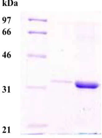
2). Yields after the column were 30-50 mg per one liter of cell culture (Table 1). The purity of the tTF is comparable to that of standard tTF as judged by SDS-PAGE analysis (Fig. 3). The slower movement of standard tTF (Fig. 3, lane 2) suggests that its commercial form may not be completely denatured, or may be slightly larger than the recombinant in real size.
3.0
2.0
1.0
5 4 3
2 1
1 10 20 30 40 50 60 70 80 Fraction number
O.D. at 280 nm Activity (units/mL) NaCl(M)
1.0
0.5
0.0 3.0
2.0
1.0
5 4 3
2 1
1 10 20 30 40 50 60 70 80 Fraction number
O.D. at 280 nm Activity (units/mL) NaCl(M)
1.0
0.5
0.0
Fig. 2. DEAE-Sephacel column chromatogram. Elution was car- ried out with a gradient of 0 - 1.0 M NaCl in 10 mM Tris-HCl buffer (pH 7.5). Symbols: ---, NaCl gradient;
■, O.D. at 280 nm, ▲, the amidolytic activity in presence of FVIIa.
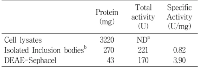
Table 1. Purification of recombinant tTF based on one liter E. coli culture
Protein (mg)
Total activity
(U)
Specific Activity (U/mg) Cell lysates
Isolated Inclusion bodiesb DEAE-Sephacel
3220 270 43
NDa 221 170
0.82 3.90
aNot determined
bThe inclusion bodies was refolded before measuring the activ- ity: The highly enriched inclusion bodies were solubilized in buffer (pH 8.0) containing 6 M guanidinium chloride, 0.5 M NaCl, and 20 mM Tris-HCl by gentle stirring for 4 h at room temperature. The refolding of recombinant tTF was accom- plished by modified procedures based on the previously de- scribed method [7].
Verification of activity of purified recombinant tTF
The catalytic activity of the recombinant tTF prepared in this study was compared with a standard tTF. The hydrol- ysis activity of FVIIa toward chromogenic substrate Spectrozyme FXa was investigated over a range of concen- trations of tTF between 1 nM and 10 µM. The assay was accomplished in the presence of 0.1 µM of FVIIa. The tTF obtained in this study was not distinguishable from the standard tTF (Fig. 4).
Discussion
We have developed a new expression system without a
fusion partner or polyhistidine tag which produces tTF at
high level in E. coli and have shown that the recombinant
protein was efficiently purified by DEAE-Sephacel column
kDa 97 66 46
31
21
Fig. 3. SDS-PAGE analysis of tTF purification. The electro- phoresis was performed on 12% polyacrylamide gels.
Lane 1, molecular weight markers; Lane 2, standard tTF;
Lane 3, final tTF product obtained by DEAE-Sephacel chromatography. The band of each lane at the bottom of the gel is trace of dye.
log [tTF]
Absorbance/min at 405 nm
-9 -8 -7 -6 -5
0.00 0.05 0.10 0.15 0.20 0.25
log [tTF]
Absorbance/min at 405 nm
-9 -8 -7 -6 -5
0.00 0.05 0.10 0.15 0.20 0.25
Fig. 4. Activity assay of recombinant tTF. Closed circle, tTF puri- fied as described in the present study; Open circle, stand- ard tTF. The X-axis is logarithm of molarity.
chromatography and successfully refolded on the column.
According to our knowledge, the tTF described here is comparable to those of the other expression systems re- ported in terms of expression level, purity, and biological activity. The expression system in this study has the ad- vantage over other tTF expression system. The tTF ex- pression level described here is one of the highest among published other tTF expression system. Our on-column re- folding procedure is simple and comparable to the method published by Gao et al. [3]. However, the present purifica- tion system does not require a protease treatment and an
additional chromatographic step to remove a fusion contaminant. Our expression and purification system allow a high level and quality production of tTF, which provides a very useful alternative to conventional expression sys- tems for the production of tTF.
Acknowledgments
This work was financially supported by the Ministry of Education, Science and Technology (MEST) and Korea Industrial Technology Foundation (KOTEF) through the Human Resource Training Project for Regional Innovation.
References
1. Bradford, M. A. 1976. Rapid and sensitive method for quan- tification of microgram quantities of protein utilizing the principle of protein-dye binding. Anal. Biochem. 72, 248-254.
2. Gao, B., S. Li, and P. E. Thorpe. 1998. A simple and rapid method for purifying the extracellular domain of human tissue factor. Thromb. Res. 91, 249-253.
3. Guan, M., B. Su, C. Ye, and Y. Lu. 2002. Production of ex- tracellular domain of human tissue factor using mal- tose-binding protein fusion system. Protein Expr. Purif. 26, 229-234.
4. Ruf, W., M. W. Kalnik, T. Lund-Hansen, and T. S.
Edgington. 1991. Characterization of factor VII association with tissue factor in solution. J. Biol. Chem. 266, 15719-15725.
5. Ruf, W., J. Shobe, S. Rao, C. D. Dickinson, and T. S.
Edgington. 1999. Importance of factor VIIa Gla-domain resi- due Arg-36 for recognition of the macromolecular substrate factor X Gla-domain. Biochemistry 38, 1957-1966.
6. Spicer, E. K., R. Horton, L. Bloem, R. Williams, A. Guha, J. Kraus, T. C. Lin, Y. Nemerson, and W. H. Konigsberg.
1987. Isolation of cDNA clones coding for human tissue fac- tor: primary structure of the protein and cDNA. Proc. Natl.
Acad. Sci. USA 84, 5148-5152.
7. Stone, M. J., W. Ruf, D. J. Miles, T. S. Edgington, and P.
E. Wright. 1995. Recombinant soluble human tissue factor secreted by Saccharomyces cerevisiae and refolded from Escherichia coli inclusion bodies: glycosylation of mutants, activity and physical characterization. Biochem. J. 310, 605-614.
8. Waxman, E., J. B. A. Ross, T. M. Laue, A. Guha, S. V.
Thiruvikraman, T. C. Lin, W. H. Konigsberg, and Y.
Nemerson. 1992. Tissue factor and its extracellular domain:
The relationship between intermolecular association with factor VIIa and enzymatic activity of the complex.
Biochemistry 31, 3998-4003.