지구성 운동이 STZ-당뇨 유발 쥐의 허혈/재 관류 손상 후 심근의 당대사 관련 표현형 단백질과 HSP-60 단백질 발현에 미치는 영향
배희숙․엄현섭․강은범․양춘열․이용로․이창국1․천우호1․전혜자1․조인호․조준용*
한국체육대학교 체육과학관 운동생화학실, 1순천향대학교 체육학과
Received February 20, 2009 /Accepted May 22, 2009
The Effect of Exercise Training (EXE) on Myocardium Glucose Metabolic Phenotypic Proteins and HSP-60 Protein Expression after Ischemia/Reperfusion Injury in STZ-induced Rats. Hee-Suk Bae, Hyun-Seob Um, Eun-Bum Kang, Chum-Yeol Yang, Yong-Ro Lee, Chang-Guk Lee
1, U-Ho Cheon
1, Hye-Ja Jeon
1, In-Ho Cho and Joon-Yong Cho*. Department of Exercise Biochemistry Laboratory, Korea National Sport University, Seoul 138-763, Korea,
1Department of Physical Education, Soon Chun Hyang University, Cheonan 336-745, Korea - The objective of this study was to identify EXE (1 hr a day at 21 m/min for 5 day/wk, at 0 % grade for 6 wk) on myocardium glucose metabolic phenotypic proteins (AMPK-PGC-1α-GLUT-4) and HSP-60 protein expression after ischemia/reperfusion injury (IRI) in STZ-induced rats. EXE was performed using STZ-induced diabetic rats on a rodent treadmill (28 m/min, 1 hr/day, 5 day/wk for 6 wk). The results of this study suggest that i) serum insulin level was not changed among groups (p>0.05). ii) the LVDP level increased significantly in the STZ-EXE-IRI group compared to the STZ-IRI group at 60 min (p<0.01), 70 min (p<0.05) and 80 min (p<0.05) after reperfusion, respectively, and iii) AMPK phosphorylation (p<0.01), PGC-1α protein (p<0.001), GLUT-4 protein ( p<0.001) and HSP-60 protein expressions (p<0.05) increased significantly in the STZ-EXE-IRI group compared to the STZ-IRI group. In conclusion, the findings of the present study reveal that EXE may provide therapeutic value to insulin dependent diabetic patients with peripheral insulin re- sistance and myocardium injury by improving glucose metabolic proteins (AMPK-PGC-1α-GLUT-4) and heat shock protein-60 (HSP-60), along with increasing LVDP levels and decreasing glucose levels.
Therefore, EXE protects the STZ-induced diabetic myocardium injury against ischemia/ reperfusion injury.
Key words : Exercise training, ischemia/reperfusion injury, AMPK, PGC-1α, GLUT-4, HSP-60
*Corresponding author
*Tel:+82-2-410-6867, Fax:+82-2-410-6700
*E-mail : [email protected]
서 론
당뇨병은 산화적 스트레스로 인한 세포내 방어 시스템 손 상을 초래하여 세포사멸을 가속화시켜 심근경색으로 인한 당 뇨환자들의 사망률을 증가시킨다[2,11,42]. 특히, 당뇨병은 심 근 세포내 단백질의 3차 구조를 유지시켜 세포손상을 최소화 하고 항상성 유지에 중요한 역할을 하는 분자 샤페론의 일종 인 열충격 단백질(heat shock proteins)합성의 감소와 당대사 관련 단백질인 AMPK (AMP-activated protein kinase), PGC-1α (peroxisome proliferator activated receptor co- activator-1α)와 GLUT-4 (glucose transporter-4)의 발현량을 감소시켜 심근의 세포손상과 당 대사 저하를 유발시킨다[14, 19,25,27,36,41,48,49,51].
심혈관질환들 중 관상동맥질환의 병리학적 증후는 허혈 (ischemia)에 의한 심근 손상으로 보고되고 있다[43]. 허혈에 의한 심근의 손상은 심장 기능의 저하로 인한 심근 세포 사
멸을 유도하며[40] 특히 당뇨병성 심장은 허혈 손상에 가장 민감하게 반응하는 것으로 당뇨 질환 모델의 동물 실험을 통 하여 보고되고 있다[11,42]. 이러한 당뇨 성 심장질환 개선을 위한 방법으로 심장의 허혈성 전 처치(ischemic pre- conditioning)에 의한 심근보호 효과에 대해 여러 학자들이 관심을 갖고 연구들을 수행해왔다. 허혈성 전 처치란 심근세 포의 허혈에 대한 방어효과로서 짧은 기간 동안 반복적인 심 근의 허혈과 재 관류처치는 이어지는 긴 시간동안의 허혈 상 황에서도 허혈에 대한 내성이 생겨 심근세포의 괴사를 막는 다고 보고하였다[32].
허혈성 전 처치에 의한 심근보호 효과는 인위적인 처치 없 이 심근에 내재되어 있는 자연적인 자기 보호기전의 발현에 의해 허혈 손상을 예방하거나 감소시키는 것으로 고혈압, 당 뇨 및 심장질환으로 인한 세포사멸과 심장의 손상방지와 같은 연구에서 볼 수 있다[31,37,44]. 특히, 허혈성 전 처치에 의해 당뇨 성 심장에서도 심근세포의 괴사 크기가 감소되었다고 보고한 Kristiansen 등[22]의 연구는 주목할 만하다.
그러나 최근에는 앞서 제시된 허혈성 전 처치에 의한 심근
보호효과 이외에 전 처치 효과로서 다른 대체 자극이 심근
보호효과를 발휘하는지에 관심을 갖게 되었다. 그 대체 자극
으로 약리적인 자극[10,14,16,18,20,46]과 고온 처지에 의한 자 극[9,30,52]이 허혈성 전 처치와 유사한 효과를 나타낸다고 보 고되어왔으며 특히, 운동이 전 처치로서 심근보호 효과를 발 휘한다고 보고되었다[39].
운동이 허혈성 전 처치 효과와 유사한 효과를 발휘한다는 이론적 근거는 운동 자체가 인체 내에서 산화질소와 과산화물 음이온(O
2-˙: superoxide) 생성 및 아데노신, 브래디키닌도 생 성한다는 사실[4]로 운동에 의해 발생하는 이러한 생성 물질들 이 허혈성 전 처치에 의해 생성되는 신호유발 단계의 물질과 동일하고 허혈성 전 처치 시 신호전달경로에서 나타나는 세포 의 적응 효과와 운동 시 나타나는 조직 및 세포의 적응효과(산 소섭취량과 항산화효소 활성 증가)가 유사하기 때문이다[38].
또한 유산소 운동은 당뇨병의 가장 핵심적인 문제인 인슐린 표적세포의 인슐린저항성과 췌장 β세포 기능장애의 신호 경 로를 조절하는 AMPK가 발현하는데 영향을 미치는 것으로 보고되고 있으며 Coven 등[8], Abel 등[1]은 운동이 인슐린 표적세포내의 AMPK의 활성화를 통해 phosphoinositides 경 로와는 별개의 독립된 경로를 통하여 PGC-1α (Peroxisome proliferator activated receptor γ/PPAR γ coactivator-1α)의 신 호전달을 통한 GLUT-4 (glucose transporter-4)의 세포막 이동 이 증가, 포도당 이용률이 증가됨을 보고하였다
그러나 아직 운동이 전 처치(exercise preconditioning)로서 허혈성 전 처치와 유사하게 고혈압, 당뇨병 및 심혈관계 질환 자들의 심장보호 효과를 발휘하는지는 명확하게 규명되지 않 은 상태이며 특히 심근의 당 대사 기능 저하와 산화적 스트레 스로 인한 세포내 방어 시스템 손상과 함께 세포사멸이 가속 화되어 심근경색 유발로 인한 당뇨환자들의 사망률이 증가하 고 있는 측면에서 볼 때 당뇨질환모델동물을 활용하여 전 처 치로서 운동에 의해 유발되는 심장 보호 효과를 규명하는 연 구는 중요하다고 하겠다.
따라서 이 연구에서는 전 처치로서 지구성 운동이 당뇨 조건 하에서 허혈/재 관류 후 심근의 손상과 당대사 기능 저하를 개 선시키는데 효과적인지를 규명하기 위해 STZ (streptozotocin)- 유도 당뇨 쥐를 활용하여 지구성 운동이 허혈/재 관류 상해 (Ischemic-reperfusion injury; IRI)후 심근의 생리적 기능 지표 인 좌심실내압과 당 대사 관련 표현형 단백질(AMPK-PGC-1α -GLUT-4) 및 심근에서 특이적으로 발현되며 조직을 보호하는 단백질인 열충격 단백질(HSP-60)의 발현량을 분석하였다.
재료 및 방법
실험 동물
이 연구에서 활용한 실험모델동물은 코아텍(KOATHCH) 실험동물센터에서 생산된 생후 6주령 된 Sprague- Dawley (SD)계 수컷 흰쥐(280±4.38 g)로 구입 후 1주간 환경적응 기간 과 1주간 운동적응 훈련을 거친 후 본 운동으로 6주간 지구성
운동을 실시하였다. 실험 6주 동안 동일한 사육 조건(온도 20±2
oC, 습도 45%, 주간전등: 07:00-19:00, 야간소등: 19:00- 07:00)을 갖춘 H대학교 동물실에서 Guiding Principles for the Care and Use of Animal (1964)의 지침에 따라 케이지 당 2마 리씩 넣어 사육하였으며 실험기간 동안 미국영양학회에서 발 표된 AIN-76A 실험동물용 식이방법에 따라 제조된 사료 (5057-Purina)를 제공하였고 식이량과 수분 섭취량은 제한 없 이 공급하였다. 집단은 당뇨 집단(STZ, n=8), 당뇨 허혈/재관 류 상해집단(STZ-IRI, n=8)과 당뇨 운동전처치 허혈/재관류 상해집단(STZ-EXE-IRI, n=8)으로 무선 배정하여 구분하였다.
당뇨 유도
환경 적응훈련 1주 중 본 실험에 들어가기 3일 전에 췌장의 β세포만을 특이적으로 파괴시키는 STZ (streptozotocin mixed anomers, SO130-SIGMA, USA)을 50 mg/kg을 10 mM citrate acid buffer (pH 4.5)에 녹인 후 복강에 1회 주사하여 당뇨를 유도하였다. 당뇨 유발 확인은 STZ 주사 후 48시간∼
72시간 사이 공복상태에서 꼬리 정맥으로부터 혈액을 채혈하 여 혈당 수준이 300 ml/dl 이상인 흰쥐를 사용하였다.
운동 방법
운동 프로그램은 [6]이 제시한 운동 프로그램을 본 실험에 맞게 수정하여 사용하였다. 먼저 트레드밀 사전 적응 훈련으 로 설치류용 트레드밀(DJ2-242, Dual Treadmill-Dae-Jong machine, Korea)을 활용하여 분당 15 m 속도로 1일 10분간 5일간 트레드밀 런닝을 실시하였으며 본 운동 시 운동방법은 6주간, 주 5일, 1일, 60분 운동량으로 분당 25 m 속도로 10분, 분당 28 m 속도로 40분, 마지막 분당 20 m 속도로 10분간 실시하였다.
허혈/재 관류
당뇨 심장의 허혈/재 관류 손상에 대한 생리적인 반응을
연구하고자 STZ-IRI와 STZ-EXE-IRI의 경우, pentobarbital (50
mg/kg)로 마취시킨 후 헤파린(1,000 U/kg i.v.)을 투여하여
[35]의 방법에 따라 심장을 적출한 후 기관(trachea)에 도관
(cannula (PE 240))를 삽입하고 설치류 호흡계(rodent ven-
tilator)를 활용해 인공 호흡시켜 in situ 상태에서 대동맥 can-
nula를 통한 역행성 관류 하에 Langendorff perfusion system
에 재빨리 매달았다. 그 후 좌심실 압 측정을 위해 압력 변압기
(pressure transducer, Power lab AD Ins, Australia)에 연결된
cannula transducer의 고무풍선에 생리식염수를 채우고 금속
cannula를 폐정맥을 통해 좌심실에 삽입시켜 좌심실 압력(left
ventricular developed pressure)은 8-10 mmHg로 주고 생리
적(고유의) 압력(physiological pressure (model, MLT 844,
AD Ins))를 이용해 정압 관류(75~80 mmHg)하여 15분 이내
안정화되는 심장만을 사용하였다. 심장을 Langendorff perfu-
cardiac extraction
↓ STZ
(n=8) STZ-injection Stabilization Ischemia Reperfusion
STZ-IRI 20 min 20 min 40 min
(n=8) STZ-injection Stabilization Ischemia Reperfusion
STZ-
EXE-IR Exercise (6 wk) 20 min 20 min 40 min
(n=8) STZ-injection Stabilization Ischemia Reperfusion
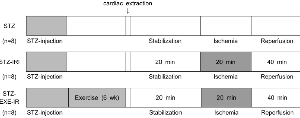
Fig. 1. Experimental protocol outlining the procedures performed in the three groups of rats. STZ-non ischemia/reperfusion control group (STZ), STZ-ischemia/reperfusion injury group (STZ-IRI), STZ-ischemia/reperfusion injury exercised group (STZ-EXE-IRI).
sion system (Power lab AD Ins, Australia)에 고정하는 순간 부터 실험을 실시하는 전체 과정 동안 생리식염수가 담긴 원 형수조에 심장이 충분히 잠기게(약 1 cm)한 후 온도가 38±0.5
oC로 철저히 유지되도록 하여 심장을 in vitro 상태에서 안정화시켜 정상적으로 심장의 기능을 발휘할 수 있도록 modified Krebs- Henseleit bicarbonate buffer (mM: NaCl, 110; KCl, 4.7; MgSO
4, 1.2; KH
2PO
4, 1.2; NaHCO
3,25; CaCl
2, 3.2; glucose, 11; Na-EDTA, 0.5)를 활용하며 실험을 진행하는 동안 O
295%, CO
25%의 혼합가스를 심장에 지속적으로 공급 해 주어 pH를 7.4로 유지시켰다. 이후 심박수, 정압(constant pressure), 관류량(flow rate, ml/min)이 안정화가 되었을 때 인위적인 허혈을 20분간 유도한 후 40분간 재 관류를 통하여 생리적 변인들을 측정하였다. 실험 프로토콜은 Fig. 1과 같다.
혈청 포도당 및 인슐린 농도 분석
모든 실험동물들은 pentobarbital sodium (50 mg/kg)을 복 강을 에 투여, 마취시킨 후 개복하여 좌심실에서 혈액 8 ml를 채혈한 후 혈청 분리 튜브로 옮겨 30분 정도 실온에서 방치 한 후 원심분리(3,000 rpm, 15분, 4
oC)하여 혈청을 분리하였다.
혈청 포도당은 생화학 분석기(EKTACHEM DT 60II, USA)를 이용하여 분석하였으며, 혈청 인슐린 농도는 enzyme im- munoassay ELISA 분석용 kit (Mercodia AB, Uppsala, Sweden)을 이용하여 분석하였다.
조직 표본 준비
모든 실험처치를 끝낸 집단들의 심장을 액화질소로 동결시 킨 후 분석 시까지 -70
oC의 초저온 냉동기(Bio-Freezer, Forma Scientific)에 보관한 후 western blot 분석을 위해 protein ex- traction solution (Cat No. 17081-iNtRON, Korea) 1 ml에 심장 의 좌심실 조직을 균질화(homogenizer, Pyrex Corning, USA) 시킨 후 14,000 rpm으로 10분간 4
oC에서 원심분리(Centrifuge
5417R- effendorf, Germany)하였다. 원심분리 후 펠렛(pellet) 을 제외한 상층부분을 따로 분리하여 분석 시까지 -70
oC 초저 온 냉동기에 보관하였다.
SDS-PAGE
총 단백질량은 BSA (bovine serum albumin, 570 nm)를 이 용하여 Bradford (1976)의 방법으로 정량하였다. 10% separa ting gel (3DW, 30% acrylamide: bisacrylamide, 1.5M tris pH 8.8, 10% SDS, TEMED, 10% ammonium persulfate)과 5%
stacking gel (3DW, 30% acrylamide: bisacylamide, 1M tris pH 6.8, 10% SDS, TEMED, 10% ammonium persul fate)을 만들어 사용하였다. 원심분리(14,000 rpm, 10 min)한 상층액 과 5XSDS Sample loading buffer (60mM tris pH 6.8, 25%
glycerol, 2% SDS, 14.4mM 2-mercaptoethanol, 0.1%
Bromophenol blue)를 잘 혼합한 후 100
oC에서 10분간 끓여 단백질을 변성시킨 후 10분간 얼음에 방치한 후 다시 14,000 rpm으로 10분간 4
oC에서 원심 분리하고 스탠다드 마커 (Pagerular Prestained Protein Ladder, #SM0671- Fermertas) 와 함께 각 샘플을 Mini-Protein Ⅱ dual-slab apparatus (Bio-Rad, CA, USA)에 준비된 stacking gel well에 총 단백질 량이 30 ug이 되도록 분주하고 80 volt에서 2시간 정도 샘플이 바닥에 닿을 때까지 전기영동 하였다.
Western blot
Methanol에 적신 PVDF membrane (Bio-rad, USA)과
Transfer buffer (190mM glycine, 50mM Tris-base, 0.05% SDS,
20% methanol)로 적신 3M paper (Whatman)를 차례로 겹쳐
Mini trans-blot cell (Bio-Rad, CA, USA)에 장치한 후 60 volt
로 2시간을 전사하고 Membrane으로 증착이 끝나면 rocker
platform 위에서 90분 동안 membrane을 3% skim milk 용액
(in TBS-T: 10 mM Tris-base, HCl-pH 7.6, 0.5 M NaCl, 0.05%
Table 1. Effect of exercise training on serum glucose, insulin in the STZ-induced diabetic rat Group Initial Body (Wt, g) Final Body (Wt, g) Change in
Wt/day, g/day Glucose (mmol/l) Insulin (Uu/ml) STZ
STZ-IRI STZ-EXE-IRI
215.40±5.55 218.17±6.37 215.00±6.12
189.80±12.64 217.17±42.93 197.40±47.22
0.61±0.02 0.02±0.05a 0.42±0.06b
44.67±2.36 45.01±1.08 29.31±1.13ab
5.03±1.04 4.07±1.09 3.91±.084 Values are means±SD for 6 animals/group, ap<0.05 vs. STZ, bp<0.05 vs. STZ-IRI
Tween-20)으로 Blocking시켰다. 이 후 1차 항체인 anti-AMPK α (#2532, Cell Signaling, USA), anti- Phospho- AMPK (#2531, Cell Signaling, USA), anti-PGC-1α (SC-13067, Santa Cruz, CA, USA), anti-GLUT-4 (#2299, Cell Signaling, USA), an- ti-HSP-60 (SC-13966, Santa Cruz, CA, USA), anti-GAPDH (SC-20357, Santa Cruz, CA, USA)를 1:1,000으로 bolcking (3%
skim milk)용액으로 각각 희석시켜 12시간 동안 흔들어 준 다음 TBS-T 용액으로 10분씩 5차례 세척한 후 2차 항체 (horseradish peroxidase-conjugated goat anti-rabbit 65- 6120, ZYMED, CA, USA; horseradish peroxidase-conjugated rab- bit anti-goat 81-1620, ZYMED, CA, USA)를 Blocking 용액으 로 1:5,000으로 희석시켜 90분 동안 흔들어 주고 난 후 TBS-T 용액으로 10분씩 5차례 세척하였다. 마지막 단계로 WBLR 용 액(Western Blotting Luminol Reagent SC-2048, Santa cruz Biotechnology, CA, USA)에 membrane을 넣고 1분간 발색하 고 얻어진 membrane을 이미지 분석 시스템(Molecular Imager ChemiDoc XRS System, Bio-Rad, USA)을 이용하여 스캔한 후 Quantity 1-D Analysis Soft ware (Bio-Rad, USA)를 이용하여 단백질량을 산출하였다.
자료 처리 방법
이 연구에서 얻어진 모든 결과는 SPSS/PC 11.0 통계 프로 그램을 이용하여, 각 변인에 대한 기술 통계치(mean±SD)를 산출하였다. 생리적 변인인 좌심실내압은 집단(3)×시간(9) 반 복측정에 의한 이원변량분석(repeated two way- ANOVA)을 실시하였으며 상호작용효과가 있을 경우 그 차이를 구체적으 로 확인하기 위하여 SNK (Student Newman- Keuls)을 이용하 여 사후검증을 하였다. 혈청 포도당, 혈청 인슐린 수준, 당대사 관련 표현형 단백질(AMPK, PGC-1α, GLUT-4)과 HSP-60 단 백질의 집단 간 차이를 확인하기 위해 일원변량분석(one way-ANOVA)을 실시하였다. 집단 간 유의한 차이가 있을 경 우 LSD (least significant difference)를 이용하여 사후 검증을 실시하였다. 이때 가설 수락 기준은 α=0.05 수준으로 하였다.
결 과
혈청 포도당, 인슐린 수준
Table 1에서 보는 바와 같이 전 처치로서 지구성 운동이
체중, 혈청 포도당과 인슐린 수준에 미치는 영향을 변량 분석 한 결과, 체중은 집단 간에 p<0.01 수준에서 차이가 있는 것으 로 나타나 사후 검증한 결과, STZ-EXE-IRI과 STZ이 각각 STZ-IRI보다 체중이 현저하게 감소한 것으로 나타났다 ( p<0.05). 혈청 인슐린 수준은 집단 간에 p>0.05 수준에서 차이 가 없는 것으로 나타난 반면 혈청 포도당 수준을 변량 분석한 결과 집단 간에 각각 p<0.05 수준에서 차이가 있는 것으로 나 타나 사후 검증한 결과 혈청 포도당 수준의 경우, STZ-EXE-IRI 은 STZ과 STZ-IRI보다 현저하게 감소(p<0.05)한 것으로 나타 났다.
좌심실 압력(LVDP) 변화
6주간 지구성 운동에 따른 좌심실 압력(left ventricular de- veloped pressure, LVDP)을 분석한 결과 집단 간과 기간 간에 상호작용 효과가 p<0.05 수준에서 있는 것으로 나타나 사후 검증한 결과, STZ-EXE-IRI은 STZ-IRI간과 각각 60분(p<0.01), 70분( p<0.05)과 80분(p<0.05)에서 차이가 있는 것으로 나타났 다(Fig. 2).
심근의 p-AMPK/AMPK 비율, PGC-1α, CLUT-4와 HSP-60 단백질 발현의 변화
Fig. 3A에서 보는 바와 같이, p-AMPK/AMPK 비율을 변량 분석한 p<0.05 수준에서 집단 간에 차이가 있는 것으로 나타나 사후 검증한 결과 STZ-EXE-IRI은 STZ과 STZ-IRI보다 p-AMPK/AMPK 비율이 더 높은 것으로 나타났으며 또한
Fig 2. Effect of 6 wk endurance exercise on LVDP in heart to ischemia for 20 min followed by 40 min reperfusion. Data are represented as means±SD. *p<0.05 vs. STZ-IC;
**p<0.01 vs. STZ-IC
Fig 3. Effect of exercise training on p-AMPK/AMPK ratio (A), PGC-1α (B), GLUT-4 (C) and HSP-60 (D) protein in the heart. Values are means±SD. STZ-non ischemia/re- perfusion control group (STZ), STZ-ischemia/reperfusion injury group (STZ-IRI), STZ-ischemia/reperfusion injury exercised group (STZ-EXE-IRI).. STZ-EXE-IRI and STZ had greater p-AMPK/AMPK ratio, PGC-1α, and GLUT-4 protein level in heart than STZ-IRI. However, STZ-EXE- IRI only had greater HSP-60 protein level in heart than STZ and STZ-IRI.
STZ은 STZ-IRI보다 p-AMPK/AMPK 비율이 더 높은 것으로 나타났다. Fig. 3B에서 보는 바와 같이, PGC-1α 단백질의 발현 량을 변량 분석한 결과 p<0.05 수준에서 집단 간에 차이가 있 는 것으로 나타나 사후 검증한 결과, STZ-EXE-IRI은 STZ과 STZ-IRI보다 PGC-1α 단백질의 발현량이 더 높은 것으로 나타 났다. 또한 STZ은 STZ-IRI보다 PGC-1α 단백질의 발현량이 더 높은 것으로 나타났다. Fig. 3C에서 보는 바와 같이, GLUT-4 단백질의 발현량을 변량 분석한 결과 p<0.05 수준에서 집단 간에 차이가 있는 것으로 나타나 사후 검증한 결과, STZ-EXE- IRI은 STZ과 STZ-IRI보다 GLUT-4 단백질의 발현량이 더 높은 것으로 나타났으며 또한 STZ은 STZ-IRI보다 GLUT-4 단백질 의 발현량이 더 높은 것으로 나타났다. Fig. 3D에서 보는 바와 같이, HSP-60 단백질의 발현량을 변량 분석한 결과 p<0.05 수 준에서 집단 간에 차이가 있는 것으로 나타나 사후 검증한 결과, STZ-EXE-IRI은 STZ과 STZ-IRI보다 HSP-60 단백질의 발현량이 더 높은 것으로 나타났다.
고 찰
당뇨병은 다양한 심장질환을 유발시키는데 영향을 미치는 대사성 질환으로 심근의 생리적 기능뿐만 아니라 심근의 에너
지 대사 이상을 초래한다. 이러한 당뇨병에 대한 예방 혹은 처치 방법으로 신체활동이 관심을 받아온 이후로 여러 학자들 이 당뇨성 심장질환 개선을 위한 방법으로 심장의 허혈성 전 처치(ischemic preconditioning)에 의한 심근보호 효과에 대해 관심을 갖고 연구들을 수행해왔으며 최근에는 지구성 운동이 전 처치로서 심근보호 효과를 발휘하는지를 검증하기 위해 많은 노력을 기울여 왔다. 따라서 이 연구에서도 당뇨실험모 델동물을 대상으로 전 처치로서 지구성 운동이 당뇨 조건하에 서 심근의 당 대사를 개선시키고 심장을 보호하는데 효과가 있는지를 검증하였다.
그 결과 STZ-IRI의 경우 전형적인 당뇨의 특성인 고혈당 증세와 함께 심장의 생리적 지표인 좌심실내압이 현저하게 낮은 것으로 나타났으며 대부분 동물조직에 존재하며 에너지 원-감지 효소(fuel-sensing enzyme)로서 심근과 골격근에서 지방산 산화와 포도당 흡수를 촉진시키는데 관여하는 AMPK, 대사적 조절인자로서 포도당과 지질 산화에 관여하는 PGC-1 α, 심근세포내로 포도당 운반 역할을 하는 포도당 수송체인 GLUT-4와 단백질 구조 변형을 억제하고 조직을 보호하는 단 백질인 HSP-60 발현량이 허혈 처치 운동집단에 비해 현저하 게 낮은 것으로 나타났다. 이러한 결과는 당뇨 조건하에서 인 슐린 결핍, 고혈당과 심장의 좌심실내압과 AMPK, PGC-1α, GLUT-4와 HSP-60 발현량 감소는 심장 기능이상, 심근의 포도 당과 대사 이상뿐만 아니라 병리학적 심근비대를 유발시킨다 고 제시한 Li 등[25], Sriwijitkamol 등[48], Chen 등[7], Garvey 등[15], Liao 등[27], Patti 등[36]과 Shan 등[45]의 결과와 일치 하는 것으로 나타났다.
최근까지도 지구성 운동은 심근 조직의 산화적 능력과 대사 적 효율성을 증가시키며 미토콘드리아 수와 양 뿐만 아니라 조직 및 세포의 대사적 기질 산화와 운반에 관련된 단백질들 의 발현량을 증가시키고[5,50] 전 처치로서도 당뇨 조건에서 심근의 당 대사 기능과 심근 보호 기능을 향상시키는데 영향 을 미친다고 보고되고 있다[2,39,47]. Table 1과 Fig. 1에서 보는 바와 같이, 이 연구에서도 전 처치로서 지구성 운동이 당뇨 조건에서 심근의 당 대사 기능과 심근 보호 기능을 향상시키 는데 영향을 미치는가를 분석한 결과 STZ-EXE-IRI의 경우 현 저하게 혈청 포도당 수준이 감소하였으며 심장의 생리적 지표 인 좌심실내압도 허혈 처치 비교집단보다 현저하게 높은 것으 로 나타나 전 처치로서 지구성 운동수행이 심장의 생리적 기 능을 향상시키고 당 대사를 개선시키는 효과적인 것으로 볼 수 있다.
특히, 전 처치로서 지구성 운동수행에 의한 좌심실 내압의
증가는 운동을 통해 당뇨 성 심근의 구조와 기능을 개선시킬
수 있다는 Diaz-Herrera 등[12]의 결과와 일치하며 혈청 포도
당 수준의 감소는 당 대사 관련 표현형 단백질(AMPK, PGC-1
α, GLUT-4) 발현량 증가와 관련이 있는데 특히, AMPK가 에너
지원-감지 효소로서 PGC-1α와 상호작용하여 심근 세포막으
로 GLUT-4의 전좌(translocation)를 자극하여 심근세포내로 포도당 운반을 촉진시킨 것에 의해 나타난 것으로 볼 수 있다 [23,48]. Fig. 3A에서 보는 바와 같이, 이 연구에서 STZ-EXE-IRI의 경우, AMPK 인산화에 따른 p-AMPK/AMPK 비율이 현저하게 증가하였다. 이러한 결과는 Liang and Matschinsky [26], Louis and Witters [28]과 Malaisse 등[29]의 결과와 일치하는 것으로 전 처치로서 지구성 운동이 당뇨 조 건에서 심근의 지방산 산화와 포도당 흡수 능력을 향상시킨 것으로 볼 수 있다. 이러한 허혈 처치 운동집단에서 p-AMPK/AMPK 비율의 증가는 Lee 등[23]과 Sriwijitkamol 등[48]이 제시한 것처럼 PGC-1α와 GLUT-4의 발현의 증가와 도 관련 있는 것으로 나타났다.
Fig. 3B, C에서 보는 바와 같이, STZ-EXE-IRI의 심근 PGC-1 α와 GLUT-4 발현량은 STZ-IRI에 비해 유의하게 증가한 것으 로 나타나 앞서 제시된 운동에 의해 유발된 AMPK 증가는 PGC-1α 발현의 잠재적 조절자로서 작용하고 특히 AMPK와 PGC-1의 상호 신호전달 작용은 세포 내 당 수송체인 GLUT-4 를 발현을 증가시킨다고 한 Barger 등[3], Knuttih 등[21], Dohm [13], Lee 등[23]과 Sriwijitkamol 등[48]의 연구결과와 일치하는 것으로 나타났다. 이러한 결과는 당뇨성 허혈에 의 한 당 대사의 감소가 전 처치로서 지구성 운동수행에 의해 개선되었음을 의미한다.
또한 당뇨 성 허혈에 의한 심근 손상은 단백질 구조의 변성 과 함께 산화적 스트레스 수준을 높이고 세포 방어시스템의 손상을 초래하여 심장 기능을 저하[34]시키는데 이러한 당뇨 성 허혈에 의한 심근 손상을 억제하거나 감소시키는데 관여하 는 단백질이 조직 특이적으로 발현되는데 이러한 단백질을 열충격 단백질(heat shock protein)이라고 부르며 특히 심근에 서는 분자량이 15 kDa에서 30 kDa에 이르는 HSP-60이 생성되 며 이 단백질은 허혈/재 관류에 의한 심근 손상과 세포사멸로 인한 미토콘드리아 손상을 억제하는 기능을 수행한다고 알려 져 있다[7,33].
Fig. 3D에서 보는 바와 같이, 이 연구에서도 6주간 실험처치 후 집단별 허혈/재 관류 후 심근의 HSP-60 단백질 발현량을 분석한 결과 STZ-EXE-IRI의 경우, STZ-IRI에 비해 HSP-60 단 백질 발현량이 유의하게 증가한 것으로 나타났다. 이러한 결 과는 전 처치로서 지구성 운동이 당뇨 조건 하에서도 허혈/재 관류로부터 발생되는 스트레스로 부터 심근 조직을 보호하는 데 긍정적인 효과가 있음을 의미하는 것으로 당뇨조건 하에서 심장의 허혈/재 관류로부터 발생되는 세포사멸을 억제시키는 것으로 보고한 Najemnikova 등[33], Chen 등[7]과 Powers 등 [40]의 연구와 일치하는 것으로 나타났다. 그러나 현재 당뇨 성 심근의 허혈 손상에 대한 전 처치로서 지구성 운동이 지닌 심근 보호 효과에 대한 기전은 명확하게 규명되지 않은 상태 이다. 따라서 당뇨 성 심근의 허혈 스트레스로 부터 전 처치로 서 운동이 지닌 심근 보호 효과를 규명하기 위해서는 지구성
운동에 의한 심근의 다양한 열충격 단백질(HSP-60, HSP-72, HSP-90, HSF-1, GRP-75, 78)의 발현 양상과 DNA 손상정도 및 항산화 기능 변화에 대한 후속 연구가 이루어져야 한다.
결론적으로 이 연구에서는 전 처치로서 지구성 운동수행이 당뇨 조건하에서 허혈/재 관류로 인한 심장기능의 저하를 개 선시켰다는 결과를 좌심실내압 회복 정도를 통해 확인하였으 며 또한 당뇨 조건하에서 허혈/재 관류로 인해 저하된 심근의 당 대사 기능이 향상되었다는 것을 혈청 포도당 감소와 함께 당 대사 관련 표현형 단백질(AMPK, PGC-1α, GLUT-4)의 발현 량 증가를 통해 확인하였다. 특히, 전 처치로서 지구성 운동에 의해 유발된 AMPK가 PGC-1α 발현의 잠재적 조절자로서 작 용하고 AMPK와 PGC-1의 상호작용은 세포내 당 수송체인 GLUT-4를 발현을 증가시킨다고 제시한 Barger 등[3], Dohm [13], Knuttih 등[21], Lee 등[23]과 Sriwijitkamol 등[48]의 결과 를 다시 확인하였다. 마지막으로 전 처치로서 지구성 운동이 HSP-60 단백질의 발현량 증가를 통해 당뇨 조건하에서도 허 혈/재 관류로부터 발생되는 스트레스로부터 심근 조직을 보 호한다는 것을 확인하였다.
References
1. Abel, E. D., O. Peroni, J. K. Kim, Y. B. Kim, O. Boss, E.
Hadro, T. Minnemann, G. I. Shulman, and B. B. Kahn. 2001.
Adipose-selective targeting of the GLUT4 gene impairs in- sulin action in muscle and liver. Nature 409, 729-733.
2. Atalay, M., N. K. Oksala, D. E. Laaksonen, S. Khanna, C.
Nakao, J. Lappalainen, S. Roy, O. Hanninen, and C. K. Sen.
2004. Exercise training modulates heat shock protein re- sponse in diabetic rats. Journal of Applied Physiology 97, 605-611.
3. Barger, P. M., A. C. Browning, A. N. Garner, and D. P.
Kelly. 2001. p38 mitogen-activated protein kinase activates peroxisome proliferator-activated receptor alpha: a potential role in the cardiac metabolic stress response. The Journal of biological chemistrym 276, 44495-44501.
4. Bolli, R. 2000. The late phase of preconditioning. Circulation research 87, 972-983.
5. Booth, F. W. and K. M. Baldwin. 1996. Muscle plasticity:
energy demand and supply processes. pp. 1075-1123, In Rowell L. B. and J. T. Shepherd (eds.), The Handbook of Physiology. Exercise: Regulation and Integration of Multiple Systems, American Physiological Society Bethesda.
6. Bradford, L. W. 1976. Problems of ethics and behavior in the forensic sciences. Journal of forensic sciences 21, 763-768.
7. Chen, H. S., Y. X. Shan, T. L. Yang, H. D. Lin, J. W. Chen, S. J. Lin, and P. H. Wang, 2005. Insulin deficiency down regulated heat shock protein 60 and IGF-1 receptor signal- ing in diabetic myocardium. Diabetes 54, 175-181.
8. Coven, D. L., X. Hu, L. Cong, R. Bergeron, G. I. Shulman, D. G. Hardie, and L. H. Young. 2003. Physiological role of AMP-activated protein kinase in the heart: graded activa-
tion during exercise. American journal of physiology Endocrinology and metabolism 285, 629-636.
9. Currie, R. W., M. Karmazyn, M. Kloc, and K. Mailer. 1988.
Heat-shock response is associated with enhanced pos- tischemic ventricular recovery. Circ. Res. 63, 543-549.
10. Dana, A., A. K. Jonassen, N. Yamashita, and D. M. Yellon.
2000. Adenosine A (1) receptor activation induces delayed preconditioning in rats mediated by manganese super-oxide dismutase. Circulation 101, 2841-2848.
11. Daviglus, M. L., J. Stamler, A. Pirzada, L. L. Yan, D. B.
Garside, K. Liu, R. Wang, A. R. Dyer, D. M. Lloyd-Jones, and P. Greenland. 2004. Favorable cardiovascular risk pro- file in young women and long-term risk of cardiovascular and all-cause mortality. The journal of the American Medical Association 292, 1588-1592.
12. Diaz-Herrera, P., A. Torres, J. A. Morcuende, J. M. Gaeci-as- tellano, J. A. Calbet, and R. Sarrat. 2001. Effect of endurance running on cardiac and skeletal muscle in rats. Histology and histopathology 16, 293-295.
13. Dohm, G. L. 2002. Regulation of skeletal muscle GLUT-4 expression by exercise. Journal of applied physiology 93, 782-787.
14. Efferth, T. 2006. Molecular pharmacology and pharmacoge- nomics of artemisinin and its derivatives in cancer cells.
Curr. Drug Targets 7, 407-421.
15. Garvey, W. T., D. Hardin, M. Juhaszova, and J. H.
Dominguez. 1993. Effects of diabetes on myocardial glucose transport system in rats: implications for diabetic cardiomyopathy. Am. J. Physiol. 264, 837-844.
16. Gross, G. J. 2003. Role of opioids in acute and delayed preconditioning. Journal of molecular cellular cardiology 35, 709-718.
17. Hamilton, K. L., J. L. Staib, T. Phillips, A. Hess, S. L. Lennon, and S. K. Powers. 2003. Exercise, antioxidants, and HSP72:
protection against myocardial ischemia/reperfusion. Free Radic. Biol. Med. 34, 800-809.
18. Han, K. H., Y. J. Jeon, Y. Athukorala, K. D. Choi, C. J. Kim, J. K. Cho, M. Sekikawa, M. Fukushima, and C. H. Lee. 2006.
A water extract of artemisia capillaris prevents 2,2'-Azobis(2-Amidinopropane) dihydro chloride-i nduced Liver Damage in Rats. Journal of Medical Food 9, 342-347.
19. Jove, M., J. Salla, A. Planavila, A. Cabrero, L. Michalik, W.
Wahli, J. C. Laguna, and M. Vázquez-Carrera. 2004.
Impaired expression of NADH dehydrogenase subunit 1 and PPARgamma coactivator-1 in skeletal muscle of ZDF rats: restoration by troglitazone. J. Lipid Res. 45, 113-123.
20. Kim, M. S., Y. P. Cheong, H. S. So, K. M. Lee, T. Y. Kim, J. Oh, Y. T. Chung, Y. Son, B. R. Kim, and R. Park. 2006.
Protective effects of morphine inperoxy nitrite-induced apoptosis of primary rat neonatal astrocytes: potential in- volvement of G protein and phosphatidylinositol 3-kinase (PI3-kinase). Biochemical Pharmacology 61, 779-786.
21. Knuttih, D., D. Kressler, and A. Kralli. 2001. Regulation of the transcriptional coactivator PGC-1 via MAPKsensitive in- teraction with a repressor. Proceedings of the National Academy of Sciences of the United States of America 98,
9713-9718.
22. Kristiansen, S. B., B. Lofgren, N. B. Stottrup, D. Khatir, J.
E. Nielsen-Kudsk, T. T. Nielsen, H. E. Botker, and A.
Flyvbjerg. 2004. Ischaemic preconditioning does notprotect the heart in obese and lean animal models of type 2 diabetes. Diabetologia 47, 1716-1721.
23. Lee, W. J., M. Kim, H. S. Park, H. S. Kim, M. J. Jeon, K.
S. Oh, E. H. Koh, J. C. Won, M. S. Kim, G. T. Oh, M. Yoon, K. U. Lee, and J. Y. Park. 2006. AMPK activation increases fatty acid oxidation in skeletal muscle by activating PPARalpha and PGC-1. Biochem. Biophys. Res. Commun. 340, 291-305.
24. Lee, Y. T., C. C. Hsu, M. H. Lin, K. S. Liu, and M. C. Yin.
2005. Histidine and carnosine delay diabetic deterioration in mice and protect human low density lipoprotein against oxidation and glycation. Eur. J. Pharmacol. 513, 145-150.
25. Li, Q., Y. Guo, W. Tan, Q. Ou, W. J. Wu, D. Sturza, B. Dawn, G. Hunt, C. Cui, and R. Bolli. 2007. Cardioprotection af- forded by inducible nitric oxide synthase gene therapy is mediated by cyclooxygenase-2 via a nuclear factor-kappaB dependent pathway. Circulation 116, 1577-1584.
26. Liang, Y. and F. M. Matschinsky. 1991. Content of CoA-es- ters in perifused rat islets stimulated by glucose and other fuels. Diabetes 40, 327-333.
27. Liao, Y., S. Takashima, N. Maeda, N. Ouchi, K. Komamura, I. Shimomura, M. Hori, Y. Matsuzawa, T. Funahashi, and M. Kitakaze. 2005. Exacerbation of heart failure in adipo- nectin-deficient mice due to impaired regulation of AMPK and glucose metabolism. Cardio. Res. 67, 705-713.
28. Louis, N. A. and L. A. Witters. 1992. Glucose regulation of acetyl-CoA carboxylase in hepatoma and islet cells. J. Biol.
Chem. 267, 2287-2293.
29. Malaisse, W. J., I. Conget, A. Sener, and P. Rorsman. 1994.
Insulinotropic action of AICA riboside. II. Secretory, meta- bolic and cationic aspects. Diabetes Res. 25, 25-37.
30. Marber, M. S., D. S. Latchman, J. M. Walker, and D. M.
Yellon. 1993. Cardiac stress protein elevation 24 hours after brief ischemia or heat stress is associated with resistance to myocardial infarction. Circulation 88, 1264-1272.
31. Maulik, N., T. Yoshida, R. M. Engelman, D. Deaton, J. E.
Flack, J. A. Rousou, and D. K. Das. 1998. Ischemic pre- conditioning attenuates apoptotic cell death associated with ischemia/reperfusion. Mol. Cell Biochem. 186, 139-145.
32. Murry, C. E., R. B. Jennings, and K. A. Reimer. 1986.
Preconditioning with ischemia: a delay of lethal cell injury in ischemic myocardium. Circulation 74, 1124-1136.
33. Najemnikova, E., C. D. Rodgers, and M. Locke. 2007.
Altered heat stress response following streptozotocin-in- duced diabetes. Cell Stress Chaperones 12, 342-352.
34. Niku, K. J., E. Oksala, E. David, J. Laaksonen, L. Savita, K. Chitose, N. O. Hanninen, C. K. Hanninen, and M. A.
Sen. 2006. Heat shock protein 60 response to exercise in dia- betes effects of a-lipoic acid supplementation. Journal of Diabetes and Its Complications 20, 257-261.
35. Parker, J. E., A. Pagliuc, A. Mijovic, J. O. Cullis, B.
Czepulkowski, S. M. Rassam, I. R. Samaratunga, R. Grace,
P. A. Gover, and G. J. Mufti. 1997. Fludarabine, cytarabine, G-CSF and idarubicin (FLAG-IDA) for the treatment of poor-risk myelodysplastic syndromes and acute myeloid leukaemia. British journal of haematology 99, 939-944.
36. Patti, M. E., A. J. Butte, S. Crunkhorn, K. Cusi, R. Cusi, S. Kashyap, Y. Miyazaki, I. Kohane, M. Costello, R. Saccone, E. J. Landaker, A. B. Goldfine, E. Mun, R. DeFronzo, J.
Finlayson, C. R. Kahn, and L. J. Mandarino. 2003.
Coordinated reduction of genes of oxidative metabolism in humans with insulin resistance and diabetes: Potential role of PGC1 and NRF1. Proc. Natl. Acad. Sci. USA 100, 8466-8471.
37. Piot, C. A., D. Padmanaban, P. C. Ursell, R. E. Sievers, and C. L. Wolfe. 1997. Ischemic preconditioning decreases apop- tosis in rat hearts in vivo. Circulation 96, 1598-1604.
38. Powers, S. K., J. Quindry, and K. Hamilton. 2004. Aging, exercise, and cardioprotection. Annals of the New York Academy of Sciences 1019, 462-470.
39. Powers, S. K., S. L. Lennon, J. Quindry, and J. L. Mehta.
2002. Exercise and cardioprotection. Exercise and car- dioprotection 17, 495-502.
40. Powers, S. K., J. C. Quindry, and A. N. Kavazis. 2008.
Exercise-induced cardioprotection against myocardial ische- mia-reperfusion injury. Free radical biology & medicine 44, 193-201.
41. Puntschart, A., M. Vogt, H. R. Widmer, H. Hoppeler, and R. 1. Billeter. 1996. Hsp70 expression in human skeletal muscle after exercise. Acta. Physiol. Scand. 157, 411-417.
42. Qi, J. S., K. W. Kam, M. Chen, S. Wu, and T. M. Wong.
2004. Failure to confer cardioprotection and to increase the expression of heat-shock protein 70 by preconditioning with a kappa-opioid receptor agonist during ischemia and re- perfusion in streptozotocin-induced diabetic rats.
Diabetologia 47, 214-220.
43. Reeve, J. L., A. M. Duffy, T. O'brein, and A. samali. 2000.
Don't lose heart therapeutic value of apoptosis prevention in the treatment of cardiovascular disease. J. Cell Mole. Med.
9, 609-622.
44. Schultz, J. J., A. K. Hsu, and G. J. Gross. 1997. Ischemic preconditioning and morphine-induced cardioprotection in-
volve the delta (delta)-opioid receptor in the intact rat heart.
J. Mol. Cell Cardio. 29, 2187-2195.
45. Shan, Y. X., T. L. Yang, R. Mestril, and P. H. Wang. 2003.
Hsp10 and Hsp60 suppress ubiquitination of insulin-like growth factor-1 receptor and augment insulin-like growth factor-1 receptor signaling in cardiac muscle: implications on decreased myocardial protection in diabetic cardiomyopathy. J. Biol. Chem. 278, 45492-45498.
46. Singh, N. P. and H. C. Lai. 2004. Artemisinin induces apop- tosis in human cancer cells. Anticancer Res. 24, 2277-2280.
47. Siu, P. M., R. W. Bryner, J. K. Martyn, and S. E. Alway.
2004. Apoptotic adaptations from exercise training in skel- etal and cardiac muscles. Official publication of the Federation of American Societies for Experimental Biology 18, 1150-1152.
48. Sriwijitkamol, A., J. L. Ivy, C. Christ-Roberts, R. A.
DeFronzo, L. J. Mandarino, and N. Musi. 2006. LKB1-AMPK signaling in muscle from obese insulin-resistant Zucker rats and effects of training. Am. J. Physiol. Endocrinol. Metab. 290, E925-932.
49. Tatsumi, T., S. Matoba, M. Kobara, N. Keira, A. Kawahara, K. Tsuruyama, T. Tanaka, M. Katamura, C. Nakagawa, B.
Ohta, Y. Yamahara, J. Asayama, and M. Nakagawa. 1998.
Energy metabolism after ischemic preconditioning in strep- tozotocin-induced diabetic rat hearts. J. Am. Coll. Cardiol.
31, 707-715.
50. Williams, R. S. and P. D. Neufer. 1996. Regulation of gene expression in skeletal muscle by contractile activity. pp.
1124-1150, In Rowell, L. B. and J. T. Shepherd (eds.), The Handbook of Physiology. Exercise: Regulation and Integration of Multiple Systems, Oxford University Press, New York, USA.
51. Yamagishi, N., K. Nakayama, T. Wakatsuki, and T.
Hatayama. 2001. Characteristic changes of stress protein ex- pression in streptozotocin-induced diabetic rats. Life Sci. 69, 2603-2609.
52. Yellon, M. D., E. Pasini, A. Carononi, M. S. Marber, D. S.
Latchman, and R. Ferrari. 1992. The protective role of heat stress in the ischemic and reperfused rabbit myocardium.
J. Mol. Cell Cardiol. 24, 895-907.