Copyright ⓒ 2013, The Microbiological Society of Korea
제주도 용암동굴 대섭이굴 미생물 막의 독특한 원핵미생물 군집
문종근1․정만영1․김종걸1․박수제1․김대신2․김종식3․이성근1
*
1충북대학교 미생물학과, 2한라산 연구소, 3경북해양바이오산업연구원
A Unique Prokaryotic Assemblage of Wall Biofilm of a Volcanic Cave (Daesubee) in Jeju
Jong-Geun Moon
1, Man-Young Jung
1, Jong-Geol Kim
1, Soo-Je Park
1, Dae-Shin Kim
2, Jong-Shik Kim
3, and Sung-Keun Rhee
1*
1Department of Microbiology, Chungbuk National University, Cheongju 361-763, Republic of Korea
2Research Institute for Mt. Halla, Jeju 690-700, Republic of Korea
3Gyeongbuk Institute for Marine Bio-Industry (GIMB), Uljin 767-813, Republic of Korea (Received April 8, 2013 / Accepted May 30, 2013)
Cave environment provides special ecosystems for evolution of lives distant from surface environments. We investigated bacterial and archaeal communities of wall biofilm obtained from of a volcanic cave (Daesubee) in Jeju, Republic of Korea. Bacterial and archaeal 16S rRNA genes were PCR-amplified and sequenced using pyrosequencing technologies. Unique prokaryotic communities with low diversities were observed. The main bacterial sequences (ca. 83% of total reads) were affiliated with Pseudonocardia mongoliensis of phylum Actinobacteria and clustered with clones obtained from various caves. Reflection of light on the wall surface of cave might be caused by formation of beads of water caused by hydrophobic filaments of actinobacterial colonies. Main archaeal sequences (ca. 65.7% of total reads) were related with those of I.1a-Associated group of phylum Thaumarchaeota. The sequences were related with that of Candidatus Nitrosotalea devanaterra which was known to oxidize ammonia under acidic condition (ca. pH 5.0). Nutrients leached through volcanic soils contribute formation of unique microbial communities of wall biofilm of cave Daesubee.
Keywords: Actinobacteria, Thaumarchaeota, biofilm, pyrosequencing technologies, volcanic cave
*For correspondence. E-mail: [email protected]; Tel.: +82-43- 261-2300; Fax: +82-43-264-9600
동굴환경은 빛이 없는 환경이며 지상으로부터 격리되어 생명 체의 진화가 일어나고 있는 독특한 육상 생태계이다. 국외에서 는 동굴지역의 다양한 물리화학적 환경으로 인해 발생하는 독특 한 미생물 생태계에 대해서 보고되고 있다(Chen et al., 2009;
Tetu et al., 2013). 특히 미생물의 에너지원으로 작용하는 물질 들인 환원된 황 및 질소(H2S, NH3) 등이 존재하는 지역은 화학 무기영양(Chemolithotroph) 세균에 의해 생태계의 일차생산이 일어나는 독특한 미생물 생태계가 생성되기도 한다(Northup et al., 2011; Borsodi et al., 2012; Tetu et al., 2013). 국내에도 다 양한 요인에 의해 생성된 많은 형태의 동굴들이 존재하며, 그 중 대부분은 석회동굴이다. 하지만 제주도는 화산활동으로 인해 생 성된 섬이며 용암의 작용으로 생성된 동굴들이 다수 보고되었 다. 그 중 제주도의 대표적인 용암지대인 곶자왈 지역은 독특한 식생을 보유하고 있다. 또한 내부의 동백동산 일대는 현무암질 용암류로 이뤄진 안산암이 분포된 곳이며 용암동굴인 대섭이굴
이 존재하고 있다.
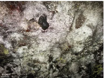
대섭이굴은 비교적 지상으로부터 얕은 깊이에 위치하고 있 다. 그래서 강우에 의해 지상으로부터 각종 무기물과 유기물 등 을 포함한 물질들이 침출을 통해 굴의 벽 안 쪽으로 스며들어 벽 표면까지 확산 될 것이라고 추정한다. 대섭이굴 탐사과정에서 동굴 벽면에 빛을 조사했을 때 특이하게도 백색의 생물막에서 빛이 반사되는 것을 육안으로 확인 할 수 있었다(Fig. 1). 본 연구 는 이러한 대섭이굴 표면에 형성 된 미생물 막의 특성을 관찰하 고자 분자생태학적인 기법(16S rRNA 유전자의 PCR 증폭 및 pyrosequencing 기술)을 이용하여 세균 및 고세균의 군집조성을 조사하였다.
본 실험을 위하여, 동굴표면의 미생물 막을 무균조작을 통해 채취하고 얼음이 존재하는 상태에서 하루 내에 실험실로 운반하 였다. 또한 미생물 막으로부터 DNA를 추출하기 전에 냉동 (-70℃) 보관하였다. 냉동 보관된 시료로부터 DNA를 추출하기 위하여 액체질소를 이용한 추출방법으로 수행 하였으며, 그 상 세한 방법은 토양에서 DNA를 추출하는 방법을 따랐다(Rhee et al., 2004). 추출된 DNA는 Sephadex를 이용한 겔 투과법(gel
Fig. 1. Light-reflecting cave biofilm on the wall in cave Daesubee.
filteration)을 통해 PCR 저해 물질을 제거하고 최종적으로 세균 및 고세균의 16S rRNA 유전자 증폭에 이용하였다. 세균의 16S rRNA 유전자를 증폭하기 위하여 사용한 primer는 세균 특이적 primer인 8F (5′-AGAGTTTGATCCTGGCTCAG-3′)와 338R (5′-TGCTGCCTCCCGTAGGAGT-3′)을 쌍으로 이용하였다(Ovreas et al., 1997; Vickerman et al., 2007). 고세균 16S rRNA 유전자 를 증폭하기 위하여 사용한 primer는 고세균 특이적인 primer인 514F (5′-GGTGBCAGCCGCCGCGRKAAHACC-3′)와 918R (5′-TGCYCCCCCGYCWATTSCTTTA-3′)을 쌍으로 이용하였 다(Kim et al., 2013). 세균 및 고세균 유전자의 증폭 반응을 위해 2× EF Taq mixture (SolGent Co., Ltd. Korea) 10 μl와 10 pmol forward와 reverse primer 각각 1 μl와 3차 증류수 7 μl를 넣고, 시료로부터 추출한 DNA를 1 μl씩 넣어 총 20 μl의 반응액을 만 들었다. 16S rRNA 유전자를 증폭시키기 위해 95℃에서 5분 동 안 변성(denaturation)시켰다. 그 다음 95℃에서 30초, 55℃에서 30초, 72℃에서 30초 동안 반응시키고 이를 30회 반복 수행하였 다. 마지막으로 72℃에서 7분간 최종 신장(extension)을 시켰다.
증폭된 세균 및 고세균의 16S rRNA 유전자는 Agarose gel 전기 영동법을 이용하여 크기를 확인하였다.
증폭된 16S rRNA 유전자는 PCR purification kit (Cosmo Genetech Co. Ltd, Korea)를 이용하여 정제하였으며, 정제된 산 물은 Pyrosequencing 기법을 이용하여 염기서열 분석을 시도하 였다. Pyrosequencing에 요구되는 모든 반응은 염기서열 제조사 인 454 Life Science (Branford, USA)의 방법에 따라 서울대학 교 농업과학공동기기센터(NICEM, Korea)에서 진행 하였으며, GS FLX Titanium system (454 Life Science)을 이용하여 염기 서열을 결정하였다. Barcode sorting에 따라 분리된 각 서열들에 서 barcode, primer 그리고 linker 서열을 제거 한 뒤 Mothur software (Schloss et al., 2009)의 Chimera-uchime (Edgar et al., 2011)을 통해 chimera로 의심되는 염기서열 제거를 수행하였다.
또한 Ribosomal database project (RDP)의 Pyrosequencing pipeline 중에 pipeline initial process를 통해 낮은 질의 염기서 열(<Q20 bp)과 작은 크기의 염기서열(<200 bp)을 제거하였다.
이렇게 얻은 염기서열들을 RDP Classifier를 이용해 세균의 계통 분류를 실시하였다(bootstrap cutoff=50%) (Cole et al., 2007). 고 세균의 경우에는 배양이 되지 않은(uncultured) 고세균 그룹의 계통 분류가 정확하지 않기 때문에 GenBank (http://ncbi.nlm.nih.gov) 의 database와 비교한 후 BLASTN program으로 상동성 및 계통 분류를 실시하였다. 그 결과 세균은 총 2663개의 raw reads 중 421 reads를 얻었으며, 단독개체(singletons)의 수는 5개였다. 고 세균의 경우 증폭효율이 낮아 총 295개의 raw reads를 얻을 수 있었으며, 낮은 질의 염기서열을 제거 후 182개의 reads를 확보 하였다. 하지만 이 중 많은 세균의 염기서열(115개의 reads)이 존재하여 이를 제거하고, 최종적으로 총 67 reads의 고세균 염기서 열을 얻을 수 있었으며 단독개체의 수는 3개였다. Phylogenetic tree는 RDP의 Pyrosequencing pipeline 중에 Complete linkage clustering (cutoff=97%)을 통해 얻어진 대표 서열을 이용하였 다. 염기서열 정리(Sequence alignment)는 CLUSTER W 알고 리즘으로 정렬한 후 MEGA 5.1 program (Tamura et al., 2011) 의 neighbor-joining을 따라 계통도를 작성하여 유전적 유사성을 검토하였다. 군집의 다양성을 분석하기 위하여 Taxonomy-Based Clustering (TBC) program (Lee et al., 2012)을 이용하고 3%
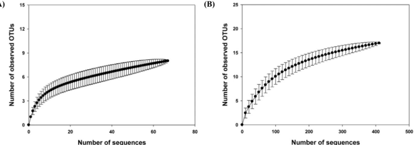
cutoff 기준으로 distance matrix를 만들었으며, 이를 PAST program (Øyvind et al., 2001)에 적용하였다. 이를 통해 Operational Taxonomic Unit (OTU) (세균: 17, 고세균: 8), Chao1 richness estimate (세균: 20.33, 고세균: 9.50) 그리고 Shannon index (세 균: 0.81, 고세균: 1.53) 등의 다양성 지수분석 값을 구하였다. 세 균과 고세균의 Rarefaction 분석은 PAST program (http://folk.uio.no/
ohammer/past)의 individual-based Coleman 방법을 사용하였으 며, 또한 SigmaPlot (V7.101, SPSS)을 통하여 rarefaction curve 의 plotting을 수행하였다(Park et al., 2008). 세균과 고세균의 정 량 PCR을 위하여 16S rRNA 유전자를 이용하였다. 또한 이에 사용한 primer와 자세한 방법은 Jung 등(2011)에서 설명한 바와 같으며 간단히 설명하면 다음과 같다. 정량 PCR은 MiniOpticon real-time PCR detection system (Bio-Rad Laboratories, USA)과 Opticon Monitor software version 3.1 (Bio-Rad Laboratories) 을 이용하여 진행하였으며 모든 유전자의 증폭은 Thermal-cycling parameters를 따랐다. 표준곡선(Standard Curve)은 이미 알려진 기준 유전자(reference gene)의 복제 개수(copy number)와 cycle threshold (CT) 값 사이의 관계를 가지고 산출하였다.
본 연구에서 Pyrosequencing법을 이용하였음에도 불구하고 적은 reads를 확보하였으나 아래에 설명한 결과로 미루어 보아 군집분석에는 심각한 영향을 미치지 않았다. 즉, 세균의 경우 관 찰된 OTU (17)와 richness (풍부도: 20.33)에 있어서 큰 차이가 없었으며, rarefaction 분석 결과 rarefaction curve가 점근선 (asymptote)에 거의 도달하는 것을 볼 수 있었기 때문에 비록 pyrosequencing을 통하여 얻은 염기서열의 수가 적으나 분석에 는 큰 문제점이 없음을 확인할 수 있었다(Fig. 2A). 또한 총 reads 수(N)와 OTU에서의 singleton 수(n)를 이용하여 만든 공 식(1-n/N)을 통해 구한 Good coverage (Good, 1953; Qian et al., 2011)의 값이 약 0.99가 나오기 때문에, 비록 적은 염기의 개수 를 얻었지만 자료 분석에는 큰 문제가 없음을 확인할 수 있었다.
(A) (B)
Fig. 2. Rarefaction analysis of the bacterial and archaeal 16S rRNA gene sequences. Rarefaction analysis of the bacterial (A) and archaeal (B) 16S rRNA gene sequences recovered from cave Daesubee. The rarefaction curve, plotting the number of observed phylotypes as a function of the number of clones, was computed by PAST. The dotted lines represent 95% confidence intervals. Phylotypes were defined as groups of sequences differing by 5% at the nucleotide sequence level.
(A) (B)
Fig. 3. Bacterial and archaeal composition of wall surface biofilm of cave. (A) Bacterial and (B) archaeal composition of wall surface biofilm of cave Daesubee in phylum level based on PCR and sequencing 16S rRNA gene.
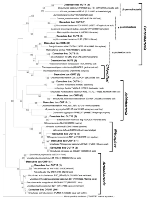
이를 통해 증폭된 세균 16S rRNA 유전자의 분석결과 대부분(약 83%)의 유전자들이 방선균(Actinobacteria) 문(phylum), 특히 그 중에서도 82.5%가 Pseudonocardineae (suborder)에 속하는 것으로 밝혀졌다(Figs. 3A and 4A). 이는 Slovenia 석회암 동굴 에서 높은 세균 다양성을 보이는 것과 대조를 이룬다(Park et al., 2008). 데이터베이스의 방선균 문 염기서열들과 가장 가까운 것 은 실제로 다양한 동굴의 벽이나 퇴적층에서 발견된 방선균 문 의 염기서열들과 유사함을 알 수 있다(Cañveras et al., 2001;
Groth et al., 2001; Chelius and Moore, 2004; Park et al., 2008).
특히, 대섭이굴 생물막의 방선균 문 염기서열들이 분리된 세균 중 에서는 Pseudonocardia mongoliensis MN08-A0270T (Ara et al., 2011)와 가장 가까운 근연관계가 있음을 확인할 수 있었다(16S rRNA 유전자 유사성 95.3%) (Fig. 4A). P. mongoliensis의 경우 성장은 pH 5–11에 걸쳐 일어나며 최적 pH는 6.0–6.8로 약산성 에 가깝다고 알려져 있다. Slovenia 석회암 동굴에서 방선균 염 기서열이 차지하는 비율은 16%에 불과하지만 다양한 방선균으 로 이루어져 있다(Park et al., 2008). Spain의 Altamira 동굴도
Pseudonocardia 속의 미생물 염기서열이 발견되긴 하나 역시 4% 이하의 비율을 보임을 알 수 있다(Portillo et al., 2008). 하지 만 대섭이굴에서는 어떻게 이들 방선균들이 같이 발견된 다른 일 반 종속영양(heterotroph) 세균인 Proteobacteria나 호산성 종속영 양세균인 Acidobacteria보다 우점하고 있는지에 대한 후속 연구 가 필요하다. 또한 아질산을 산화하는 것으로 알려진 미생물을 포함하는 phylum인 Nitrospirae와 관련된 염기서열이 약 1.9%
존재하고 있었다(Fig. 3A). 일반적으로 Pseudonocardia를 포함 하는 많은 방선균들이 고체배지 위에서 집락(colony)을 이룰 경 우 기균사(aerial hyphae) 및 포자(spore)들은 보통 소수성 (hydrophobicity)을 나타내는 것으로 알려져 있다(Clark et al., 1985; Ding and Lammler, 1992). Pseudonocardia 속의 미생물 들이 동굴 벽의 표면에 집락을 형성하며 생물막을 이룰 경우 물 을 흡수하지 않으므로 물방울들이 형성되며, Fig. 1에서 보는 바 와 같이 빛의 반사를 일으킬 것으로 추정된다. Pašić 등 (2010) 의 경우에서도 석회암 동굴이지만 역시 동굴 벽에서 빛이 반사 되는 특성을 보고하였다. 하지만, 이 석회암 동굴에서 빛이 적게
(A)
Fig. 4. Phylogenetic tree show the relationship between bacteria (A) and reference sequences. Phylogenetic tree show the relationship between archaea (B) and reference sequences. Neighbor-joining algorithm was used for construction of the phylogenetic tree based on 16S rRNA gene sequences. Names with bold were from this study. One representative sequence was selected among a group of sequences with more than 97%
similarity. Parenthesis after the name of the representative sequence is the number of sequences of the group. Numbers at the nodes are bootstrap values obtained from 1,000 replicates. Nucleotide sequence accession numbers are shown in parentheses. Scale bar indicates 50 inferred nucleotide substitutions per 1,000 nucleotide.
반사되는 것은 낮은 방선균의 양(약 16% 방선균 염기서열)과 관 계가 있음을 추정 할 수 있다. 이는 앞으로 생물막 자체에 대한 in situ 실험(Fluorescence in situ hybridization 및 방선균 세포 벽 구성물질 정량 및 정성분석)을 통해 증명 할 수 있을 것이다.
고세균의 16S rRNA 유전자는 고세균 특이적 primer를 사용 하였음에도 불구하고 많은 세균의 16S rRNA 유전자가 증폭 되 었다. 이는 본 실험에 사용한 primer가 다양한 고세균 증폭을 위 해 degeneracy를 높였기 때문이며, 고세균의 비율이 세균에 비
해 상대적으로 낮은 점 때문이라 추정된다. 실제로 고세균 16S rRNA 유전자의 높은 다양성으로 인하여 전체 고세균에 공통으 로 포함하는 염기서열을 찾는 것은 세균에 비해 어려움이 있는 것으로 보고되고 있다(Tetu et al., 2013). 일례로, Pašić 등 (2010)의 실험결과를 보면 석회암동굴에서 세균의 16S rRNA 유전자 증폭은 문제가 없었으나, 고세균 유전자의 증폭에는 실 패하였다. 또한 아래의 정량 PCR 결과 세균에 비해 고세균이 약 1% 정도로 낮게 분포함을 알 수 있었다. 하지만 대섭이굴에서
(B)
Fig. 4. continued.
관찰된 고세균 OTU (8)와 추정되는 richness (풍부도: 9.50)가 비슷하게 나타나고 있으며, rarefaction 분석 결과(Fig. 2B)에서 도 같은 결과를 관찰할 수 있었다. 또한 Good coverage의 경우 약 0.96의 수치가 나왔다. 이러한 결과들은 비록 pyrosequencing을 통해 적은 수의 염기서열을 확보하였으나 본 실험의 목적을 위 한 군집분석에 이용할 수 있음을 지지해주고 있다. 증폭된 고세 균 16S rRNA 유전자의 대부분(전체 염기서열 개수의 93%)은 놀랍게도 최근 제안된 Thaumarchaeota (Brochier-Armanet et al., 2008) 문이며 토양에서 광범위하게 발견되는 고세균이었다 (Fig. 3B). Altamira 동굴에서 Thaumarchaeota 문의 고세균 활 성을 검출하고 다양한 그룹(I.1a, I.1b, I.1a-Associated)들이 관 여하고 있음을 보고하였다(Gonzalez et al., 2006). 이들 고세균
은 토양에서 암모니아를 산화하는데 필요한 단백질인 AMO (ammonia monooxygenase) 유전자를 함유하는 것으로 알려지 고 있어 토양질소순환에 중요한 작용을 할 것으로 기대되고 있 다 (Leininger et al., 2006). 대섭이굴 생물막의 고세균중 대부분 (약 66%)은 Thaumarchaeota 중에서도 1.1a-Associated 그룹에 속하는 고세균으로 주로 산성(pH<5)의 토양에서 암모니아 산화 에 관여하는 것으로 알려진 고세균 Candidatus Nitrosotalea devanaterra (Lehtovirta-Morley et al., 2011)와 높은 유사성을 가 지고 있음이 확인 되었다(Fig. 4B). 대섭이굴의 1.1a-Associated 그룹은 크게 두 개의 소그룹으로 세분화됨을 알 수 있었다. 또한 암모니아를 산화하는 고세균 그룹 중 하나인 I.1a 그룹의 염기서 열이 약 27%를 차지하고 있었다(Jung et al., 2011). 정량 PCR을
통한 분석 결과 추출된 DNA의 ng당 고세균 16S rRNA 유전자 가 7.2 × 10³ (± 2.74 × 10²), 세균의 16S rRNA 유전자가 5.2 × 10⁵ (± 3.73 × 10⁴)으로 고세균이 세균에 비해 1.4%에 불과하므 로 대섭이굴에서 상대적인 고세균의 생지화학적 순환에 대한 기 여도가 높지 않을 것으로 추정된다. 하지만 질소순환의 측면에 서 보면 대섭이굴의 동굴 벽의 질산화 과정에 Nitrospira 속의 아 질산-산화 세균과 함께 암모니아-산화 고세균이 매우 중요한 기 능을 함을 추정할 수 있다. Nitrospira 속의 염기서열이(세균 전 체 염기서열의 1.9%) 정량 PCR로 확인된 세균에 대한 고세균의 16S rRNA 유전자 비율(1.4%)과 매우 유사함을 알 수 있다. 이 를 통해 지표면에서 암모니아가 침출되어 암모니아 산화 고세균 이 자라게 되고 그 부산물로 생긴 아질산을 산화하는 Nitrospira 속의 세균이 생장하였을 것으로 추정된다. 토양 환경에서 일반 적으로 우점하는 것으로 알려진 그룹 I.1a에 속하는 암모니아 산 화 고세균(Jung et al., 2011)이 발견되었으나, I.1b 그룹(Kim et al., 2012)은 발견되지 않았다. 또한 산성토양에서 일반적으로 많이 발견되는 I.1c 그룹의 고세균(Lehtovirta et al., 2009)의 염 기서열도 발견되지 않는 것은 매우 특이한 점이라 할 수 있다. 그 외에 토양환경에서 발견되지만 기능이 알려지지 않는 고세균의 염기서열인 DHVE6 그룹도 일부 발견되었지만, 절대혐기성 고 세균인 메탄생성균(methanogen)에 속하는 유전자는 발견되지 않았다.
수 미터(m) 정도의 비교적 깊지 않은 대섭이굴에서는 지표수 (빗물)가 용암층을 통과하는 동안 지표면의 영양분이 침출되는 과정을 통하여 걸러진 후 남은 영양분이 용암동굴의 벽 표면에 도달하게 된다. 이러한 영양분이 미생물의 성장에 기여하는 것 으로 추정되지만, 어떻게 Pseudonocardia와 Thaumarchaeota I.1a-Associated 그룹이 우점하는 다양성이 낮은 세균 및 고세균 의 군집을 가진 독특한 생물막의 형성에 기여하는 지는 알 수 없 다. 앞으로 곶자왈 지역의 투수과정에서 여과된 유기 및 무기 영 양분의 조성에 대한 정밀한 연구를 통하여 대섭이굴의 독특한 미생물 막의 군집조성에 미치는 영향을 규명할 수 있을 것으로 사료된다.
적 요
동굴환경은 표면 토양환경과는 다른 독특한 생태계를 이루는 것으로 알려져 있다. 본 연구를 통하여 제주도 용암동굴(대섭이 굴)의 생물막(biofilm)으로부터 얻은 시료를 pyrosequencing 기 술을 통해 16S rRNA 유전자를 증폭하여 세균과 고세균의 군집 을 조사하였다. 생물막에 우점하는 세균은 Actinobacteria문 (phylum)의 Pseudonocardia mongoliensis (전체 세균 reads수 의 82.5%)와 깊은 근연관계가 있었으며, 동굴유래의 다양한 세 균들과 같이 무리(cluster)를 형성하였다. 동굴 벽면에 빛을 조사 하였을 때 반사되어 빛나는 것은 아마도 방선균의 균사(hypha) 들로 이루어진 생물막이 수분을 흡수하지 못하기 때문으로 추정 된다. 우점하는 고세균은 Thaumarchaeota문의 I.1a-Associated group (전체 archaeal reads수의 약 66%)에 속한다. 이 고세균 염 기서열은 산성 환경(약 pH 5.0)에서 암모니아를 산화하는 고세
균으로 알려진 Candidatus Nitrosotalea devanaterra와 높은 근 연관계에 있어 동굴환경에서의 질산화에 중요한 기능을 하고 있 을 것으로 추정된다. 표층수가 용암토양을 투과하는 과정에서 침 출(Leaching)되는 영양분이 대섭이굴 벽면에 다양성이 낮은 독 특한 미생물 군집을 형성하는데 기여하고 있는 것으로 추정된다.
감사의 말
본 연구는 2011년도 충북대학교 교내 연구비에 의해 지원되 었습니다.
참고문헌
Ara, I., Tsetseg, B., Daram, D., Suto, M., and Ando, K. 2011.
Pseudonocardia mongoliensis sp. nov. and Pseudonocardia khuvsgulensis sp. nov., isolated from soil. Int. J. Syst. Evol.
Microbiol. 61, 747–756.
Borsodi, A.K., Knáb, M., Krett, G., Makk, J., Márialigeti, K., Erőss, A., and Mádl-Szőnyi, J. 2012. Biofilm bacterial communities inhabiting the cave walls of the buda thermal karst system, hungary.
Geomicrobiol. J. 29, 611–627.
Brochier-Armanet, C., Boussau, B., Gribaldo, S., and Forterre, P. 2008.
Mesophilic Crenarchaeota: proposal for a third archaeal phylum, the Thaumarchaeota. Nat. Rev. Microbiol. 6, 245–252.
Cañveras, J.C., Sanchez-Moral, S., Soler, V., and Saiz-Jimenez, C. 2001.
Microorganisms and microbially induced fabrics in cave walls.
Geomicrobiol. J. 18, 223–240.
Chelius, M.K. and Moore, J.C. 2004. Molecular phylogenetic analysis of Archaea and Bacteria in wind cave, South Dakota. Geomicrobiol.
J. 21, 123–134.
Chen, Y., Wu, L., Boden, R., Hillebrand, A., Kumaresan, D., Moussard, H., Baciu, M., Lu, Y., and Colin Murrell, J. 2009. Life without light:
microbial diversity and evidence of sulfur- and ammonium-based chemolithotrophy in Movile Cave. ISME J. 3, 1093–1104.
Clark, W.B., Lane, M.D., Beem, J.E., Bragg, S.L., and Wheeler, T.T.
1985. Relative hydrophobicities of Actinomyces viscosus and Actinomyces naeslundii strains and their adsorption to saliva-treated hydroxyapatite. Infect. Immun. 47, 730–736.
Cole, J.R., Chai, B., Farris, R.J., Wang, Q., Kulam-Syed-Mohideen, A.S., McGarrell, D.M., Bandela, A.M., Cardenas, E., Garrity, G.M., and Tiedje, J.M. 2007. The ribosomal database project (RDP-II):
introducing myRDP space and quality controlled public data.
Nucleic Acids Res. 35, D169–172.
Ding, H. and Lammler, C. 1992. Cell surface hydrophobicity of Actinomyces pyogenes determined by hexadecane adherence- and salt aggregation studies. Zentralb.l Veterinarmed. B 39, 132–138.
Edgar, R.C., Haas, B.J., Clemente, J.C., Quince, C., and Knight, R. 2011.
UCHIME improves sensitivity and speed of chimera detection.
Bioinformatics 27, 2194–2200.
Gonzalez, J., Portillo, M.C., and Saiz-Jimenez, C. 2006. Metabolically active Crenarchaeota in Altamira Cave. Naturwissenschaften 93, 42–45.
Good, I.J. 1953. The population frequencies of species and the estimation of population parameters. Biometrika 40, 237–264.
Groth, I., Schumann, P., Laiz, L., Sanchez-Moral, S., Canaveras, J.C., and Saiz-Jimenez, C. 2001. Geomicrobiological study of the Grotta dei Cervi, Porto Badisco, Italy. Geomicrobiol. J. 18, 241–258.
Jung, M.Y., Park, S.J., Min, D., Kim, J.S., Rijpstra, W.I., Sinninghe Damste, J.S., Kim, G.J., Madsen, E.L., and Rhee, S.K. 2011.
Enrichment and characterization of an autotrophic ammonia-oxidizing archaeon of mesophilic crenarchaeal group I.1a from an agricultural soil. Appl. Environ. Microbiol. 77, 8635–8647.
Kim, J.G., Jung, M.Y., Park, S.J., Rijpstra, W.I., Sinninghe Damste, J.S., Madsen, E.L., Min, D., Kim, J.S., Kim, G.J., and Rhee, S.K. 2012.
Cultivation of a highly enriched ammonia-oxidizing archaeon of thaumarchaeotal group I.1b from an agricultural soil. Environ.
Microbiol. 14, 1528–1543.
Kim, G.J., Park, S.J., Cha, I.T., Kim, S.J., Kim, K.H., Yang, E.J., Kim, Y.N., Lee, S.H., and Rhee, S.K. 2013. Unveiling abundance and distribution of planktonic bacteria and archaea in a Polynya in Amundsen Sea, Antarctica. in review.
Lee, J.H., Yi, H., Jeon, Y.S., Won, S., and Chun, J. 2012. TBC: a clustering algorithm based on prokaryotic taxonomy. J. Microbiol. 50, 181–
185.
Lehtovirta, L.E., Prosser, J.I., and Nicol, G.W. 2009. Soil pH regulates the abundance and diversity of Group 1.1c Crenarchaeota. FEMS Microbiol. Ecol. 70, 367–376.
Lehtovirta-Morley, L.E., Stoecker, K., Vilcinskas, A., Prosser, J.I., and Nicol, G.W. 2011. Cultivation of an obligate acidophilic ammonia oxidizer from a nitrifying acid soil. Proc. Natl. Acad. Sci. USA 108, 15892–15897.
Leininger, S., Urich, T., Schloter, M., Schwark, L., Qi, J., Nicol, G.W., Prosser, J.I., Schuster, S.C., and Schleper, C. 2006. Archaea predominate among ammonia-oxidizing prokaryotes in soils.
Nature 442, 806–809.
Northup, D.E., Melim, L.A., Spilde, M.N., Hathaway, J.J., Garcia, M.G., Moya, M., Stone, F.D., Boston, P.J., Dapkevicius, M.L., and Riquelme, C. 2011. Lava cave microbial communities within mats and secondary mineral deposits: implications for life detection on other planets. Astrobiology 11, 601–618.
Ovreas, L., Forney, L., Daae, F.L., and Torsvik, V. 1997. Distribution of bacterioplankton in meromictic Lake Saelenvannet, as determined by denaturing gradient gel electrophoresis of PCR-amplified gene fragments coding for 16S rRNA. Appl. Environ. Microbiol. 63, 3367 –3373.
Øyvind, H., Harper, D.A.T., and Ryan, P.D. 2001. Past: paleontological
statistics software package for education and data analysis.
Palaeontologia Electronica. 4, 9.
Park, S.-J., Park, B.-J., and Rhee, S.-K. 2008. Comparative analysis of archaeal 16S rRNA and amoA genes to estimate the abundance and diversity of ammonia-oxidizing archaea in marine sediments.
Extremophiles 12, 605–615.
Pašić, L., Kovče, B., Sket, B., and Herzog-Velikonja, B. 2010. Diversity of microbial communities colonizing the walls of a Karstic cave in Slovenia. FEMS Microbiol. Ecol. 71, 50–60.
Portillo, M.C., Gonzalez, J.M., and Saiz-Jimenez, C. 2008. Metabolically active microbial communities of yellow and grey colonizations on the walls of Altamira Cave, Spain. J. Appl. Microbiol. 104, 681–691.
Qian, P.Y., Wang, Y., Lee, O.O., Lau, S.C., Yang, J., Lafi, F.F., Al-Suwailem, A., and Wong, T.Y. 2011. Vertical stratification of microbial communities in the Red Sea revealed by 16S rDNA pyrosequencing. ISME J. 5, 507–518.
Rhee, S.K., Liu, X., Wu, L., Chong, S.C., Wan, X., and Zhou, J. 2004.
Detection of genes involved in biodegradation and biotransformation in microbial communities by using 50-mer oligonucleotide microarrays. Appl. Environ. Microbiol. 70, 4303–4317.
Schloss, P.D., Westcott, S.L., Ryabin, T., Hall, J.R., Hartmann, M., Hollister, E.B., Lesniewski, R.A., Oakley, B.B., Parks, D.H., Robinson, C.J., and et al. 2009. Introducing mothur: open-source, platform-independent, community-supported software for describing and comparing microbial communities. Appl. Environ.
Microbiol. 75, 7537–7541.
Tamura, K., Peterson, D., Peterson, N., Stecher, G., Nei, M., and Kumar, S. 2011. MEGA5: molecular evolutionary genetics analysis using maximum likelihood, evolutionary distance, and maximum parsimony methods. Mol. Biol. Evol. 28, 2731–2739.
Tetu, S.G., Breakwell, K., Elbourne, L.D., Holmes, A.J., Gillings, M.R., and Paulsen, I.T. 2013. Life in the dark: metagenomic evidence that a microbial slime community is driven by inorganic nitrogen metabolism. ISME J. 7, 1227–1236.
Vickerman, M.M., Brossard, K.A., Funk, D.B., Jesionowski, A.M., and Gill, S.R. 2007. Phylogenetic analysis of bacterial and archaeal species in symptomatic and asymptomatic endodontic infections. J.
Med. Microbiol. 56, 110–118.