Isolation of Anaerobic Cellulolytic Bacteria from the Rumen of Holstein Dairy Cows to Develop
Feed Additives for Ruminants
1)
Choi, Nag-Jin*
†․Lee, Gi-Young**
†․Jeong, Kwang-Hwa***․Kim, Chang-Hyun****
반추동물용 사료첨가제개발을 위한 홀스타인 젖소의 반추위로부터 분리한 혐기성 섬유소 분해균의 특성연구
최낙진․이기영․정광화․김창현
In order to develop a high cellulolytic direct-fed microorganism (DFM) for ruminant productivity improvement, this study isolated cellulolytic bacteria from the rumen of Holstein dairy cows, and compared their cellulolytic abilities via DM degradability, gas production and cellulolytic enzyme activities. Twenty six bacteria were isolated from colonies grown in Dehority’s artificial (DA) medium with 2%
agar and cultured in DA medium containing filter paper at 39 ℃ for 24h. 16s rDNA gene sequencing of four strains from isolated bacteria showed that H8, H20 and H25 strains identified as Ruminococcus flavefaciens, and H23 strain identified as Fibrobacter succinogenes. H20 strain had higher degradability of filter paper compared with others during the incubation. H8 (R. flavefaciens), H20 (R.
flavefaciens), H23 (F. succinogenes), H25 (R. flavefaciens) and RF (R. flavefaciens sijpesteijn, ATCC 19208) were cultured in DA medium with filter paper as a single carbon source for 0, 1, 2, 3, 4 and 6 days without shaking at 39 ℃, respectively. Dry matter degradability rates of H20, H23 and H25 were relatively higher than those of H8 and RF since 2 d incubation. The cumulative gas pro-
* The Institute of Rare Earth for Biological Application, Department of Animal Science, Chonbuk National University
** Graduate School of Biotechnology, Environmental and Information Technology, Hankyong National University
*** National Institute of Animal Science, RDA
**** Corresponding Author, School of Animal Life and Environment Science, Biogas Research Center, Hankyong National University ([email protected])
†
These authors contributed equally to this work.
duction of isolated cellulolytic bacteria increased with incubation time. At every incubation time, the gas production was highest in H20 strain. The activities of carboxymethylcellulase (CMCase) and Avicelase in the culture supernatant were significantly higher in H20 strain compared with others at every incubation time (p<0.05). Therefore, although further researches are required, the present results suggest that H20 strain could be a candidate of DFM in animal feed due to high cellulolytic ability.
Key words : cellulolytic bacteria, rumen, dry matter degradability, gas production, enzyme activity
Ⅰ. Introduction
There are a lot of interests in manipulating the microbial ecosystem in the rumen to improve feed utilization and production efficiency by ruminants. Various attempts have been used to optimize rumen microbial ecosystem. Direct-fed microorganisms (DFM) are defined as a source of live, naturally occurring microorganism (Krehbiel et al., 2003). In addition, the term DFM has included specific and nonspecific yeast, fungi, bacteria, cell fragments, and filtrates (Beharka and Nagaraja, 1993; Sullivan and Martin, 1999). Primary microorganisms that have been used as DFM for ruminants are fungal cultures including Aspergillus oryzae (AO) and Saccharomyces cerevisiae (SC) and lactic acid bacteria such as Lactobacillus or Streptococcus. Modes of action are variable and include native rumen microbiota enhancement through increased substrate breakdown, enhancement of nutrient uptake, and provision of growth factors (Martin and Nesbit, 1992).
Although anaerobic bacteria, protozoa and fungi degrade cellulose in the rumen, cellulolytic
bacteria among them play the most important role for fibrolytic activity and they are dominant
in the rumen (Windham and Akin, 1984). Particularly, Fibrobacter succinogenes, Ruminococcus
flavefaciens and R. albus are mostly important cellulolytic bacteria in the rumen (Hungate,
1963). These anaerobic bacteria in the rumen show strong cellulase and hemicellulase activity
(Bryant, 1973; Forsberg et al., 1981; Greve et al., 1984; McDermid et al., 1990; Pettipher and
Latham, 1979). The cellulolytic anaerobic bacteria originated from host animals could be a new
candidate of dietary DFM supplements for the ruminant. Therefore, the objective of this study
was to compare cellulolytic abilities of isolated rumen cellulolytic bacteria with their filter paper
degradability, gas production and cellulolytic enzyme activities.
Ⅱ. Materials and Methods
1. Isolation and identification of cellulolytic anaerobic bacteria
1) Inoculum and culture conditions
Rumen contents used to isolate cellulolytic bacteria were collected from the rumen of three Holstein dairy cows (average body weight of 580kg). The animals were prepared with a permanent cannula into the rumen and were housed in a free-stall pen. Timothy hay and corn silage were fed at 2% of body weight twice daily (09:00h and 18:00h). Water was available ad libitum, and animals were received proprietary mineral and vitamin supplements in the form of licks. Rumen fluid and contents were collected 300ml and 20g from each animal at 3h after the morning ration had been consumed. Collected samples were strained through four layers of cheesecloth and brought immediately to the laboratory being gassed with oxygen-free CO
2and kept at 39 ℃.
The anaerobic culture techniques of Hungate as described by Bryant (1973) were used for all incubations. For isolating cellulolytic bacteria from the rumen contents, 10g of rumen contents containing proportions of solid and liquid material were inoculated into 90ml of Dehority’s artificial (DA) medium (Dehority, 1963) containing cellulose powder (Sigmacell, No. 5504, Sigma, USA) as carbon and energy sources. After incubating for 2 days at 39 ℃, the culture medium was shaken for 15min at 4 ℃ for separating cellulolytic bacteria strongly attached on the surface of feed particles. The culture medium was then serially diluted, in 10-fold steps, in the anaerobic mineral solution by making the method of Bryant and Burkey (1953a), and then, the diluted samples were seeded in roll tubes containing DA medium with 2.0% agar and incubated between 2 and 4 days at 39 ℃.
2) Isolation and preservation of colony
Colony was picked from roll tubes with a bent platinum- iridium wire, and was inoculated
into DA broth medium added with a filter paper strip (Whatman No. 1, Whatman, England) as
a single carbohydrate source. Colonies were selected from roll tubes as randomly as possible,
and as many colonies as possible were picked from the same roll tubes. Inoculated cultures were
incubated at 39 ℃ and growth was obtained in most of the cultures within 3 or 4 days by
degradation of filter paper strip. The cultures of bacteria showing significant degradation of filter
paper were selected for further research. To be secure of pure strains, the roll tube procedure
repeated two times with the isolated high cellulolytic bacteria. And then the pure strains were
preserved in DA medium containing 20% glycerol at -70 ℃ (Teather, 1982).
3) 16s rDNA analysis of isolated cellulolytic bacteria.
Isolated cellulolytic bacterial 16s rDNA was extracted by using G-spin
TMGenomic DNA Extraction Kit (No. 17121, (C)iNtRon, Korea). Extracted cellulolytic bacterial DNA was amplified by using Quick PCR premix (containing Taq DNA Polymerase, dNTPs, reaction buffer and tracking dye). The thermocycler profile was 2 min at 94 ℃ (initial denaturation) and then 30 cycles of 20 sec at 94 ℃ (denaturation), 20 sec at 50℃ (annealing), 1 min at 72℃ (extention) and followed by 5min final extension at 72 ℃. PCR products were analyzed by electrophoresis (1.0% agarose gel) in 40 mM Tris-acetate containing 1 mM EDTA (50×TAE). It was stocked at 4 ℃ in a cold chamber until using. PCR Clean-up Kit (No. 17091, (C) iNtRON, Korea) was designed to extract and purify DNA fragments from normal agarose gel in Tris acetate.
Amplicated DNA and marker were loaded to well, and then they were purificated by using PCR Clean-up Kit (No.17091, (C) iNtRON, Korea). After finishing the purification, pGEM ⓡ-T and pGEM ⓡ-T Easy Vector Systems was used for transforming PCR products. DNA vector finished transforming was transformed to competent cell (No. 15041, DH5 α, (C) iNtRON, Korea).
Transformed cell was incubated for 1h at 37 ℃. The incubated competent cell was spreaded on LB Agar (Sigma Cat. No L-7025, Sigma, USA) contained X-gal/IPTG. If white-color colony was grown after incubating for 12h, ampicillin was added to E-coli LB broth. Plasmid DNA Extraction Kit ((C) iNtRON Cat. No 17093) was used for purification after incubating for 12h at 37 ℃ and then DNA was sequenced.
2. Cellulolytic characteristics of pure cultured anaerobic bacteria
1) Microorganisms
The isolated pure strains, H8 (R. flavefaciens), H20 (R. flavefaciens), H23 (F. succinogenes) and H25 (R. flavefaciens) and RF (R. flavefaciens sijpesteijn, ATCC 19208) as type culture were used to compare their fibrolytic properties of in vitro gas production, cellulose degradation and polysaccharidase and monosaccharidase enzyme activities.
2) Culture conditions
The anaerobic culture techniques of Hungate as described by Bryant (1973) were used for all
incubations. The soluble carbon and energy sources were replaced with substrates of Whatman
No. 1 cellulose filter paper. Approximately 75mg of punched filter paper (0.6 mm diameter)
were added to 30ml triplicate serum vials. Anaerobic liquid medium (14ml of DA medium lacking a carbon source) was transferred into the triplicate serum vials that had been purged with oxygen-free CO
2and the vials were subsequently sterilized by autoclaving at 121 ℃ for 15 min with fast exhaust. The pH after autoclaving was between 6.68 and 6.70 (6.69±0.02). One milliliter of each bacterial strain was anaerobically inoculated to each vial. Incubations were performed anaerobically at 39 ℃ without shaking for 0, 1, 2, 3, 4 and 6 days. At each sampling time, gas production, filter paper (FP) cellulose degradation and enzyme activities were deter- mined in triplicate for each treatment.
3) Determination of degradation rate of filter paper cellulose
The degradation rate of FP cellulose was determined in triplicate for each treatment. Cultures were collected after 0, 1, 2, 3, 4 and 6 days of incubation. Supernatant from each culture was separated from sedimentable material by centrifugation at 500×g for 20 min. To determine the extent of FP cellulose degradation, residual FP samples were boiled in 1mol NaOH (to remove adherent bacterial matter), rinsed three times with absolute alcohol (60 ℃), twice with running distilled water, and dried until a constant weight was achieved (approximately 24h at 78 ℃).
Apparent FP degradation rate (%) was calculated from the differences of initial FP as a substrate and final FP collected after each incubation time using the equation : FP degradation rate = (initial FP - final FP) / initial FP ×100.
4) Determination of enzyme activities
Supernatants from three replicate cultures were analyzed for enzyme activity. Activities of CMCase, Avicelase and xylanase were assayed by using the method described in Mandels et al.
(1976). Extracellular enzyme activity against carboxymethylcellulose (CMC) was determined by
incubating 0.5ml aliquots of supernatant from the growing cultures (grown under different
conditions) with 0.5ml of 10g CMC (No. C-4888, carboxymethyl cellulose, Sigma, USA) dis-
solved in the 1L of 0.05M citrate buffer (pH 5.5). The reaction proceeded at 45 ℃ for 1h
without shaking and the reaction was stopped by boiling for 5min. Samples were centrifuged at
12,000×g for 5 min and reducing sugars in the supernatants were assayed colorimetrically using
the DNS (dinitrosalicylic acid) method of Miller et al. (1960). For determination of extracellular
Avicelase activity, 0.5ml of the supernatant was mixed with 0.5ml of 10g Avicel (No. 11365,
Avicel
®PH-101, Fulka, USA) dissolved in the 1L of 0.05M citrate buffer (pH 5.5). After
incubating, method of analysis was same as it of CMCase. For determination of extracellular
xylanase activity, 0.5ml of the supernatant was mixed with 0.5ml of 10g oat spelt xylan (No.
X0627, Sigma, USA) dissolved in the 1 L of 0.05M potassium phosphate buffer (0.05M, pH 7.0). The reaction proceeded for at 45 ℃ 1h with shaking. After incubating, method of analysis was same as it of CMCase activity. One unit of enzyme activity was defined as the amount of enzyme which liberated lµmol of glucose (for CMCase and Avicelase) and xylose (for xylanase) equivalent per min. Monosaccharide degradation enzyme, β-D-glucosidase (No. N-7006, Sigma, USA) activity was assayed by method of Kohchi and Tohe (1986). Fifty microliter of 10mM p-NP glucopyranoside (No. N7006, p-Nitrophenyl β-D-glucopyranoside, Sigma, USA) was mixed with 10µl of potassium phosphate buffer (0.5M, pH 6.5), 20µl of distilled water and 20µl of the supernatant. The reaction proceeded at 50 ℃ for 30 min. Fifty microliter of 1M Na
2CO
3made the reaction stopped and then analyzed by ELISA reader (Sunrise
TMRemote/Touch Screen F039300, Tecan, Switzeland). One unit of enzyme activity was defined as the amount of enzyme that produced burstsize (µmol/ml/min) equivalent of p-NP (paranitrophenyl) per minute.
5) Total gas production
At the end of the each incubation time, a glass syringe needle was inserted through the butyl rubber stopper, and the volume of gas exceeding 1 atm was measured through the displacement of the syringe plunger using the techniques of Fedorak and Hrwdey (1983) with modifications (Callaway and Martin, 1996).
3. Statistical analysis
SAS (1996) was used to calculate the standard errors and perform the analysis of variance and statistical significance (P<0.05) among treatment means was determined by Duncan’s multiple range test.
Ⅲ. Results and Discussion
1. Isolation of cellulolytic bacteria from the rumen of Holstein dairy cows
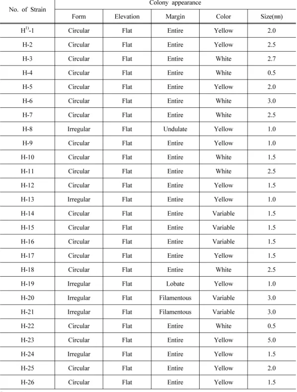
Twenty-six cellulolytic bacteria were isolated by roll-tube method from the rumen of Holstein dairy cows. The colonies of the isolated cellulolytic bacteria grown on DA medium with 2%
agar were observed (Table 1). The sizes of isolated cellulolytic bacteria were ranged from one
to three millimeters in diameter. The colonies of H8, H13, H19, H20, H21 and H24 strains were
No. of Strain
Colony appearance
Form Elevation Margin Color Size( ㎜)
H
1)-1 Circular Flat Entire Yellow 2.0
H-2 Circular Flat Entire Yellow 2.5
H-3 Circular Flat Entire White 2.7
H-4 Circular Flat Entire White 0.5
H-5 Circular Flat Entire Yellow 2.0
H-6 Circular Flat Entire White 3.0
H-7 Circular Flat Entire White 2.5
H-8 Irregular Flat Undulate Yellow 1.0
H-9 Circular Flat Entire Yellow 1.0
H-10 Circular Flat Entire White 1.5
H-11 Circular Flat Entire White 2.5
H-12 Circular Flat Entire Yellow 1.5
H-13 Irregular Flat Entire Yellow 1.0
H-14 Circular Flat Entire Variable 1.5
H-15 Circular Flat Entire Variable 1.5
H-16 Circular Flat Entire Variable 1.5
H-17 Circular Flat Entire Yellow 1.5
H-18 Circular Flat Entire White 2.5
H-19 Irregular Flat Lobate Yellow 1.0
H-20 Irregular Flat Filamentous Variable 3.0
H-21 Irregular Flat Filamentous Variable 3.0
H-22 Circular Flat Entire White 0.5
H-23 Circular Flat Entire Yellow 5.0
H-24 Irregular Flat Entire Yellow 1.5
H-25 Circular Flat Entire Yellow 2.0
H-26 Circular Flat Entire Yellow 1.5
1)
H: Bacteria from the rumen of Holstein cows.
Table 1. Growth characteristics of strains isolated from the rumen of Holstein dairy cows
on Dehority’s artificial agar medium
irregular shapes, and the others were in a round shape. Especially, the center of irregular shaped colonies of H20 and 21 strains were appeared in yellow color and their edges were in white radial form. The colony of H25 strain was in double round shape with thick yellow color.
Although it is difficult to identify bacterial strain with colonial shapes and colors, yellow colony is known to contain bacterium that has an ability to degrade both cellulose and cellobiose when Rumincoccus flavefaciens is incubated in pure culture (Sijpesteijn, 1951; Hungate, 1957).
In the case of R. albus, white colony was formed and degradation rate of cellulose was high (Hungate, 1957). In general, bacteria colonies isolated from cellulose agar roll tubes are predominantly ruminococci along with lesser numbers of Butyrivibrio (Dehority, 2003). From observing the shapes and colors of the colonies cultured in roll tubes it seems that they could be ruminococi and other cellulolytic bacteria.
2. Identification and cellulose degradation properties of isolated bacteria
After incubation for 24h, H25 strain only degraded a strip of FP which was transformed to shrunken shape. Up to 48h for further incubation, H8, H20 and H23 strains degraded FP. Strains of H7, 16 and 21 also had relatively higher growing activities comparing with the others, but filter paper was not degraded by those strains. Seven strains were finally selected from 26 isolated strains based on their cellulolytic abilities and growth rates (data not shown here) in DA medium. In order to identify the selected strains, 16s rDNA analysis was carried out. As the electrophoresis of 16s rDNA applications of the cellulolytic bacteria are shown in Figure 1, six strains among seven isolated ones were observed normal PCR bands, but the band of H23 strain is not shown. Thus, partial sequence of 1.5kbp was obtained for strains H7, H8, H16, H20, H21 and H25, but H23 was not sequenced. After reanalysis of 16s rDNA, partial sequence of 900 bp was obtained for H23. Data of partial sequences of the isolated strains were compared with available 16S RNA gene sequences in GenBank by use of the NCBI (National Center for Biotechnology Information) Blast program and then, similarity analysis placed all strains within Megasphaera elsdenii, R. flavefaciens and F. succinogenes (Table 2). Sequence similarity (1.5 kbp) of H7, H16 and H21 was at least 94.8% among M. elsdenii, non cellulolytic bacterium.
H8, H20 and H25 strains were more closely related to R. flavefaciens (mean similarity, 91.9%).
H23 strain was closely related to F. succinogenes with sequence similarity of 94.0%.
Figure 1. Electrophoresis of 16s rDNA amplications of isolated cellulolytic bacteria from the rumen of Holstein dairy cows.
Table 2. Identification of cellulolytic bacteria isolated from Holstein dairy cows in the experiment using rDNA analysis
Bacterial
isolates Species and genus Strains Mean
similarities (%)
H7 Megasphaera elsdenii 5T, T81, La03, JL1, AW106,
ATCC25940, YJ-4, S2, S3, 7-11 96.3
H16 Megasphaera elsdenii YJ-4, 5T, T81, AW106, JL1, 4-13, La03,
7-11, 2-9 94.8
H20 Ruminococcus flavefaciens LP9155, AR69, 4, LP-RI-Adx, FD-1,
AR72, V1, 17, JM1, 007 91.2
H21 Megasphaera elsdenii YJ-4, 5T, La03, T81, 2-9, AW106, JL1,
7-11, 4-13 95.7
H25 Ruminococcus flavefaciens LP-R1-Adx, LP-C14-Adx, R13e2, AR69,
LP9155, ATCC49949, 4, Y1, AR72 91.8
H8 Ruminococcus flavefaciens LP9155, R13e2, LP-R1-Adx, 4, AR72,
LP-C14-Adx, ATCC 49949, 17, FD-1, V1 92.6
H23 Fibrobactor succinogenes
Partial S85, scucinogenes BL2, succinogenes B1, succinogenes A3C, HM2, MC1, MM4, MB4
94.0
Degradation of FP cellulose by H8, H20, H23, H25 and R. flavefaciens Sijpesteijn (ATCC
19208) is shown in Table 3. At 1 d incubation, there were no significant differences of DM
degradation by different strains. H20 dramatically increased (p<0.05) DM degradation of FP at
Incubation
Time (day) H8 H20 H23 H25 RF SEM
1 1.39 0.30 0.00 0.00 5.22 1.202
2 0.00
c14.36
a1.52
bc5.34
b0.67
bc1.703
3 4.92
c37.82
ab32.74
b43.45
a4.43
c4.607
4 12.65
c48.49
a27.12
b54.15
a0.00
d6.287
6 17.08
d58.03
b64.57
ab66.21
a31.72
c5.858
1
DA medium: Dehority’s artificial medium.
H8: Ruminococcus flavefaciens H8.
H20: Ruminococcus flavefaciens H20.
H23: Fibrobacter succinogenes H23.
H25: Ruminococcus flavefaciens H25.
RF: Ruminococcus flavefaciens Sijpesteijn (ATCC 19208).
a,b,c,d
Means in the same row with different superscripts differ (p<0.05).
Table 3. Dry matter degradation rates (%) of filter paper cellulose by pure cultured cellulolytic bacteria isolated from the rumen of Holstein dairy cows and cultured with DA medium1 with filter paper as a carbohydrate source
2 d incubation compared with the other strains. After 3 d incubation, the degradation rate of H25 strain was significantly higher than others (p<0.05). At the last day for the incubation, H25 was in the highest DM degradability compared with other microorganisms (p<0.05) except for H23. In addition, H25 strain had higher DM degradation rate compared with others during whole incubation, while, RF strain barely degraded filter paper until 4 d incubation. In the very early study by Halliwell and Bryant (1963), the degradability of ground cellulose powder and dewaxed cotton fibers by one strain (S-85) of F. succinogenes and two strains (C-94 and FD-1) of R.
flavefaciens was compared. For five day incubation, the ground cellulose powder digestibility of
F. succinogenes S-85, R. flavefaciens C-94 and FD-1 were 88%, 72% and 90%, respectively,
while, for seven day incubation, the cotton fibers degradability of those 3 strains showed 97%,
0% and 55%, respectively. The values for the ground cellulose powder degradability from the
work by Halliwell and Bryant (1963) were higher than the degradability FP cellulose disk in the
present experiment. The higher degradability could be caused by more degradable cellulose type
and smaller particle size of substrate used than the present experiment. However, the very low
degradability of cotton fibers by the strains of R. flavefaciens from the work of Halliwell and
Bryant (1963) can explain the reason of the lower filter paper degradability of H8 and RF
compared with the other strains of R. flavefaciens in the present experiment. That is, although
Incubation
Time (Day) H8 H20 H23 H25 RF SEM
1 2.82
c4.21
a3.13
bc3.30
b2.39
d0.092
2 5.08
c9.21
a6.89
b7.09
b3.63
d0.238
3 6.70
d12.92
a9.88
c11.22
b4.67
e0.413
4 7.58
c15.41
a12.17
b14.48
a5.92
d0.592
6 8.47
c17.93
a14.98
b16.87
a7.55
c0.841
1
DA, H8, H20, H23, H25 and RF: Same as in Table 4.
a,b,c,d,e
Means in the same row with different superscripts differ (p<0.05).
Table 4. Cumulative gas production (ml/0.1g DM substrate) by pure cellulolytic bacteria isolated from the rumen of Holstein dairy cows and cultured with DA medium1 with filter paper as a carbohydrate source
cellulolytic bacteria are within a same species, the cellulolytic activity could be variable by different types of cellulose. As only one strain of F. succinogenes was available for the present study, it is not known whether this species would, in general, more extensively degrade fibrous forms of cellulose as compared to ruminococci. It is of interest in this respect that F.
succinogenes was found in much greater numbers than ruminococci in the rumen of a cow fed wheat-straw, but ruminococci were more numerous when good quality alfalfa hay was fed (Bryant and Burkey, 1953b).
3. Cumulative gas production
Cumulative gas production by isolated cellulolytic bacteria cultured with DA medium
containing FP as a carbohydrate source is shown in Table 4. Cumulative gas production from
H20 strain was significantly higher in comparison with others up to 3 d incubation (p<0.05),
while at 4 and 6 d incubation the cumulative gas production from H20 and H25 was higher than
other microorganisms (p<0.05). The cumulative gas production from H8 and RF strains was
lower than others during the whole incubation time reflecting the results of DM degradation
rates. Thus, cumulative gas production of H20 and H25 strains was higher than others, and this
could be due to its higher microbial activity and growth rate. Since the late 1970s, measurement
of in vitro gas production has become increasingly popular for determining forage digestion
characteristics and the kinetics of fermentation (Theodorou et al., 1994, 1998). The amount gas
produced depends on the amount of substrate fermented and the amount and molar proportions
of the VFA produced (Beuvink and Spoelstra, 1992). Rymer and Givens (2002) suggested that the gas production technique predicts dynamic parameters of rumen fermentation, particularly the short chain fatty acids (SCFA). Again, Theodorou et al. (1995) demonstrated that measuring the accumulation of fermentation gases during rumen anaerobic fungal growth is a useful method for the rapid and precise determination of microbial growth on soluble and particulate substrates. In the present experiment, although the relationship between gas volume and DM degradability was not observed exactly in a same manner, the gas production technique seems useful for an indirect tool to assess cellulolytic activity of microorganisms.
4. Enzyme activity
The time courses of extracellular CMCase, Avicelase, xylanase and β-D-glucosidase acitivi- ties in the different strains are shown in Table 5. Activities of CMCase and xylanase gradually increased with incubation time up to 4 d in all bacteria in the experiment, while activities of Avicelase and β-D-glucosidase were not influenced by incubation time in the most of microor- ganisms. Extracellular CMCase activity was significantly higher in H20 strain compared with other strains during whole incubation time (p<0.05), while RF strain had relatively lower in its activity compared with others up to 6 d incubation. Again, H20 strain showed higher CMCase activity at very early incubation time (1h) compared with other microorganisms. Extracellular Avicelase activity was also significantly higher in H20 strain compared with other strains during the whole incubation time, except for 6 d (p<0.05). During the incubation, xylanase activity was highest in H25 and second highest in H20 indicating that R. flavefaciens strains have higher xylanase activities than F. sucssinogenes. β-D-glucosidase activity was not significantly different at 1 d, but its activity was highest in H23 from 3 to 6 d among the strains.
Compared with other enzymes, CMCase activity was highly related to DM degradation in the experiment. Silva et al. (1987) reported that CMCase activity after 24h was highly correlated with DM degradability of the same samples at 24h (r = 0.98) and 48h (r = 0.94) mentioning that cellulolytic activity as measured by CMCase was a good indicator of DM degradation. Xylanase enzyme activity showed highest values among the other enzyme activities in Table 5. However, the activity values of xylanase in H23 were not representative for DM degradation of H23. This might be explained from Hungate’s works (Hungate 1950, 1957) in which he observed that F.
succinogenes could not attack hemicelluloses which mainly contains polysaccharide xylan ( β-1-4
linked xylose with occasional branches formed by 1-3 linkages), while some strains of cellul-
olytic cocci could utilize hemicelluloses. It is likely that enzyme activities of cellulolytic bacteria
Incubation
Time (Day) H8 H20 H23 H25 RF SEM
CMCase activities (glucose μmol/ml/min)
1 36.22
c69.44
a31.82
d48.12
b13.48
e4.963
2 36.82
d73.89
a48.40
c61.68
b17.24
e5.357
3 47.67
d84.73
a59.04
c65.80
b24.13
e5.265
4 45.75
d76.15
a62.31
c69.38
b31.52
e4.417
6 40.77
d74.31
a69.83
b65.70
c39.07
d4.029
Avicelase activities (glucose μmol/ml/min)
1 7.42
b9.06
a6.35
c8.07
b5.92
c0.316
2 10.36
ab10.87
a10.32
ab10.14
b10.74
ab0.106
3 12.59
b13.47
a12.37
b13.67
a12.27
b0.186
4 9.93
bc11.18
a9.29
c10.80
ab9.42
c0.234
6 9.88
a9.38
b9.25
b9.68
ab9.62
ab0.081
Xylanase activities (xylose μmol/ml/min)
1 218.67
c324.58
b35.52
d347.49
a22.55
d37.003
2 215.51
c364.25
b60.16
d469.71
a69.02
d43.488
3 248.12
c427.14
b127.47
d520.95
a133.79
d42.484
4 268.09
c444.73
b141.43
d576.23
a151.07
d45.648
6 154.96
c206.28
b141.53
c255.84
a147.97
c12.232
β-D-glucosidase activities (pNP μmol/ml/min)
1 34.26 37.98 42.71 44.48 33.56 1.737
2 25.48
ab30.61
ab34.74
ab37.58
a19.06
b2.602
3 26.88
b29.42
ab34.80
a20.50
c20.26
c1.639
4 21.07
bc32.06
ab39.19
a35.39
ab15.46
c3.049
6 24.25
b22.58
b78.01
a9.99
c13.69
c6.631
1
DA, H8, H20, H23, H25 and RF: Same as in Table 4.
a,b,c,d,e