Copyrightⓒ 2008, The Korean Academy of Oral Biology
205
Journal of Oral Biology
Cyclosporin A-induced Gingival Overgrowth is Closely Associated with Regulation Collagen Synthesis by the Beta Subunit of Prolyl 4-hydroxy- lase and Collagen Degradation by Testican 1- mediated Matrix Metallo- proteinase-2 Expression
Seong-Hee Park1, Jae-Yoen Kim2,3, Hyun-Jeong Kim2, Kwang-Kyun Park2,3, Kyoo-Sung Cho1,3, Seong-Ho Choi1,3, and Won-Yoon Chung2*
1Department of Periodontology, Yonsei University College of Dentistry, 134 Shinchon-Dong, Seodaemoon-Ku, Seoul 120-752, South Korea
2Department of Oral Biology, Yonsei University College of Dentistry, 134 Shinchon-Dong, Seodaemoon-Ku, Seoul 120-752, South Korea
3Brain Korea 21 Project, Yonsei University College of Dentistry, 134 Shinchon-Dong, Seodaemoon-Ku, Seoul 120-752, South Korea
(Received December 4, 2008 ; Revised December 12, 2008 ; Accepted December 19, 2008)
Gingival overgrowth can cause dental occlusion and seriously interfere with mastication, speech, and dental hygiene. It is observed in 25 to 81 % of renal transplant patients treated with cyclosporine A (CsA). CsA-induced gingival overgrowth (CIGO) is caused by quantitative alteration of the extracellular matrix components, particularly collagen. However, the molecular mechanisms involved in the pathogenesis of CIGO remain poorly understood, despite intense clinical and laboratory investigations. The aim of the present work is to identify differentially expressed genes closely associated with CIGO.
Human gingival fibroblasts were isolated by primary explant culture of gingival tissues from five healthy subjects (HGFs) and two patients with the CIGO (CIGO-HGFs).
The proliferative activity of CsA-treated HGFs and CIGO- HGFs was examined using the MTT assay. The identification of differentially expressed genes in CsA- treated CIGO-HGF was performed by differential display reverse transcriptase-polymerase chain reaction (RT-PCR) followed by DNA sequencing. CsA significantly increased the proliferation of two HGFs and two CIGO-HGFs,
whereas three HGFs were not affected. Seven genes, including the beta subunit of prolyl 4-hydroxylase (P4HB) and testican 1, were upregulated by CsA in a highly proliferative CIGO-HGF. The increased P4HB and testican-1 mRNA levels were confirmed in CsA-treated CIGO-HGFs by semiquantitative RT-PCR. Furthermore, CsA increased type I collagen mRNA levels and suppressed MMP-2 mRNA levels, which are regulated by P4HB and testican-1, respectively. These results suggest that CsA may induce gingival overgrowth through the upregulation of P4HB and testican-1, resulting in the accumulation of extracellular matrix components.
Key words: cyclosporine A; gingival overgrowth; beta subunit of proline 4-hydroxylase; testican-1
Introduction
Cyclosporin A (CsA) is the most frequently used immunosuppressor for the prevention of the organ transplant rejection because of its low toxicity and potential application in the management of a variety of systemic disorders (Wysocki et al., 1983). Despite its considerable success as a truly selective immunosuppressant drug, CsA therapy is also associated with various adverse side effects
*Corresponding author: Won-Yoon Chung, PhD. Department of Oral Biology Yonsei University College of Dentistry 134 Shinchon-Dong, Seodaemoon-Ku, Seoul 120-752, KOREA.
Tel.: +82-2-2228-3057, Fax.: +82-2-364-7113
such as nephropathy, hypertension, hepatotoxicity, neurotoxicity, and gingival overgrowth (Tyldesley and Rotter, 1984).
Gingival overgrowth is observed between 25 to 81 % of renal transplant patients treated with CsA. The enlargement of gingival tissue can allow the accumulation of oral bacteria into the pockets followed by the marginal periodontitis and systemic infection in some cases.
Therefore, the better understanding of the pathogenesis of the drug-induced gingival overgrowth is one of the important subjects in clinical periodontology.
Clinical and cellular studies associated with CsA-induced gingival overgrowth (CIGO) have demonstrated that CsA affects the gingival fibroblast proliferation (Bartold, 1989;
Park et al., 2007) and connective tissue extracellular matrix (ECM) accumulation, resulting from the abnormal synthesis of collagen and other ECM molecules (Tipton et al., 1991) and the decreases of the activity of collagen degrading enzymes (Barber et al., 1992; Schincaglia et al., 1992). In addition, it has been suggested that the impaired ability of matrix degradation may play an important role in CIGO rather than the increase of the number of fibroblasts (Nares et al., 1996). Therefore, studies have focused on interstitial collagens (Barber et al., 1992; Gagliano et al., 2004) and matrix metalloproteinases (MMPs) as the main targets of CsA (Bolzani et al., 2000).
Type I collagen is the main collagen species in all layers of gingival connective tissue. Its content is determined by a dynamic balance between synthesis and degradation, and disturbances in collagen turnover may be important in the development of GO. While a stimulating effect of CsA on type I collagen synthesis has been reported (Schincaglia et al., 1992; Gagliano et al. 2004), some workers demon- strated that collagen synthesis was not affected (Bartold et al., 1989) or inhibited by CsA (Barber et al., 1992). Analysis of immunohistological preparations in cases of GO demonstrated that type IV collagen was present in significantly higher amounts in the CsA-treated gingiva than in healthy gingiva, but type III collagen was not significantly greater (Bonnaure-Mallet et al., 1995).
MMP family of zinc-dependent endopeptidases is responsible for the remodeling of ECM in both physiological and pathological processes (Cotrim et al., 2002). Although MMPs are broadly divided into interstitial collagenases, gelatinases, stromelysins, and membrane- type MMPs, each group can degrade essentially the ECM components. Evidence has been shown that CsA inhibits significantly the activity of MMP-1, MMP-2 and MMP-3 (Bolzani et al., 2000; Yamada et al., 2000; Cotrim et al., 2002; Hyland et al., 2003; Gagliano et al., 2004; Kim et al., 2008), thereby contributing to the extracellular matrix accumulation found in CIGO. However, the molecular mechanisms involved in the pathogenesis of CIGO remain poorly understood despite intense clinical and laboratory investigation. The aim of the present work is to explore the molecular mechanisms of CIGO by identifying the
differentially expressed genes closely associated with CIGO.
Materials and Methods
Chemicals
Cyclosporin A (CsA), 3-[4,5-dimethylthiazol-2-yl]-2,5- diphenyl-tetrazolium bromide (MTT), dimethylsulfoxide (DMSO), isopropanol and chloroform were purchased from Sigma-Aldrich (St. Louis, MO, USA). Dulbecco’s modified Eagle’s medium (DMEM), fetal bovine serum (FBS), phosphate buffered saline (PBS), antibiotic-antimycotic mixture (10,000 units/ml penicillin G sodium, 10,000µg/ml streptomycin sulfate, and 25µg/ml amphotericin B) and trypsin-EDTA solution (0.25 % trypsin, 1 mM EDTA) were purchased from Invitrogen Corporation (Grand Island, NY, USA).
Cell cultures
Human gingival fibroblasts were obtained using primary explant culture of gingival tissues from 5 healthy subjects without evidences of inflammation, hyperplasia or the history of taking drugs associated with gingival overgrowth, and from 2 patients with the CsA-induced gingival overgrowth, respectively. The donors were from 27 to 65 years old. Informed consent was obtained from each subject. The protocol for the study was approved by Institutional Review Board of Yonsei University College of Dentistry, Seoul, Korea. Gingival biopsies were washed with sterile PBS and plated in 25 cm2 culture flasks, and incubated with DMEM supplemented with 20 % heat- inactivated FBS and 2 % antibiotic-antimycotic mixture at 37oC in a humidified atmosphere of 5 % CO2. When reaching confluence, the cells were trypsinized with 0.25 % trypsin containing 1 mM EDTA. All experiments were performed with cells between the third and sixth passages.
Cell proliferation assay
Human gingival fibroblasts from healthy subjects (HGFs) or patients with CIGO (CIGO- HGFs) were seeded at 1 × 10³ cells/well in DMEM containing 10 % FBS in 96-well plates.
24 h later, the medium was replaced to fresh 2 % FBS- DMEM containing the various concentrations of CsA (0, 0.1, 1.0, 10, 100 ng/ml), and the cells were cultured for 3 days and 5 days. CsA (1 mg/ml) was dissolved in DMSO and diluted with 2 % FBS-DMEM. Control cells received 0.01 % DMSO alone. When cells reached confluence, a MTT solution (final concentration: 0.5 mg/ml) was added to each well and incubated for 4 h at 37oC. The MTT solution was removed and the remaining formazan products were dissolved by 100µl of DMSO. Absorbance was measured at 570 nm using spectrophotometric microplate reader (Bio- Rad, Hercules, CA, USA).
Differential display reverse transcriptase-polymerase chain reaction (DDRT-PCR)
Total cellular RNA was isolated from CIGO-HGFs treated with CsA (0, 0.1, 1.0, 10 ng/ml) using TRIzol reagent (Life technologies, Austria). DDRT-PCR was performed using the RNAimage kit (GenHunter Corp., Nashville, TN, USA).
The DNase I-treated total RNA pools (200 ng per each group) were subjected to reverse transcription in reverse transcriptase buffer (25 mM Tris-HCl, pH 8.3, 37.6 mM KCl, 1.5 mg MgCl2 and 5 mM DTT) with 5 unit/µl of MMLV-reverse transcriptase, 20µM dNTP mix and 0.2 µM of guanosine-anchored oligo(dT) primer (HT11-G). The cDNA samples were used for PCR in dilution of 1:10. PCR (20µl) was performed in PCR buffer (10 mM Tris-HCl, pH 8.4, 50 mM KCl, 1.5 mM MgCl2 and 0.001 % gelatin) containing 2µM dNTP, 0.2 µM of HT11-G, 0.2µM of arbitrary primer (from H-AP1 to H-AP10), 0.2µl of α- [33P]dATP (2000 Ci/mmole), and 0.05 unit/µl of AmpliTaq DNA Polymerase (Perkin Elmer Inc., Wellesley, MA, USA). The thermocycler (GeneAmp PCR System 9700, Applied Biosystems, Australia) was programmed as follows: 40 cycles at 94oC for 30 sec, 40oC for 2 min, and 72oC for 30 sec, and terminated with a final extension at 72oC for 5 min. Radiolabeled PCR products were separated on 6 % denaturing polyacrylamide gel and the bands visualized by autoradiography.
Cloning and DNA sequencing
The differentially expressed cDNA fragments identified on the autoradiogram were excised from the gel, eluted by boiling in water and reamplified by PCR with the same set of primers and conditions used in DD-PCR. The reamplified PCR products were cloned in PCR-TRAP vector using PCR-TRAP cloning system (GenHunter Corp.) according to the manufacturer’s instructions. Sequencing of cloned cDNAs was performed at Takara Korea Biomedical Inc.
(Suwon, Korea) and the sequence alignment was performed in GenBank of National Center for Biotechnology Information (NCBI) using standard nucleotide-nucleotide BLAST (blastn) program (http://www.ncbi.nlm.nih.gov/
BLAST/) and all EMBL libraries using Fasta3 program (http://www.ebi.ac.uk/fasta3/).
RT-PCR
Semi-quantitative RT-PCR was performed to confirm the results from DDRT-PCR and to investigate mRNA expression levels of type I collagen and metalloproteinase-2 (MMP-2) in CIGO-HGFs treated by CsA. First-strand cDNA was synthesized with 1µg of total RNAs and 1 µM of oligo-dT15 primer using MMLV Reverse Transcriptase (Promega, Medison, CA, USA). Using the recombinant Taq DNA polymerase kit (Takara, Shiga, Japan), PCR was performed with 0.5µg of first-strand cDNA and 20 pmole of primers (Table 1). PCR reaction consisted of initial denaturation at 94oC for 3 min, 30 cycles at 94oC for 40 sec, 52oC for 40 sec (61oC for type I collagen), and 72oC for 1 min, and final extension at 72oC for 10 min. The amplified PCR products were electrophoresed on a 1.5 %-2 % agarose gel and visualized by staining the gel with ethidium bromide. The mRNA expression level of each gene was normalized with that of glyceraldehyde-3-phsophate dehydrogenase (GAPDH).
Statistical Analysis
Statistical analysis of the data was performed using the unpaired Student’s t test. The p value less than 0.05 was considered to be statistically significant.
Results
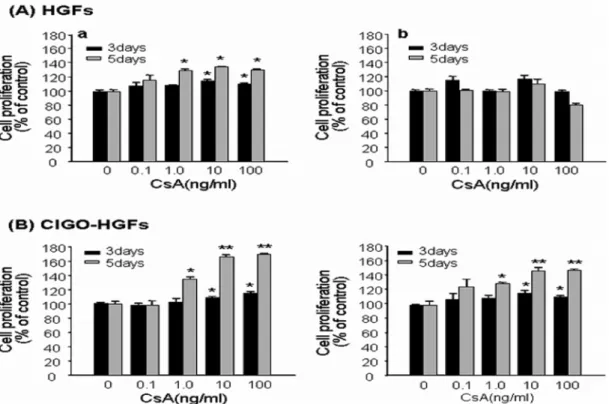
Among 5 HGFs isolated from 5 healthy subjects, the proliferation of 2 HGFs was increased meaningly by CsA treatment for 3 and 5days (Fig. 1A-a), whereas that of 3 HGFs were not changed (Fig.1A-b). In contrast, all CIGO- HGFs from 2 patients with gingival overgrowth showed significantly increased proliferation when cultured with CsA for 3 and 5 days (Fig. 1B).
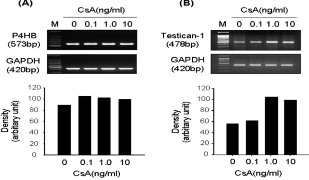
To identify the differentially expressed genes associated with CsA-induced proliferation in CIGO-HGFs, DDRT- PCR was performed. As described in Table 2, seven genes were upregulated by CsA treatment. Among them, the increased mRNA expression levels of β subunit of prolyl 4- hydroxylase (P4HB; Fig. 2A) and testican-1 (Fig. 2B) were confirmed in CsA-induced highly proliferated CIGO-HGFs Table 1. Primers.
Target genes Sequences Product size(bp)
Testican 1 Forward 5’-TGTGTGACCCAGGACTACCA-3’
Reverse 5’-ACTTGTTGAACATCCAGCCC-3’ 478
P4HB Forward 5’-GGAGATGACCAAGTACAAGC-3’
Reverse 5’-GGCTTTGCGTATTACAGTTC-3’ 573
MMP-2 Forward 5’-GTCGCCCATCATCAAGTTC-3’
Reverse 5’-CTCCCAAGGTCCATAGCTCA-3’ 557
Type I collagen Forward 5’-GGCGGCCAGGGCTCCGAC-3’
Reverse 5’-CCACGGGGTCTGGTCCTTAA-3’ 347
GAPDH Forward 5’-GTCAGTGGTGGACCTGACCT-3’
Reverse 5’-AGGGGTCTACATGGCAACTG-3’ 420
by semiquantitive RT-PCR. Furthermore, mRNA expression levels of type I collagen (Fig. 3A) and MMP-2 (Fig. 3B) were increased in CsA-induced highly proliferated CIGO- HGFs.
Discussion
Gingival overgrowth (GO), seriously interfering with occlusion, mastication, speech and dental hygiene, is Fig. 1. The effect of CsA on the proliferation of human gingival fibroblasts. Five HGFs and two CIGO-HGFs were stimulated with CsA at the indicated doses for 3 and 5 days, respectively. Cell proliferation was measured by MTT assay. Graph A shows the representatives of two HGFs indicating the increased proliferation by CsA treatment (a), and three HGFs which were not affected by CsA treatment (b). The graph B represents CsA-induced proliferation of two CIGO-HGFs. The results are expressed as the mean± SE of triplicate assays. *P < 0.05,
**P < 0.01.
Table 2. Genes upregulated by CsA treatment in CIGO-HGFs.
Clone
No. Acession No. Definition Homology
3 NM_000918.2
Homo sapiens procollagen-proline, oxoglutarate 4-dioxygenase
(proline 4-hydroxylase), beta polypeptide (protein disulfide isomerase; thyroid hormone binding protein p55) (P4HB), mRNA
100
15 AF231124.1 Homo sapiens testican 1 mRNA, complete cds 96
17 NM_000366.3 Homo sapiens tropomyosin 1 (alpha) (TPM1), mRNA 100
24 AAN37426.1
NADH dehydrogenase (ubiquinone) (EC1.6.5.3) chain 2-human mitochondrion, partial (84 %)
94
33 M94314.1 Homo sapiens ribosomal protein L30, mRNA,
complete cds 95
34 BC000690.1
Homo sapiens ribosomal protein L24, mRNA (cDNA clone MGC:2240 IMAGE:3349215), complete cds
100
36 BC040354.1
Homo sapiens, similar to caldesmon 1, clone MGC:21352 IMAGE:4753285, mRNA, complete cds
100
observed in a significant number of cases treated with immunosuppressants (CsA, etc), antiepileptics (phenytoin, etc) and calcium channel antagonists (nifedipine, etc). It has been known that the drugs resulting in GO directly or indirectly influence both the growth and function of the gingival fibroblasts, which play an important role in ECM
turnover through the production of both matrix macromolecules and matrix-degrading enzymes such as MMPs (Nares et al., 1996; Atilla and Kutukculer, 1998;
Swarga et al., 2001). Up to date, various cell culture studies have focused on connective tissue extracellular matrix accumulation to elucidate the pathogenesis of gingival
Fig. 2. mRNA expression of P4HB and testican 1 in CsA-stimulated CIGO-HGFs. Semi-quantitative RT-PCR was performed to investigate mRNA expression levels of P4HB and testican I as described in Materials and Methods.
Fig. 3. mRNA expression of type I collagen and MMP-2 in CsA-stimulated CIGO-HGFs. Semi-quantitative RT-PCR was performed to investigate mRNA expression levels of P4HB and type I collagen as described in Materials and Methods.
overgrowth.
In this study, CsA treatment increased the proliferation of two HGFs and all CIGO-HGFs, but that of three HGFs was not changed by CsA, when cultured between 0.1 ng/ml and 100 ng/ml of CsA for 3 and 5 days. Two CIGO-HGFs, rather than HGFs, proliferated more significantly by CsA stimulation. These results suggest that the priming of the gingival tissue with cyclosporin in human can lead to the increased susceptibility of the fibroblasts on CsA in vitro.
To investigate the molecular mechanisms by which CsA increases the proliferation of human gingival fibroblasts and extracellular matrix accumulation, the differentially expressed genes following CsA treatment were identified in a highly proliferating CIGO-HGF by CsA stimulation using DDRT-PCR. Among seven genes upregulated by CsA treatment, P4HB is a multifunctional protein regarded as the protein disulfide isomerase (PDI) (Koivu et al., 1987). PDI catalyzes the formation of disulfide bonds in type I and II procollagens (Koivu and Myllylä, 1987) and prolyl 4- hydroxylase (Koivu and Myllylä, 1986), a key enzyme of collagen synthesis. The function of PDI as a component of prolyl 4-hydroxylase appears to be to maintain the catalytic subunits in a soluble form rather than directly participating in catalysis, and thereby participates during proline hydroxylation (John et al., 1993). PDI also acts as a molecular chaperone assisting the folding of polypeptides (Cai et al., 1994). More recently, the association of PDI with type I procollagen has been proposed to lead to endoplasmic reticulum retention of type I procollagen, which is composed of two proα1(I) chains and one proα2(I) chain and intracellularly degraded immediately after its synthesis, in corneal endothelial cells (Ko et al., 2004). Furthermore, it has been reported that the proyl 4-hydroxylase inhibitor is more effective for the inhibition of proliferation than for inhibition of collagen synthesis of rat hepatic stellate cells (Aoyagi et al., 2002). Therefore, our data demonstrate that the overexpression of PDI mRNA by CsA in CIGO-HGFs may increase cell proliferation while producing collagen, by stabilizing prolyl 4-hydroxylase and delaying the intracellu- lar degradation of type I procollagen. In addition, these results can provide the molecular basis of other in vitro studies that CsA causes a significant increase in the level of type I procollagen and cell proliferation (Bolzani et al., 2000).
Testican-1 is a highly conserved chimeric proteoglycan carrying both chondroitin sulfate and heparan sulfate chains (Alliei et al., 1993). Testican-1 has been shown to inhibit the activation of pro-MMP-2 by either MT1-MMP or MT3- MMP (Nakada et al., 2001) as well as the activity of lysosomal enzyme cathepsin L (Bocock et al., 2003), regulating the degradation of extracellular matrices (Nakada et al., 2003). Recent studies have reported that the reduced activity of cathepsin-L (Yamada et al., 2000) and matrix metalloproteinases (Bozani et al., 2000; Cotrim et al., 2002;
Hyland et al., 2003; Gangliano et al., 2004) by CsA,
resulting in the inhibition of protein degradation in gingival connective tissues, plays important roles in the pathogenesis of CIGO. Our result suggests that the suppression of MMP- 2 expression in CIGO can be due to the overexpression of testican-1 by CsA. Furthermore, alterations of type I collagen and MMP-2 expression following the upregulation of P4HB and testican-1 mRNA levels were assessed in CIGO-HGF cells treated with CsA by RT-PCR. We confirmed that CsA increased type I collagen mRNA levels and suppressed MMP-2 mRNA levels.
In conclusion, CsA may induce the gingival overgrowth through the upregulation of P4HB and testican-1, resulting in the increase of collagen synthesis and the accumulation of extracellular matrix components by inhibiting MMP-2 activity, in patients receiving CsA therapy. This is the first report demonstrating that the accumulation of collagen in CIGO regulates by P4HB and testican-1.
Acknowledgement
This study was supported by a grant of the Korea Health 21 R&D Project, Ministry of Health & Welfare, Republic of Korea (01-PJ5-PG3-20507-0020).
References
Alliel PM, Perin JP, Jolles P, Bonnet F. Testican, a multidomain testicular proteoglycan resembling modulators of cell social behaviour. Eur J Biochem. 1993:214:347-50.
Aoyagi M, Sakaida I, Suzuki C, Segawa M, Fukumoto Y, Okita K. Prolyl 4-hydroxylase inhibitor is more effective for the inhibition of proliferation than for inhibition of collagen synthesis of rat hepatic satellite cells. Hepatol Res.
2002;23:1-6.
Atilla G, Kutukculer N. Cervicular fluid interleukin-1β, tumor necrosis factor-α, and interleukin-8 levels in renal transplant patients receiving cyclosporin A. J Periodontol. 1998;69:784-90.
Barber MT, Savage NW, Seymour GJ. The effect of cyclosporin and lipopolysaccharide on fibroblasts: implications for cyclosporin-induced gingival overgrowth. J Periodontol.
1992;63:397-404.
Bartold PM. Regulation of human gingival fibroblast growth and synthetic activity by cyclosporin-A in vitro. J Periodont Res. 1989;24:314-21.
Bocock JP, Edgell CS, Marr HS, Erickson AH. Human proteoglycan testican-1 inhibits the lysosomal cysteine protease cathepsin L. Eur J Biochem. 2003;270:4008-15.
Bolzani G, Della Coletta R, Martelli Junior H, Martelli Junior H, Graner E. Cyclosporin A inhibits production and activity of matrix metalloproteinases by gingival fibroblasts. J Periodont Res. 2000;35:51-8.
Bonnaure-Mallet M, Tricot-Doleux S, Godeau GJ. Changes in extracellular matrix macromolecules in human gingival after treatment with drugs inducing gingival overgrowth. Arch
Oral Biol. 1995;40:393-400.
Cai H, Wang CC, Tsou CL. Chaperone-like activity of protein disulfide isomerase in the refolding of a protein with no disulfide bonds. J Biol Chem. 1994;269:24550-2.
Cotrim P, de Andrade CR, Martelli-Junior H, Graner E, Sauk JJ, Coletta RD. Expression of matrix metalloproteinases in cyclosporin-treated gingival fibroblasts is regulated by transforming growth factor (TGF)-beta1 autocrine stimulation.
J Periodontol. 2002;73:1313-22.
Gagliano N, Moscheni C, Dellavia C, Torri C, Stabellini G, Ferrario VF, Gioia M. Effect of cyclosporin A on human gingival fibroblast collagen turnover in relation to the development of gingival overgrowth: an in vitro study.
Biomed Pharmacother. 2004;58:231-8.
Hyland PL, Traynor PS, Myrillas TT, Marley JJ, Linden GJ, Winter P, Leadbetter N, Cawston TE, Irwin CR. The effects of cyclosporin on the collagenolytic activity of gingival fibroblasts, J Periodontol. 2003;74:437-45.
John DC, Grant ME, Bulleid NJ. Cell-free synthesis and assembly of prolyl 4-hydroxylase: the role of the beta- subunit (PDI) in preventing misfolding and aggregation of the alpha-subunit. EMBO J. 1993;12:1587–95.
Kim JY, Park SH, Cho KS, Kim HJ, Lee CK, Park KK, Choi SH, Chung WY. Mechanism of azithromycin treatment on gingival overgrowth. J Dent Res. 2008;87:1075-9.
Ko MK, Kay EP. PDI-mediated ER retention and proteasomal degradation of procollagen I in corneal endothelial cells.
Exp Cell Res. 2004;295:25-35.
Koivu J, Myllylä R. Protein disulfide-isomerase retains procollagen prolyl 4-hydroxylase structure in its native conformation. Biochemistry. 1986;25:5982-6.
Koivu J, Myllylä R. Interchain disulfide bond formation in types I and II procollagen. Evidence for a protein disulfide isomerase catalyzing bond formation. J Biol Chem.
1987;262:6159-64.
Koivu J, Myllylä R, Helaakoski T, Pihlajaniemi T, Tasanen K, Kivirikko KI. A single polypeptide acts both as the beta subunit of prolyl 4-hydroxylase and as a protein disulfide- isomerase. J Biol Chem. 1987;262:6447-9.
Nakada M, Miyamori H, Yamashita J, Sato H. Testican 2
abrogates inhibition of membrane-type matrix metallopro- teinases by other testican family proteins. Cancer Res.
2003;63:3364-9.
Nakada M, Yamada A, Takino T, Miyamori H, Takahashi T, Yamashita J, Sato H. Suppression of membrane type I matrix metalloproteinase (MMP)-mediated MMP-2 activation and tumor invasion by testican 3 and its splicing variant gene product, N-Tes. Cancer Res. 2001;61:8896-902.
Nares S, Na MG, Dill RE, Cutler CW, Lacopino AM.
Cyclosporin A upregulates platelet-derived growth factor B chain in human hyperplastic gingiva. J Periodontol.
1996;67:271-8.
Park JI, Lee GS, Jeong YJ, Kim BK, Kim JH, Lim HS, Kim SH, Kim WJ, Jung JY. Involvement of antiapoptotic signals in rat PC12 cells proliferation by cyclosporin A treatment.
Int J Oral Biol. 2007;32:51-7.
Schincaglia GP, Forniti F, Cavallini R, Piva R, Calura G, Del Senno L. Cyclosporin-A increases type I procollagen production and mRNA level in human gingival fibroblasts in vitro. J Oral Pathol Med. 1992;21:181-5.
Swarga JD, Mohamed HP, Irwin O. Upregulation of keratinocyte growth factor in cyclosporin A-induced gingival overgrowth. J Periodontol. 2001;72:745-52.
Tipton DA, Stricklin GP, Dabbous MK. Fibroblast hetero- geneity in collagenolytic response to cyclosporine. J Cell Biochem. 1991;46:152-65.
Tyldesley WR, Rotter E. Gingival hyperplasia induced by cyclosporin A. Brit Dent J. 1984;157:305-9.
Wysocki GP, Gretzinger HA, Laupacis A, Ulan RA, Stiller CR. Fibrous hyperplasia of the gingiva: A side effect of cyclosporin A therapy. Oral Surg Oral Med Oral Pathol.
1983;55:274-8.
Yamada H, Nishimura F, Naruishi K, Chou HH, Takashiba S, Albright GM, Nares S, Lacopino AM, Murayama Y.
Phenytoin and cyclosporin A suppress the expression of MMP-1, TIMP-1, and cathepsin L, but not cathepsin B in cultured gingival fibroblasts. J Periodontol. 2000;71:955-60.