231
<원례보저>
Cytokine expression pattern in milk somatic cells of subclinical mastitis-affected cattle analyzed by real time PCR
Vaibhav D. Bhatt
1, Prasad S. Khade
2, Sagar B. Tarate
3, Ajai K. Tripathi
4, Dev S. Nauriyal
2, Dharamshi N. Rank
3, Anju P. Kunjadia
1,*, Chaitanya G. Joshi
41
Ashok and Rita Patel Institute of Integrated Study and Research in Biotechnology and Allied Sciences, Anand 388120, Gujarat, India
Departments of
2Veterinary Medicine, and
3Animal Genetics and Breeding, and
4
Animal Biotechnology, College of Veterinary Science and Animal Husbandry, Anand Agricultural University, Anand 388001, Gujarat, India
(Received: March 20, 2012; Revised: October 19, 2012; Accepted: October 25, 2012)
Abstract : The expression profiles of inflammatory cytokines viz. interleukins (IL)-6, IL-8, IL-12, granulocyte macrophage-colony stimulating factor, interferon-γ and tumor necrosis factor-α in response to subclinical mastitis in indigenous cattle breed Kankrej (n = 6), Gir (Bos indicus) (n = 12) and crossbred (Bos taurus × Bos indicus) (n = 7) were investigated using quantitative real time PCR. Significant correlation (p < 0.05) was observed between total bacterial load and somatic cell count (SCC) in all three breeds of cattle. All the cytokines were observed to be up-regulated compared to cows with healthy quarters, however, level of their expression varied among three breeds of cattle. In Kankrej most cytokines were found to be transcribed to higher levels than in other two breeds; the milk had higher load of bacteria but not so high SCC, implying that Kankrej has a higher inherent resistance against mastitis. The results of present study indicated that mammary glands of crossbred cattle are more sensitive to bacterial infection than indigenous breed of cattle as they elicit immune response at lower bacterial load and result into higher SCC. Research on identification of factors responsible for differentially expressed cytokines profiles and use of cytokines as immunomodulatory tools can pave way for formulating control strategies against bovine mastitis.
Keywords : cytokines, interleukins, interferon, quantitative real time PCR
Introduction
Mastitis is one of the most prevalent and costly diseases affecting the dairy industry worldwide [5, 30, 38]. Costs mainly arise from decreased milk production and poor milk quality, therapeutic interventions, loss of antibiotic contami- nated milk and the cost incurred on extra labour [11].
Kankrej cattle are a breed of Zebu cattle from Gujarat state located on the west coast of India [24]. Another native breed of cattle from Gujarat is Gir which is a famous milk breed of India. During the last few years, Brazil - a South American nation - has exploited the production potential of Gir cows and consequently has emerged as world’s biggest supplier of improved embryos and semen of cattle of Indian origin. Of the two indigenous breeds, Kankrej cattle are known to be sturdy and aggressive whereas Gir cattle appear predisposed to prolapse of prepucial sheath in male animals [14], and cer- vico-vaginal prolapse mastitis in female animals [35].
Of the two forms of mastitis, subclinical mastitis is subtle,
causes huge economic losses, and is difficult to detect as the cow appears healthy, the udder does not show any signs of inflammation and the milk appears normal. However, micro- organisms and somatic cells are found in elevated numbers in the milk. The cows that have subclinical mastitis serve as reservoir of organisms and may result in infection to healthy cows. Most clinical cases start as subclinical and therefore, control of subclinical mastitis is extremely important to check it’s development to clinical cases and curtailing heavy economic losses.
In recent years, the role of cytokines in the patho-physiol- ogy of bovine mastitis has been the subject of many studies.
Cytokines are immunoregulatory mediators that play a cen- tral role in the regulation of immune responses against differ- ent infections [7]. Several studies have indicated that variations in cytokine expression are associated with disease activity in immune-mediated or inflammatory disorders [16, 26]. The use of cytokines in immunotherapy of bovine mastitis or as adjuvants in the immunopotentiation of mammary gland has
*Corresponding author
Tel: +91-9426-583535, Fax: +91-2692-229189 E-mail: [email protected]
revealed their important role in the regulation of the mam- mary gland defences [15]. Understanding the regulation of the immune response during infections of the mammary gland is important for designing prophylactic vaccines and for optimi- zation of therapeutic protocols. Certain aspects of immune mechanisms in cattle and their regulation by cytokines and growth factors have already been addressed [33, 34]. How- ever, the interplay of immune compartments involved in the generation of protective immunity against specific bacterial infections is yet to be fully understand. Therefore, the objective of this study was to define the cytokine profile of diseased and healthy cows during lactation and investigate the correlation between somatic cell count (SCC) and total bacterial load as well as levels of transcription of different cytokines in Gir, Kankrej and crossbred cattle using quantitative real time PCR.
Materials and Methods
Animals and preparation of milk samples
The milk samples were collected from the cattle farms in and around the Anand city of Gujarat state, India. Milk sam- ples were collected for three consecutive days aseptically in sterile wide mouth glass stopper bottles. Before sample col- lection the udder was thoroughly washed with potassium per- manganate solution (1 : 1,000) and the teats were swabbed with 70% ethyl alcohol.
Ten crossbred (Holstein Friesian × Jersey × Kankrej), 30 Kankrej (Bos indicus) and 30 Gir (Bos indicus) cows from each breed were taken as experimental material. The animals were selected based on milk SCC and the presence of bacte- ria in the milk for three consecutive days.
Microbiological cultures
All milk samples were evaluated by the 1 µL calibrated loop method for microbiological growth in sheep blood agar medium and McConkey’s medium. The staining and cellular morphological features of organisms were ascertained by microscopic examination of Gram stained smears. By follow- ing standard laboratory policy, the inoculated plates were examined daily for the growth of potential organisms.
Total DNA Extraction
Thirty mL of milk from each sample was diluted ten times with sterile water. Milk was strained to remove the somatic cells from the milk and centrifuged at 1,000 ×g for 15 min in 50 mL tubes. The pellet was collected after discarding fat layer and the supernatant and washed twice in sterile normal saline solution (pH 7.2) prepared in diethylpyrocarbonate (DEPC) treated water. The pellet was suspended in 150 µL of normal saline solution. DNA was extracted using the pro- tocol described by Cremonesi et al. [10].
Preparation of DNA standards for determining bac- terial load
Bacterial specific universal primers described by Muyzer
et al. [28] were used for amplification of target region from pooled DNA. PCR amplified products were cloned using InsT/Aclone PCR product cloning kit (Fermentas, Lithuania) and the vector containing the insert was propagated in Escherichia (E.) coli host (DH5- α). Plasmid was extracted using QIAprep Miniprep kit (Qiagen, Germany) and copy number of plasmid in the solution were calculated using fol- lowing formula:
Where, 660 = Molecular weight of nucleotide base pair 6.022 × 10
23= Avogadro’s number
Recombinant plasmids were diluted 10
2- 10
8to prepare standard curve for determining bacterial count by Real time PCR.
Isolation of milk somatic cells
Aliquots of 25 mL milk were used to determine SCC using a Fossomatic milk cell counter (Foss, Denmark). The remain- der of the milk was centrifuged at 1,000 ×g for 15 min at room temperature. Fat layer and the supernatant were dis- carded and the cell pellets were washed twice in 50 mL phos- phate buffered saline.
Extraction of RNA and cDNA Synthesis
Somatic cells were collected by centrifugation and RNA was extracted as described by Chomczynski and Sacchi [9].
The RNA template was qualitatively assessed and quantified using 2100 Bioanalyzer (Agilent technologies, USA) with the RNA 6000 Nano Labchip kit (Agilent technologies).
Reverse transcription reactions were performed following the manufacturer’s instructions using Sensiscript Reverse Tran- scriptase kit (Qiagen, USA) for 20- µL reactions using a fixed amount of input RNA for each cDNA reaction.
Real-Time PCR
All PCR reactions were performed in optical 96 well plates using the ABI Prism 7500 Fast Sequence Detection System (Applied Biosystems, USA). In each reaction, approximately 20 ng of reverse-transcribed RNA (based on the initial RNA concentration) was used for real time PCR. Glyceraldehyde 3-phosphate dehydrogenase (GAPDH) and β-actin was used as an endogenous control. The primers were synthesized as described by Lee et al. [22]. The reactions were performed as initial step 95
oC for 5 min, followed by 45 cycles of 95
oC for 30 sec, 58
oC for 30 sec and 72
oC for 30 sec in a 20- µL reac- tion volume. For each sample a dissociation curve was gen- erated after completion of amplification and analyzed to determine the specificity of PCR reaction. All reactions were run in triplicate and quantification cycle (Cq) values for tar- get genes were normalized by the internal positive control.
Copy No. / µL =
Concentration of plasmids (g/µL)
× 6.022 × 10
23Length of recombinant plasmid (bp) × 660
To determine the amplification efficiencies of reference gene and the cytokine cDNAs, six dilutions of cDNA prepara- tions in triplicate were amplified to obtain standard curves.
Differences of the slopes between standard curves obtained from reference gene the cytokines were plotted against the dilution of input total RNA and the regression line was cal- culated.
Cytokine quantification was achieved using the cycle threshold (Ct) comparative method, and is expressed as “n- fold up regulation of cytokine transcription” in relation to a calibrator which is represented by the smallest signal detect- able for that specific cytokine. The expression of each gene was analyzed using the relative quantification method. For relative quantitation by the comparative CT method, values are expressed relative to a reference sample, called the cali- brator (Samples from healthy cattle). The CT for the target gene and the CT for the internal control were determined for each sample and the calibrator. The expression of selected cytokine genes was calibrated by that of the reference gene, bovine GAPDH and β-actin, at each time point and con- verted to the relative expression ratio (fold of induction).
Statistical analysis
The data were analyzed using SAS (ver. 4.1; SAS insti- tute, USA) software. The methods of analysis of variance (ANOVA) and Pearson’s correlation analysis were used in the statistical analysis.
Results
On screening ten crossbred, thirty Kankrej and thirty Gir cows, 7, 6 and 12, respectively were identified harbouring subclinical mastitis. Individual colony characteristics and haemolytic patterns on blood agar were noted. The isolated bacteria were identified on the basis of their cultural, mor- phological and biochemical characteristics. The results revealed that E. coli, an environmental mastitis pathogen, was the commonest organism among the milk samples from Kankrej and Gir cattle. In case of crossbred cows, however, Staphylococcus spp. and Streptococcus spp. were identified as predominant contagious pathogens.
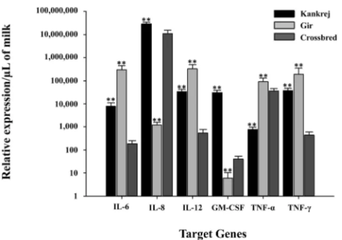
The real time PCR system was used to assess cytokine pro- files in somatic cells extracted in the milk samples from healthy and infected quarters with the amplification effi- ciency of 79% to 105%. GAPDH showed least expression variations among different samples and breeds, however, β- actin exhibited unstable expression pattern among all the samples, Based on this, GAPDH was considered as potential endogenous control. Compared to the healthy quarters, all the cytokines were observed to be up-regulated in quarters affected with subclinical mastitis in all three breeds (Fig. 1). The average fold induction of all targeted cytokines was altered in response to bacterial challenges. In crossbred cattle the over- all apparent expression of cytokines was lower than in Kankrej and Gir cattle.
The total bacterial load was found to be 1.49 ± 0.036 × 10
9, 6.41 ± 0.382 × 10
8and 1.12 ± 0.055 × 10
8cells/mL in Kankrej, Gir and crossbred cattle, respectively. The average SCC count in three breeds was found to be 2.37 ± 0.49 × 10
6, 2.74 ± 0.31 × 10
6and 2.79 ± 0.74 × 10
6cells/mL, respectively.
The geometric mean of SCC and total bacterial load in the milk of Kankrej, Gir and crossbred cattle is given in Fig. 2.
Data of transcriptional activity of all the cytokines, SCC and total bacterial load were analyzed to assess breed varia- tion among Kankrej, Gir and crossbred cattle. Homologous means were compared using one-way ANOVA analysis.
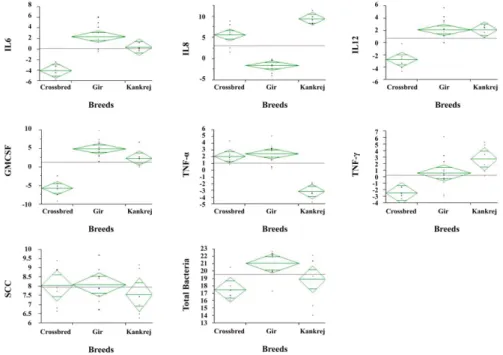
Highly significant difference (p < 0.01) was observed with regard to all the cytokines and total bacterial count, however, the difference in SCC among all three breeds of cattle was non-significant (Fig. 3). The highly significant positive corre- lation between SCC and total bacterial count indicated that
Fig. 1. Relative Quantification (means± SE) of transcriptional level of target genes in Kankrej, Gir and crossbred cattle during subclinical mastitis compared to healthy. Fold expression/cell was converted to fold expression/µL of milk by multiplying fold expression/cell value with the somatic cell count (SCC)/µL. **p < 0.01.
Fig. 2. SCC and total bacteria present in the milk of Kankrej, Gir and crossbred cattle.
Fig. 3. The continuous by nominal/ordinal scatter plot with means diamonds shows significant difference in all the cytokines and total bacteria, whereas SCC showed non-significant difference between all the breeds. Line across each diamond represents the group mean.
The vertical span of each diamond represent the 95% confidence interval for each group. The data were log transformed for ANOVA.
Table 1. Pearson’s correlations between and within cytokine transcriptional activity, total bacteria and somatic cell count (SCC) present in milk of Kankrej, Gir and crossbred cattle
IL-6 IL-8 IL-12 GM-CSF TNF-á IFN-ã Total Bacte-
ria SCC
IL-6 Kankrej 1 -0.032 .996** .979** 0.703 .946* 0.845 .992**
Gir 1 0.189 -0.131 -0.206 0.469 0.279 -0.211 -0.244
Crossbred 1 .939** 0.445 0.245 0.311 0.29 .945** .824*
IL-8 Kankrej -0.032 1 -0.075 -0.149 0.662 -0.125 -0.17 -0.017
Gir 0.189 1 .900** -0.16 0.198 -0.192 0.566 .764**
Crossbred .939** 1 0.622 0.356 0.55 0.577 .808* 0.656
IL-12 Kankrej .996** -0.075 1 .986** 0.667 .971** 0.866 .987**
Gir -0.131 .900** 1 -0.067 -0.029 -0.157 0.573 .797**
Crossbred 0.445 0.622 1 0.065 0.424 0.64 0.308 0.256
GM-CSF Kankrej .979** -0.149 .986** 1 0.577 .969** 0.789 .952*
Gir -0.206 -0.16 -0.067 1 -0.056 -0.143 0.112 0.239
Crossbred 0.245 0.356 0.065 1 .856* 0.574 0.024 -0.141
TNF-α Kankrej 0.703 0.662 0.667 0.577 1 0.572 0.573 0.731
Gir 0.469 0.198 -0.029 -0.056 1 -0.127 -0.051 -0.037
Crossbred 0.311 0.55 0.424 .856* 1 .905** 0.015 -0.146
IFN-γ Kankrej .946* -0.125 .971** .969** 0.572 1 0.841 .926*
Gir 0.279 -0.192 -0.157 -0.143 -0.127 1 -0.41 -0.35
Crossbred 0.29 0.577 0.64 0.574 .905** 1 -0.008 -0.062
Total Bacteria Kankrej 0.845 -0.17 0.866 0.789 0.573 0.841 1 .893*
Gir -0.211 0.566 0.573 0.112 -0.051 -0.41 1 .895**
Crossbred .945** .808* 0.308 0.024 0.015 -0.008 1 .893**
SCC Kankrej .992** -0.017 .987** .952* 0.731 .926* .893* 1
Gir -0.244 .764** .797** 0.239 -0.037 -0.35 .895** 1
Crossbred .824* 0.656 0.256 -0.141 -0.146 -0.062 .893** 1
The significant correlations are expressed in bold. *Correlation is significant at the 0.05 level (2-tailed). **Correlation is significant at the 0.01 level (2-tailed).
the intensity of mastitis (as reflected by SCC) increased with the level of bacterial load in all the three breeds (Table 1).
The correlation of transcriptional activity of cytokines, total bacterial load and SCC in Kankrej, Gir and crossbred cattle revealed that in Kankrej cattle SCC was significantly (p < 0.05) and positively correlated to interleukins (IL)-6, IL- 12, granulocyte macrophage-colony stimulating factor (GM- CSF), and interferon (IFN)- γ, whereas total bacterial load exhibited non-significant positive correlation with all the cytokines, except IL-8. As for remaining cytokines, IL-6, IL- 12, GM-CSF, tumor necrosis factor (TNF)- α and IFN-γ showed significant and positive correlation among each other (Table 1).
In Gir cattle SCC was significantly (p < 0.01) and posi- tively correlated to IL-8 and IL-12 only, whereas total bacte- rial load did not show significant correlation with any of the cytokines. Correlations among the cytokines were mostly non-significant and negative, except between IL-8 and IL-12 which was significant and positive (Table 1). In crossbred cattle, SCC showed significantly (p < 0.01) positive correla- tion only with IL-6, whereas total bacterial load was found to have significantly positive correlation with IL-6 and IL-8.
Significant and positive correlations were also observed between IL-6 and IL-8, GM-CSF and TNF- α, and TNF-α and IFN-γ.
Discussion
Mastitis not only causes inflammation in the parenchyma of the mammary gland, but it also affects the composition of milk and the degree of changes depends on the infective agent and the inflammatory response. Indicators of inflam- mation in the milk can be determined by using rapid, pre- cise, reliable and easy to perform markers for early detection of mastitis. SCC is one such indicator which showed highly significant correlation with total bacterial load in the present study. Cytokines are a sensitive means to study the immune response of mammary gland and serve as a suitable tool to monitor udder health to evaluate efficacy of treatment of mastitis or to test efficiency of a vaccine against mastitis pathogens. The immunoregulatory role of cytokines in mobi- lizing innate and specific immunity of bovine mammary gland is well documented [33]. Cytokines of bovine mammary gland have been considered useful markers in defining mam- mary gland defenses [15].
Many cellular functions are regulated by changes in gene expression. Thus, quantification of transcription levels of genes plays a central role in the understanding of gene func- tion and of abnormal alterations in regulation that may result in a disease state. In the present study, transcriptional activ- ity of bovine cytokines was measured using quantitative real- time PCR systems in milk cells of Kankrej, Gir and cross- bred cattle. The overall apparent transcriptional activity of cytokines in the milk cells of crossbred cattle was lower than in Kankrej and Gir cattle.
Detection of IL-6 in milk can be used as a marker for sub-
clinical mastitis. The average IL-6 concentration has been reported to be significantly higher in mastitic milk than in milk from healthy quarters [32]. In our study the expression of IL-6 was observed to be highest in Gir, followed by Kankrej, and the least in crossbred cattle. The activity of IL-6 increases with response to coliform infection [19, 29]. IL-6 is also induced by other gram negative bacterial infections. In case of Gir and Kankrej cattle, the gram negative bacterial (E. coli) infections were predominant whereas in crossbred cattle gram positive organisms (Staphylococcus spp. and Streptococcus spp.) prevailed. Perhaps this explains as to why IL-6 was probably not expressed in crossbred cattle as profoundly as in Kankrej and Gir. The role of IL-6 as an anti- inflammatory cytokine has been shown to mediate through its inhibitory effects on TNF- α [39] which, however, was not observed in the present study. In Kankrej, Gir and crossbred cattle, IL-6 and TNF- α showed positive, though non-signifi- cant correlation indicating no inhibitory effect of IL-6 on TNF- α. IL-6 also acts synergistically with GM-CSF [8]. IL-6 showed positive and significant correlation with GM-CSF only in Kankrej, whereas in other two breeds it was non-sig- nificant.
IL-8 is a chemokine produced by macrophages and epithe- lial cells in response to bacterial invasion. IL-8 mediates neu- trophil function by allowing neutrophils to resolve bacterial infections by migrating through blood vessel walls to the site of infection [21]. The expression of IL-8 is induced by both, exogenous (e.g. bacteria, viruses, fungi, parasite and prod- ucts derived from these pathogens) and endogenous (e.g.
TNF- α, and IL-1β) proinflammatory stimuli [25, 27]. In the present experiment, IL-8 and TNF- α showed positive but non-significant correlation in all three breeds. The transcrip- tional level of IL-8 mRNA was found to be higher in Kankrej and crossbred than in Gir cattle, while Kankrej cat- tle exhibited high total bacterial load. Crossbred cattle showed higher IL-8 expression in spite of low total bacterial load suggesting the response of the breed at lower bacterial load by attracting more neutrophils at the site of infection.
Kankrej, on the other hand, showed elevated IL-8 expression at higher bacterial load.
Interleukin-12 is naturally produced by dendritic cells and
macrophages. It has an anti-angiogenic activity which it elic-
its by increasing production of IFN- γ [36]. In a study IL-12
has been reported to have shown a significant elevation in
mammary gland experimentally infected with Staphylococ-
cus (S.) aureus 24 h post-infection [2]. The elevation of IL-
12 transcriptional activity probably indicates its vital role in
enhancing innate immunity response in mammary gland,
especially the activation of natural killer cells through the
augmentation of IFN- γ synthesis. In the present study, aug-
mentation of IFN- γ activity with IL-12 was significant in
Kankrej but not in crossbred and Gir cattle. In Kankrej IL-8,
and in crossbred IL-12, did not show significant correlation
with any other cytokines, whereas in Gir, IL-8 and IL-12
showed significant correlation (p < 0.01). This perhaps indi-
cates to the response of individual breed to infection. Tran- scriptional activity of IL-12 was observed to be high in Gir, followed by Kankrej, and the least in crossbred cattle.
In vitro, GM-CSF has been shown to increase the effecter activities of phagocytic cells (neutrophils, macrophages, eosi- nophils, and basophils). Because an early and rapid regres- sion of functionally competent neutrophils from the blood stream is critical for the control of new intra-mammary infec- tion, enhancement of chemotaxis and phagocytosis of neutro- phils with GM-CSF, as well as the kinetics of their induction, could increase resistance of mammary gland against invad- ing pathogens. In the present study, Kankrej and Gir cattle showed higher level of transcriptional activity of GM-CSF as compared to crossbred cattle. In an earlier study GM-CSF was reported to be significantly correlated with TNF- α in late and mid lactation period [1]. Interestingly GM-CSF was positive and significantly correlated to IFN- γ and TNF-α in Kankrej and crossbred, respectively but not in Gir cattle.
Similarly, correlations among IL-6, IL-8 and IL-12 were also not consistent between breeds.
TNF- α is a cytokine involved in systemic inflammation which stimulates acute phase reaction. Increased concentra- tion of TNF- α in milk cells has been reported in earlier stud- ies on E. coli mastitis [4, 18]. It was also reported in earlier studies [4] that TNF- α concentration did not peak before the manifestation of clinical signs but was detected at the time of appearance of systemic signs and later than the local signs. In case of mastitis caused by gram-negative bacteria, patho- physiological responses to endotoxins are thought to occur indirectly through the release and subsequent absorption of endogenous mediators from infected mammary glands into the circulation [23]. Cytokine TNF- α is induced by endot- oxin and plays an important role in the development of acute inflammatory response [13]. In the present study, the level of TNF- α expression was higher in Gir and crossbred cattle as compared to Kankrej cattle which may have occurred as a consequence of E. coli infection. The morbidity associated with acute coliform mastitis may have been reduced because of the less activity of TNF- α in Kankrej cattle. TNF-α showed positive correlations (p > 0.05) with all the targeted cytokines in Kankrej cattle, however, the correlation was sig- nificant (p < 0.05) with only GM-CSF and IFN- γ in cross- bred cattle.
Increases in IFN- γ mRNA have been detected in cells iso- lated from the milk of mammary glands infected with E. coli [22] and S. aureus [31]. During the course of naturally occur- ring mastitis [17], as well as in the induction of experimental mastitis by E. coli, Mycoplasma bovis, S. aureus, Pseudomo- nas aeruginosa, Serratia marcescens, and Streptococcus uberis [3, 20], increase in milk protein concentrations of IFN- γ have also been detected. In addition, IL-12 induces production of IFN- γ [36]. In our study, the transcriptional activity of IFN- γ showed positive and significant correlation with IL-12 in Kankrej, whereas the correlation was positive and non-significant in crossbred, and negative and non-sig-
nificant in Gir cattle.
SCC was significantly and positively correlated to all the cytokines except IL-8 studied in Kankrej, with IL-8 and IL- 12 in Gir, and with IL-6 in crossbred cattle. Total bacterial load was significantly and positively correlated to IL-6 and IL-8 in crossbred, however, in indigenous breeds none of the cytokines exhibited significant correlation with total bacte- rial load. The ability of bacteria to establish infection is mediated in part by the ability of host to respond to the invading organism [6]. It is difficult to associate the increase in milk SCC with individual bacterial species, however, a close association between total bacteria and milk SCC was evident in the study. In Kankrej and Gir cattle the total bac- terial load was much higher than corresponding SCC in the same quarters. Kankarej and Gir breeds are well adapted to local conditions and possess good disease resistance. It can be inferred that crossbred cattle are much more sensitive to bacterial infection of mammary glands than indigenous breeds of cattle, elicit immune response at lower bacterial load, and result into higher SCC which is substantiated by the reports of considerably high incidence of subclinical mas- titis in crossbred cows [12, 37].
In conclusion, milk cell RNA preparations can be used to study mammary gland dynamics. This provides a convenient and improved alternative to collection of biopsy samples and allows easy and repetitive sampling without damaging mam- mary tissue. Quantitative real-time PCR system has been used successfully to study immunological status of bovine udder during mastitis. In the three cattle breeds included in the present investigation, all the cytokines were observed to be up-regulated in subclinical mastitis compared to healthy quarters, however, the level of their expression varied among the breeds. In Kankrej, most cytokines were found to be tran- scribed to higher levels than in other breeds, with the milk showing higher bacterial load and low SCC possibly imply- ing that Kankrej has a higher inherent resistance against mas- titis. The current study adds to the understanding of specific and nonspecific immune mechanisms involved in the mam- mary gland defense against invading bacteria which, in turn, may lead to use of cytokines in the therapy and development of new vaccines against mastitis pathogens thus paving way for effective control of bovine mastitis.
Acknowledgments
Farmers and field Veterinarians of Anand Agricultural Uni- versity (India) are thankful for their cooperation in this study.
We thankful to AMUL for providing the facility of somatic cell counter.
References
1. Alluwaimi AM, Cullor JS. Cytokines gene expression patterns of bovine milk during middle and late stages of lactation. J Vet Med B Infect Dis Vet Publich Health 2002, 49, 105-110.
2. Alluwaimi AM, Leutenegger CM, Farver TB, Rossitto PV, Smith WL, Cullor JS. The cytokine markers in Staphylococcus aureus mastitis of bovine mammary gland. J Vet Med B Infect Dis Vet Public Health 2003, 50, 105-111.
3. Bannerman DD, Chockalingam A, Paape MJ, Hope JC.
The bovine innate immune response during experimentally- induced Pseudomonas aeruginosa mastitis. Vet Immunol Immunopathol 2005, 107, 201-215.
4. Blum JW, Dosogne H, Hoeben D, Vangroenweghe F, Hammond HM, Bruckmaier RM, Burvenich C. Tumor necrosis factor-alpha and nitrite/nitrate responses during acute mastitis induced by Escherichia coli infection and endotoxin in dairy cows. Domest Anim Endocrinol 2000, 19, 223-235.
5. Bradley A. Bovine mastitis: an evolving disease. Vet J 2002, 164, 116-128.
6. Burvenich C, Van Merris V, Mehrzad J, Diez-Fraile A, Duchateau L. Severity of E. coli mastitis is mainly determined by cow factors. Vet Res 2003, 34, 521-564.
7. Campos M, Godson DL, Hughes HPA, Babiuk LA.
Cytokine application in infectious diseases. In: Goddeeris BML, Morrison WI (eds.). Cell-mediated Immunity in Ruminants. 1st ed. pp. 229-37. CRC Press, Boca Raton, 1994.
8. Caracciolo D, Clark SC, Rovera G. Human interleukin-6 supports granulocytic differentiation of hematopoietic progenitor cells and acts synergistically with GM-CSF. Blood 1989, 73, 666-670.
9. Chomczynski P, Sacchi N. The single-step method of RNA isolation by acid guanidinium thiocyanate-phenol- chloroform extraction: twenty-something years on. Nat Protoc 2006, 1, 581-585.
10. Cremonesi P, Castiglioni B, Malferrari G, Biunno I, Vimercati C, Moroni P, Morandi S, Luzzana M. Technical note: Improved method for rapid DNA extraction of mastitis pathogens directly from milk. J Dairy Sci 2006, 89, 163-169.
11. DeGraves FJ, Fetrow J. Economics of mastitis and mastitis control. Vet Clin North Am Food Anim Pract 1993, 9, 421-434.
12. Devi BK, Shukla PC, Bagherwal RK. Incidence of sub- clinical mastitis in cows. Indian J Dairy Sci 1997, 50, 477- 478.
13. Dinarello CA. Molecular mechanisms in endotoxin fever.
Agents Actions 1983, 13, 470-486.
14. Gaur GK, Kaushik SN, Garg RC. The Gir cattle breed of India - characteristics and present status. Anim Genet Resour Inf 2003, 33, 21-29.
15. Godson DL, Baca-Estrada ME, Babiuk LA. Application of bovine cytokines. In: Schijns VECJ, Horzinek MC (eds.). Cytokines in Vetetinary Medicine. 1 st ed. pp. 49, CAB International, New York, 1977.
16. Hansen PJ, Soto P, Natzke RP. Mastitis and fertility in cattle - possible involvement of inflammation or immune activation in embryonic mortality. Am J Reprod Immunol 2004, 51, 294-301.
17. Hisaeda K, Hagiwara K, Eguchi J, Yamanaka H, Kirisawa R, Iwai H. Interferon-gamma and tumor necrosis factor-α levels in sera and whey of cattle with naturally occurring coliform mastitis. J Vet Med Sci 2001, 63, 1009-
1011.
18. Hoeben D, Burvenich C, Trevisi E, Bertoni G, Hamann J, Bruckmaier RM, Blum JW. Role of endotoxin and TNF-alpha in the pathogenesis of experimentally induced coliform mastitis in periparturient cows. J Dairy Res 2000, 67, 503-514.
19. Kaplanski G, Marin V, Montero-Julian F, Mantovani A, Farnarier C. IL-6: a regulator of the transition from neutrophil to monocyte recruitment during inflammation.
Trends Immunol 2003, 24, 25-29.
20. Kauf AC, Rosenbusch RF, Paape MJ, Bannerman DD.
Innate immune response to intramammary Mycoplasma bovis infection. J Dairy Sci 2007, 90, 3336-3348.
21. Kehrli ME Jr, Harp JA. Immunity in the mammary gland. Vet Clin North Am Food Anim Pract 2001, 17, 495- 516.
22. Lee JW, Bannerman DD, Paape MJ, Huang MK, Zhao X. Characterization of cytokine expression in milk somatic cells during intramammary infections with Escherichia coli or Staphylococcus aureus by real-time PCR. Vet Res 2006, 37, 219-229.
23. Lohuis JA, Verheijden JH, Burvenich C, van Miert AS.
Pathophysiological effects of endotoxins in ruminants. 2.
Metabolic aspects. Vet Q 1988, 10, 117-125.
24. Mason IL. A World Dictionary of Livestock Breeds, Types and Varieties. 4th ed. pp. 273, CAB International, Wallingford, 1996.
25. Matsukawa A, Hogaboam CM, Lukacs NW, Kunkel SL.
Chemokines and innate immunity. Rev Immunogenet 2000, 2, 339-358.
26. Mosmann TR, Sad S. The expanding universe of T-cell subsets: Th1, Th2 and more. Immunol Today 1996, 17, 138-146.
27. Mukaida N. Pathophysiological roles of interleukin-8/
CXCL8 in pulmonary diseases. Am J Physiol Lung Cell Mol Physiol 2003, 284, L566-577.
28. Muyzer G, De Waal EC, Uitterlinden AG. Profiling of complex microbial populations by denaturing gradient gel electrophoresis analysis of polymerase chain reaction-amplified genes coding for 16S rRNA. Appl Environ Microbiol 1993, 59, 695-700.
29. Nakajima Y, Mikami O, Yoshioka M, Motoi Y, Ito T, Ishikawa Y, Fuse M, Nakano K, Yasukawa K. Elevated levels of tumor necrosis factor-alpha (TNF-alpha) and interleukin-6 (IL-6) activities in the sera and milk of cows with naturally occurring coliform mastitis. Res Vet Sci 1997, 62, 297-298.
30. Petrovski KR, Trajcev M, Buneski G. A review of the factors affecting the costs of bovine mastitis. J S Afr Vet Assoc 2006, 77, 52-60.
31. Riollet C, Rainard P, Poutrel B. Differential induction of complement fragment C5a and inflammatory cytokines during intramammary infections with Escherichia coli and Staphylococcus aureus. Clin Diagn Lab Immunol 2000, 7, 161-167.
32. Sakemi Y, Tamura Y, Hagiwara K. Interleukin-6 in quarter milk as a further prediction marker for bovine subclinical mastitis. J Dairy Res 2011, 78, 118-121.
33. Sordillo LM, Shafer-Weaver K, DeRosa D. Immunobiology of the mammary gland. J Dairy Sci 1997, 80, 1851-1865.
34. Taylor BC, Keefe RG, Dellinger JD, Nakamura Y, Cullor JS, Stott JL. T cell populations and cytokine expression in milk derived from normal and bacteria- infected bovine mammary glands. Cell Immunol 1997, 182, 68-76.
35. Tiwari A, Sisodia RS, Sharma RK, Misraulia KS, Garg UK. Incidence of sub-clinical mastitis in cows of Malwa region of Madhya Pradesh. Indian J Dairy Sci 2000, 53, 328-331.
36. Trinchieri G. Interleukin-12 and the regulation of innate resistance and adaptive immunity. Nat Rev immunol 2003,
3, 133-146.
37. Verma Ak, Nauriyal DS. Prevalence of bovine sub-clinical mastitis in organized dairy farms in central Gujrat. Indian J Vet Med 2009, 29, 13-16.
38. Viguier C, Arora S, Gilmartin N, Welbeck K, O'kennedy R. Mastitis detection: current trends and future perspectives.
Trends Biotechnol 2009, 27, 486-493.
39. Xing Z, Gauldie J, Cox G, Baumann H, Jordana M, Lei XF, Achong MK. IL-6 is an antiinflammatory cytokine required for controlling local or systemic acute inflammatory responses. J Clin Invest 1998, 101, 311-320.