大韓獸醫學會誌 (2014) 第 54 卷 第 1 號 Korean J Vet Res(2014) 54(1) : 55~57 http://dx.doi.org/10.14405/kjvr.2014.54.1.55
55
<Short Communication>
Effective DNA extraction method to improve detection of Mycobacterium avium subsp. paratuberculosis in bovine feces
Hong-Tae Park1, Min-Kyoung Shin1, Kyung Yong Sung1, Hyun-Eui Park1, Yong-Il Cho2, Han Sang Yoo1,3,*
1Department of Infectious Diseases, College of Veterinary Medicine, Seoul National University, Seoul 151-742, Korea
2Department of Animal Resources Development, National Institute of Animal Science, Rural Development Administration, Cheonan 331-801, Korea
3Institute of Green Bio Science and Technology, Seoul National University, Pyeongchang 232-916, Korea (Received: December 13, 2013; Revised: February 12, 2014; Accepted: March 14, 2014)
Abstract : Paratuberculosis caused by Mycobacterium avium subsp. paratuberculosis (MAP) has extended latent periods of infection. Due to this property, difficulties in the detection of fecal shedder have been raised. A newly designed method for DNA extraction from fecal specimens, mGITC/SC was evaluated in terms of diagnostic efficiency. The detection limit of IS900 real-time PCR was about 50 MAP (1.5 cfu) in 250 mg of feces (6 cfu per g). Also, this DNA extraction method was faster and cheaper than that using commercial kit or other methods. Consequently, the mGITC/
SC is an economical DNA extraction method that could be a useful tool for detecting MAP from fecal specimens.
Keywords : DNA extraction, feces, Mycobacterium avium subsp. paratuberculosis, real-time PCR
Paratuberculosis (PTB), or Johne’s disease, caused by Mycobacterium avium subsp. paratuberculosis (MAP) is one of the most widespread and economically significant dis- eases in cattle [10]. Because PTB has long incubation period, infected cows without clinical signs of the disease might excrete feces containing MAP for months and years, contam- inating their environment [14]. Therefore, finding a fecal shedder in the subclinical stage is the most important and efficient strategy for controlling PTB.
To detect subclinical fecal shedders, bacterial culture has always been used as the gold standard [13]. However, MAP is a slow-growing bacteria and identifying the colony would take a long time (up to 16 weeks) [4]. Indeed, the culture method has relatively low sensitivity and sometimes yields false negative results in cattle that are shedding low levels of bacteria [4].
In the case of MAP diagnosis, a PCR method using DNA extracted from fecal specimens has the advantage of identify- ing infected cattle more rapidly than the bacterial culture method. The insertion sequence IS900 has been used to detect MAP because of presence of multicopy (12~20) of the gene [3].
Because MAP has a thick and lipid-rich cell wall, bacte- rial cell lysis is more difficult when using lysing methods typically used for other bacteria [1]. Additionally, many PCR inhibitors are inherently present in feces [7]. Therefore, many
studies have also focused on obtaining high yields of DNA from fecal specimens, as well as on its purity, in order to diagnose MAP effectively [1, 7, 15].
In the present study, we designed a new method for extracting MAP DNA from fecal specimens. The diagnostic efficiency of this new DNA extraction method was com- pared with a commercial kit and other published DNA extraction methods [5-7, 15].
Mycobacterium avium subsp. paratuberculosis ATCC19698 was cultured in modified Herrold’s egg yolk medium (HEYM) supplemented with 2 mg/L of mycobactin J (ID-Vet; Mont- pellier, France) and three antibiotics (nalidixic acid, vanco- mycin, amphotericin B) conditioned at 37oC for 8 weeks.
Colonies were harvested by swab and transferred into 5 mL of phosphate buffered saline (PBS) supplemented with 0.25% Tween 80, and then vortexed for 1 min to minimize the clumping of cells [15]. Optical density (OD600) of sus- pended cells was OD600 1.012 and cell concentration was estimated by direct microscopic count and confirmed by col- ony forming unit (cfu) enumeration on HEYM slant. Con- centration of MAP at OD600 1.012 was about 1.02 × 10⁹cells/
mL, HEYM enumeration resulted in 3.03 × 10⁷cfu/mL.
DNA was extracted from pure cultured MAP and MAP- spiked fecal samples. DNA extraction was composed of two major steps, bacterial cell lysis and DNA purification. The guanidinium thiocyanate (GITC) buffer was used in lysis
*Corresponding author
Tel: +82-2-880-1263, Fax: +82-2-874-2738 E-mail: [email protected]
56 Hong-Tae Park, Min-Kyoung Shin, Kyung Yong Sung, Hyun-Eui Park, Yong-Il Cho, Han Sang Yoo
step previously described by Boom et al. [2] and spin col- umn (SC) was used for purification of DNA [2]. This method was named as mGITC/SC. First, one mL of GITC L6 lysis buffer [5.25 M GuSCN, 50 mM Tris-HCl (pH 6.4), 20 mM EDTA, 1.3% Triton X-100, distilled water] [2] was added to the MAP suspensions (100 µL of pure culture and 250 mg of MAP-spiked feces) in a 2 mL tube, vortexed for 30 sec, and then incubated at 95oC for 15 min. The tube was vortexed again and centrifuged at 13,000 × g for 2 min. The superna- tant (300 µL) was transferred into a new 1.5 mL tube con- taining 700 µL of L6 lysis buffer and incubated at 70oC for 5 min, after which 250 µL of 100% ethanol were added; the mixture was then incubated at 56oC for 5 min. After incuba- tion, the mixture was passed through a mini spin column fit- ted with a silica membrane (Epoch Biolab, Sugarland, TX), washed with 700 µL of L2 washing buffer (5.25 M GuSCN, 50 mM Tris-HCl, distilled water) [2] and washed again with 700 µL of 70% ethanol twice. Finally, DNA was eluted with 40 µL of nuclease-free water.
The extracted DNA samples were analyzed by real-time PCR targeting the IS900 elements specific for MAP. The primer sequences were SF214; 5’-ATGACGGTTACGGAG- GTGGTT-3’ (forward primer), SR289; 5’-TGCAGTAATG- GTCGGCCTTAC-3’ (reverse primer), and PR265; 5’-FAM CGACCACGCCCGCCCAGATAMRA-3’ (probe) as described previously [11]. The real-time PCR reaction was conducted using a Rotor-Gene Q real-time PCR cycler (Qiagen, USA) and a reaction mixture consisting of 1× Rotor-Gene Probe PCR master mix (Qiagen), 400 nM primers, 100 nM probe, 4 µL of template DNA, and nuclease-free water to give a total volume of 20 µL. Samples were amplified according to the following conditions: 1 cycle at 95oC for 5 min, 45 cycles at 95oC for 15 sec, 60oC for 1 min. The positive control con- sisted of 10 ng of MAP (ATCC 19698) genomic DNA, and nuclease-free water was used as no template control for this reaction.
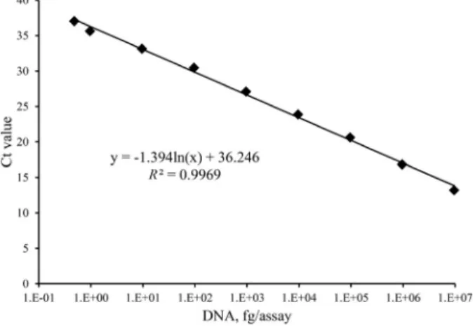
Real-time PCR sensitivity was evaluated. A pure cultured 3.03 × 10⁷ cfu of MAP’s DNA was extracted using mGITC/
SC method and yield of the DNA was measured by spectro- photometer (ND-1000; Thermo Fisher Scientific, USA). The concentration of the DNA harvested from 3.03 × 10⁷cfu of MAP was 2.3± 0.15 ng/uL (SE), corresponds to 3.04 fg of DNA extracted per cfu. Serial dilutions of template DNA were assayed to determine the detection limit of IS900 real- time PCR. Minimum detectable limit was 4.6 fg of DNA, Ct value of 37.03 (Fig. 1).
Diagnostic efficiency of the mGITC/SC method was ana- lyzed using MAP spiked fecal samples. 10-fold diluted MAP ranged from 1.5 × 10⁶ to 0.15 cfu were spiked into 250 mg of feces. Negative control of feces was treated with PBS supple- mented with 0.25% Tween 80.
The average yield, concentration and purity of extracted DNA were estimated. In the 250 mg of feces, the average concentration of DNA was 7.22± 0.38 ng/µL, yield per sam- ple was 116± 6.0 ng/100 mg. The average purity of DNA
(Abs260/Abs280) was 1.67± 0.03. Quantitative analysis was conducted by real-time PCR. Ct values for the real-time PCR ranged from 18.9± 0.43 to 38.3 ± 0.1 and linear correlation between the Ct values and MAP concentration was analyzed using standard curve analysis (Fig. 2). Experimental detec- tion limit was 1.5 cfu in 250 mg of feces (6 cfu/g). No ampli- fication signal was observed with the all negative controls (no template controls, negative fecal samples).
In this study, DNA extraction method for the detection of MAP in bovine feces was described. To lyse MAP’s thick and lipid-rich cell wall, the bead beating method has been used in MAP DNA extraction [9]. However, heating method combined with guanidine lysis was used in mGITC/SC and this could also provide high yield and PCR sensitivity.
Diagnostic efficiency of this new method was evaluated.
DNA purity is major consideration in fecal DNA extraction because of many inhibitors in feces affecting PCR reaction [8, 12]. Purity of DNA with mGITC/SC recorded the fourth Fig. 1. Real-time PCR assay of serial dilutions of DNA extracted from pure cultured MAP ATCC19698. R2 (the coef- ficient of determination) are indicated on the graph.
Fig. 2. The detection limit and amplification efficiency of mGITC/SC in MAP-spiked fecal samples. Ct values are the average of 3 independent experiments. R2 (the coefficient of determination) are indicated on the graph.
Effective DNA extraction method for MAP detection 57
highest value compared to other methods described in Leite et al. [6]. According to et al. [6], there were no significant correlation between DNA purity and PCR sensitivity due to the inaccuracy of measuring DNA purity by spectrophotom- etry.
In quantitative analysis, minimum detectable amount of DNA extracted from pure cultured MAP was about 4.6 fg, corresponds to 1.5 cfu (3.04 fg per cfu). Detection limit of real-time PCR on MAP-spiked fecal samples was also appeared similar level to pure cultured MAP. This result suggests that mGITC/SC can detect the lowest level of MAP in the MAP- spiked feces.
Major advantageous feature of the mGITC/SC method is that there is no need for pre-treatment of the fecal specimen.
The mGITC/SC method uses 250 mg of feces mixed directly with 1 mL of GITC buffer to lyse the MAP cells. Other in- house DNA extraction methods use about 1 g of feces mixed with large amounts of water or buffers followed by a settle down and washing step; the time required for this step is more than 1 h [5, 15]. Because of differences between the initial extraction steps in the mGITC/SC method and the other methods, mGITC/SC has the advantage of extraction time. Another feature of mGITC/SC method is relatively low cost compared to other in house methods or commercial kits.
These two features allow to extraction of MAP DNA from bovine feces more quickly and economically.
Acknowledgments
This work was supported by Grant No. PJ00897001 2012 from the Rural Development Administration, and Research Institute of Veterinary Science, Seoul National Uni- versity, Korea.
References
1. Amaro A, Duarte E, Amado A, Ferronha H, Botelho A.
Comparison of three DNA extraction methods for Mycobacterium bovis, Mycobacterium tuberculosis and Mycobacterium avium subsp. avium. Lett Appl Microbiol 2008, 47, 8-11.
2. Boom R, Sol CJA, Salimans MMM, Jansen CL, Wertheim- van Dillen PME, van der Noordaa J. Rapid and simple method for purification of nucleic acids. J Clin Microbiol 1990, 28, 495-503.
3. Bull TJ, Hermon-Taylor J, Pavlik I, El-Zaatari F, Tizard M. Characterization of IS900 loci in Mycobacterium avium subsp. paratuberculosis and development of multiplex
PCR typing. Microbiology 2000, 146, 2185-2197.
4. Collins MT. Diagnosis of paratuberculosis. Vet Clin North Am Food Anim Pract 1996, 12, 357-371.
5. Irenge LM, Walravens K, Govaerts M, Godfroid J, Rosseels V, Huygen K, Gala J-L. Development and validation of a triplex real-time PCR for rapid detection and specific identification of M. avium sub sp. paratuberculosis in faecal samples. Vet Microbiol 2009, 136, 166-172.
6. Leite FL, Stokes KD, Robbe-Austerman S, Stabel JR.
Comparison of fecal DNA extraction kits for the detection of Mycobacterium avium subsp. paratuberculosis by polymerase chain reaction. J Vet Diagn Invest 2013, 25, 27-34.
7. Logar K, Kopin R, Bandelj P, Stari J, Lapanje A, Ocepek M. Evaluation of combined high-efficiency DNA extraction and real-time PCR for detection of Mycobacterium avium subsp. paratuberculosis in subclinically infected dairy cattle: comparison with faecal culture, milk real-time PCR and milk ELISA. BMC Vet Res 2012, 8, 49.
8. Monteiro L, Bonnemaison D, Vekris A, Petry KG, Bonnet J, Vidal R, Cabrita J, Mégraud F. Complex polysaccharides as PCR inhibitors in feces: Helicobacter pylori model. J Clin Microbiol 1997, 35, 995-998.
9. Odumeru J, Gao A, Chen S, Raymond M, Mutharia L.
Use of the bead beater for preparation of Mycobacterium paratuberculosis template DNA in milk. Can J Vet Res 2001, 65, 201-205.
10. Ott SL, Wells SJ, Wagner BA. Herd-level economic losses associated with Johne’s disease on US dairy operations. Prev Vet Med 1999, 40, 179-192.
11. Ravva SV, Stanker LH. Real-time quantitative PCR detection of Mycobacterium avium subsp. paratuberculosis and differentiation from other mycobacteria using SYBR Green and TaqMan assays. J Microbiol Methods 2005, 63, 305-317.
12. Thornton CG, Passen S. Inhibition of PCR amplification by phytic acid, and treatment of bovine fecal specimens with phytase to reduce inhibition. J Microbiol Methods 2004, 59, 43-52.
13. Whitlock RH, Wells SJ, Sweeney RW, Van Tiem J.
ELISA and fecal culture for paratuberculosis (Johne’s disease): sensitivity and specificity of each method. Vet Microbiol 2000, 77, 387-398.
14. Whittington RJ, Marshall DJ, Nicholls PJ, Marsh IB, Reddacliff LA. Survival and dormancy of Mycobacterium avium subsp. paratuberculosis in the environment. Appl Environ Microbiol 2004, 70, 2989-3004.
15. Zhang MZ, Zhang S. An efficient DNA extraction method for polymerase chain reaction-based detection of Mycobacterium avium subspecies paratuberculosis in bovine fecal samples.
J Vet Diagn Invest 2011, 23, 41-48.
c ê
c ê