A Retrospective Study of Luxation Injuries in Primary Teeth:
Prognosis with Splinting
Kiun Song1, Okhyung Nam1, Misun Kim2, Hyoseol Lee1, Kwangchul Kim2, Sungchul Choi1
1Department of Pediatric Dentistry, School of Dentistry, Kyung Hee University
2Department of Pediatric Dentistry, Kyung Hee University Dental Hospital at Gangdong
This study investigated the prognosis of luxation injuries in primary teeth treated with splinting. This study retrospectively analyzed 92 children with luxation injuries to their primary teeth who were treated with splints between 2010 and 2015. Prognoses were analyzed in patients who had been followed for more than 6 months. The prognoses with splinting were based on clinical and radiographic evaluations performed during the follow-up examinations.
The mean patient age was 42.1 months, and 67.4% were male. The most common cause of luxation injury was falling, and the mean splint duration was 2.4 weeks. The success rate of splinting was 58.9%. The highest rate of success was achieved following subluxation, while repositioning and splinting of lateral luxation had the lowest rate of success. Pulp necrosis was the most common unfavorable prognosis in the luxation injuries.
Depending upon the type of luxation, splint therapy had acceptable prognoses and might be a feasible treatment option.
Key words : Splints, Primary teeth, Tooth injuries, Luxation injury Abstract
Corresponding author : Sungchul Choi
Department of Pediatric Dentistry, School of Dentistry, Kyung Hee University, 26, Kyungheedae-ro, Dongdaemun-gu, Seoul, 02447, Korea Tel: +82-2-958-9371 / Fax: +82-2-965-7247 / E-mail: [email protected]
Received August 29, 2016 / Revised November 15, 2016 / Accepted October 26, 2016
I. Introduction
Luxation injuries comprise a large proportion of traumatic dental injuries (TDIs) in primary teeth, due to the resilience of alveolar bone[1]. Various luxation injuries are possible depend- ing upon the force and direction of impact. Treatment meth- ods vary according to the type of injury[1].
One of the treatments for stabilizing traumatized teeth is splinting, which allows periodontal tissue regeneration while providing protection from further injury. Flexible or semi-rigid fixation is sufficient for healing pulp and periodontium in most
TDIs, and rigid fixation is recommended for cervical root frac- tures[1-3]. When ankylosis is not a significant risk, a fixation period of 2 - 3 weeks is recommended[1]. Following injuries to the bone, however, 1 - 2 additional weeks are required[4].
The International Association for Dental Traumatology (IADT) guidelines recommend splinting of the primary teeth only for alveolar bone fractures and root fractures[5]. Moreover, repo- sitioning and splinting of traumatized primary teeth requires caution regarding the permanent successors[1].
Despite the significance of primary tooth injuries, few stud- ies have examined this topic. Recently, however, trials have
examined the splinting of traumatized primary teeth following alveolar process fracture, horizontal root fracture, and lateral luxation[6-9]. To advance our knowledge of TDIs, such trials are important, and should be continued. The purpose of this study was to evaluate the prognoses of luxation injuries fol- lowing the splinting of primary teeth.
II. Materials and Methods
This study was performed in the Department of Pediatric Dentistry, School of Dentistry, Kyung Hee University, Seoul, Korea. The study proposal was reviewed and approved by the Ethics Committee (KHD IRB 1606-4). This study retrospectively analyzed the medical records of children who were treated us- ing splints on their primary teeth between 2010 and 2015. A total of 838 patients (1534 teeth) visited our department for treatment of luxation injuries of their primary teeth. Of these, 92 patients (202 teeth) were treated using splints and selected for reevaluation. Among the 92 patients, 79 patients were treated with flexible round stainless steel wire (SS-GFS; Ormco, Orange, CA, USA) and flowable composite resin. Nine patients were treated with monofilament nylon fishing line (FL-GFS; HL- 1553, HDF, Busan, Korea) and flowable composite resin. In two patients, the teeth were splinted with flexible tri-flex stainless steel wire (TF-RS; RMO, Denver, CO, USA) and flowable com- posite resin. In the others, the teeth were fixed with flowable composite resin.
Clinical records, intraoral photographs, and radiographs were assessed. Radiographic data were collected to evaluate the prognoses with splinting. Data were analyzed according to age, gender, cause of injury, type of TDI, splinting method, and fixation period. Patients with incomplete records or radio- graphic examinations were excluded.
Luxation injuries were classified into concussion, subluxation, intrusive luxation, extrusive luxation, lateral luxation, and avul- sion[1,10]. Treatment prognoses were analyzed in 57 patients (124 teeth), with a follow-up period exceeding 6 months. Prog- noses were categorized based on the presence of associated complications in the clinical and radiographic examinations.
Favorable prognoses had total or partial pulp canal oblitera- tion, without complications[5]. Unfavorable prognoses included pulp necrosis, pathological root resorption, loss of teeth, anky- losis, and pathological bone resorption[1]. Success rates were calculated as the ratio of teeth with favorable prognoses to the total number of traumatized teeth in each group.
III. Results
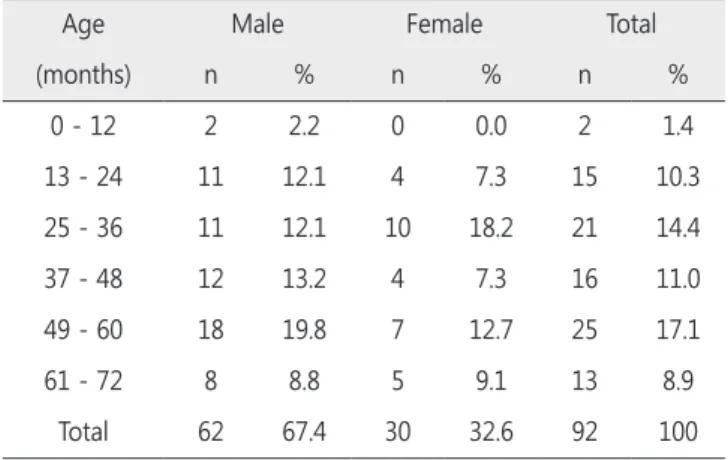
The study population consisted of 62 boys and 30 girls (sex ratio 2.07 : 1). The average age was 42.1 ± 17.4 months, and the most prevalent age was 4 - 5 years, followed by 2 - 3 years. Table 1 shows the distribution of splinting in primary teeth by age and gender.
The main cause of injury was falling (53.3%; Fig. 1); the most common place of injury was in the home (42.4%). All teeth treated by splinting were anterior teeth; the primary maxillary central incisor (67.7%) was the most commonly injured tooth.
Table 2 shows the rates of splinting among the primary teeth.
The highest rate of splinting was in extrusive luxation (28.9%), followed by lateral luxation (22.2%). The mean duration of splinting was 2.4 weeks (Table 2).
Fig. 1. Distribution of splinting in primary teeth according to cause and age.
Table 1. Distribution of splinting in primary teeth by age and gender
Age Male Female Total
(months) n % n % n %
0 - 12 2 2.2 0 0.0 2 1.4
13 - 24 11 12.1 4 7.3 15 10.3
25 - 36 11 12.1 10 18.2 21 14.4
37 - 48 12 13.2 4 7.3 16 11.0
49 - 60 18 19.8 7 12.7 25 17.1
61 - 72 8 8.8 5 9.1 13 8.9
Total 62 67.4 30 32.6 92 100
The total number of teeth analyzed was 124, and complica- tions were diagnosed within an average follow-up period of 22.1 ± 14.5 months. The success rate of splinting was 58.9%;
subluxation injuries had the highest success rate. Conversely, repositioning and splinting of lateral luxation injuries resulted in the lowest success rate (Fig. 2).
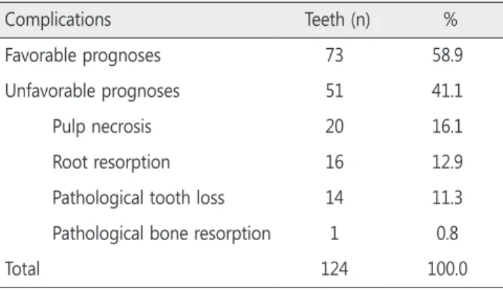
Pulp necrosis (16.1%) was the most common complication.
The distribution of complications is shown in Table 3.
IV. Discussion
Traumatic dental injuries of primary teeth have a prevalence of approximately 30 - 40%[11-13]. In addition, luxation injuries are more frequent in primary teeth than are fractures[1,14].
Despite the prevalence of TDIs, few studies have examined splinting treatment in primary teeth. Therefore, this study fo- cused on luxation injuries in primary teeth treated using splint- ing.
The predominant age group in this study was 4 - 5 years (Table 1). This was in agreement with studies that found an increased incidence of TDIs in children between the ages of 3 and 5 years[15-17]. However, this finding disagreed with other studies reporting that the peak age of TDI incidence in chil- dren is 2 - 4 years [1,15,18,19].
Boys had a higher incidence of injury than girls (2.07 : 1), which was consistent with other studies[11,18,20,21] and may reflect the observation that boys are more energetic and more likely to take part in outdoor activities than girls are[22]. The predominant age of boys in the series was 4 - 5 years, while that of girls was 2 - 3 years. This was in agreement with study that found the predominant age of boys was older than that of girls[17].
The most common cause of the injuries was accidental falls (Fig. 1). Falls and collisions accounted for 90% of cases, consistent with previous reports[18,21,23-25]. The most com- mon location for TDIs to occur was the home, followed by the kindergarten classroom. This was also consistent with previous studies[18,23,24].
The rate of splinting of luxation injuries was 13.2% (Table 2). One study reported that 9% of luxated primary teeth were treated using splinting[26]. Splinting can decrease the initial mobility of teeth and related pain, and may prevent children from touching their mobile teeth[8].
The current guidelines recommend splinting of traumatized primary teeth only following root displacement and alveolar bone fractures[5]. Andreasen et al.[1] recommended a splint for 2 - 3 weeks in the case of luxation with occlusal interfer- ence. In agreement with this suggestion, the rates of splinting in extrusive and lateral luxation injuries were higher than those Table 2. Rate of splinting in luxation injuries and mean splint durations
Type of Injury Luxated
Teeth Splinted
Teeth % Weeks
Extrusive luxation 38 11 28.9 2.4
Lateral luxation 171 38 22.2 3.1
Subluxation 818 124 15.2 2.0
Avulsion 88 12 13.6 1.5
Intrusive luxation 147 14 9.5 2.9
Concussion 272 3 1.1 0.0
Total 1534 202 13.2 2.4
Fig. 2. Treatment prognoses for splinting of primary teeth.
Table 3. Distribution of complications
Complications Teeth (n) %
Favorable prognoses 73 58.9
Unfavorable prognoses 51 41.1
Pulp necrosis 20 16.1
Root resorption 16 12.9
Pathological tooth loss 14 11.3
Pathological bone resorption 1 0.8
Total 124 100.0
in other luxation injuries.
Traumatic dental injuries in the primary teeth can cause clinical complications, including color changes, pulp necrosis, pulp canal obliteration, external root resorption, and internal root resorption[1]. Favorable and unfavorable prognoses were identified based on the literature[5,19]. The frequency of sec- ondary pulp necrosis among teeth with pulp canal oblitera- tions is low[1,14,27-29]. Consequently, partial or total pulp ca- nal obliteration was considered a clinical success. Unfavorable prognoses included pulp necrosis, pathologic root resorption, loss of tooth, and pathologic bone resorption.
The average success rate for splinting was 58.9% (Table 3, Fig. 2). As reported, severe periodontal tissue injuries resulted in complications[19,30]. Lateral, intrusive, and extrusive luxation injuries had lower success rates than other injuries. The replan- tation of avulsed primary teeth is usually not recommended based on the potential damage to permanent successors[1,5].
However, the success rate with avulsed teeth was high in this study, although this finding may have been biased due to the small sample size.
V. Conclusions
In this study, the highest rate of splinting following luxation injuries occurred in cases of extrusive luxation. The success rate of splinting luxated primary teeth was 58.9%, with subluxations having the highest success rates. This was the first study to in- vestigate the clinical prognoses of splinting in luxated primary teeth, and could be the basis for additional studies investigat- ing TDIs in primary teeth.
References
1. Andreasen JO, Andreasen FM, Andersson L : Textbook and Color Atlas of Traumatic Injuries to the Teeth, 4th ed. Black- well Munksgaard, Copenhagen, 516-538, 2007.
2. Liu X, Huang J, Bai Y, et al. : Conservation of root-fractured primary teeth-report of a case. Dent Traumatol, 29:498-501, 2013.
3. Filippi A, Pohl Y, Kirschner H : Replantation of avulsed pri- mary anterior teeth: treatment and limitations. ASDC J Dent Child, 64:272-275, 1997.
4. Oikarinen K: Tooth splinting : a review of the literature and consideration of the versatility of a wire-composite splint.
Endod Dent Traumatol, 6:237-250, 1990.
5. Malmgren B, Andreasen JO, Flores MT, et al. : International Association of Dental Traumatology guidelines for the management of traumatic dental injuries: 3. Injuries in the primary dentition. Dent Traumatol, 28:174-182, 2012.
6. Akin A, Uysal S, Cehreli ZC : Segmental alveolar process fracture involving primary incisors: treatment and 24-month follow up. Dent Traumatol, 27:63-66, 2011.
7. Bonanato K, Sardenberg F, Santos ER, et al. : Horizontal root fracture with displacement in the primary dentition.
Gen Dent, 57:31-34, 2009.
8. Kim GT, Sohn M, Ahn HJ, et al. : Intra-alveolar root fracture in primary teeth. Pediatr Dent, 34:215-218, 2012.
9. Holan G : Conservative treatment of severely luxated max- illary primary central incisors: case report. Pediatr Dent, 21:459-462, 1999.
10. Application of the International Classification of Diseases to Dentistry and Stomatology (ICD-DA). Available from URL:
http://www.who.int/classifications/icd/en/ (Accessed on February 9, 2016).
11. Bastone EB, Freer TJ, McNamara JR : Epidemiology of den- tal trauma: a review of the literature. Aust Dent J, 45:2-9, 2000.
12. Flores MT : Traumatic injuries in the primary dentition.
Dent Traumatol, 18:287-298, 2002.
13. Oliveira LB, Marcenes W, Ardenghi TM, et al. : Traumatic dental injuries and associated factors among Brazilian pre- school children. Dent Traumatol, 23:76-81, 2007.
14. Boorum MK, Andreasen JO : Sequelae of trauma to pri- mary maxillary incisors. I. Complications in the primary dentition. Dent Traumatol, 14:31-44, 1998.
15. Kramer PF, Zembruski C, Ferreira SH, Feldens CA : Trau- matic dental injuries in Brazilian preschool children. Dent Traumatol, 19:299-303, 2003.
16. Granville-Garcia AF, De Menezes VA, De Lira PIC : Dental trauma and associated factors in Brazilian preschoolers.
Dent Traumatol, 22:318-322, 2006.
17. Skaare AB, Jacobsen I : Primary tooth injuries in Norwegian children (1-8 years). Dent Traumatol, 21:315-319, 2005.
18. Choi SC, Park JH, Pae A, Kim JR : Retrospective study on traumatic dental injuries in preschool children at Kyung Hee Dental Hospital, Seoul, South Korea. Dent Traumatol, 26:70-75, 2010.
19. Ritwik P, Massey C, Hagan J : Epidemiology and outcomes of dental trauma cases from an urban pediatric emergency department. Dent Traumatol, 31:97-102, 2015.
20. Bae JH, Kim YK, Choi YH : Clinical characteristics of dental emergencies and prevalence of dental trauma at a univer- sity hospital emergency center in Korea. Dent Traumatol, 27:374-378, 2011.
21. Kim Y, Kim S, Choi N : A retrospective study of the pattern and treatment of traumatic dental injury to primary and permanent teeth. J Korean Acad Pediatr Dent, 41:314-321, 2014.
22. Rajab LD : Traumatic dental injuries in children present- ing for treatment at the Department of Pediatric Dentistry, Faculty of Dentistry, University of Jordan, 1997-2000. Dent Traumatol, 19:6-11, 2003.
23. Galea H : An investigation of dental injuries treated in an acute care general hospital. J Am Dent Assoc, 109:434-438, 1984.
24. Onetto JE, Flores MT, Garbarino ML : Dental trauma in children and adolescents in Valparaiso, Chile. Endod Dent Traumatol, 10:223-227, 1994.
25. Liew VP, Daly CG : Anterior dental trauma treated after- hours in Newcastle, Australia. Community Dent Oral Epide- miol, 14:362-366, 1986.
26. Assunção LRdS, Ferelle A, Iwakura MLH, et al. : Luxation injuries in primary teeth: a retrospective study in children assisted at an emergency service. Brazilian Oral Research, 25:150-156, 2011.
27. Santos BZ, Cardoso M, Almeida IC : Pulp canal obliteration following trauma to primary incisors: a 9-year clinical study.
Pediatr Dent, 33:399-402, 2011.
28. Oginni AO, Adekoya-Sofowora CA, Kolawole KA : Evalua- tion of radiographs, clinical signs and symptoms associated with pulp canal obliteration: an aid to treatment decision.
Dent Traumatol, 25:620-625, 2009.
29. Robertson A, Andreasen FM, Bergenholtz G, et al. : Inci- dence of pulp necrosis subsequent to pulp canal oblitera- tion from trauma of permanent incisors. J Endod, 22:557- 560, 1996.
30. Sae-Lim V, Yuen KW : An evaluation of after-office-hour dental trauma in Singapore. Endod Dent Traumatol, 13:164- 170, 1997.
국문초록
유치의 탈구성 손상에 관한 후향적 분석: 고정술의 치료 결과
송기언1ㆍ남옥형1ㆍ김미선2ㆍ이효설1ㆍ김광철2ㆍ최성철1
1경희대학교 치의학전문대학원 소아치과학교실
2강동경희대학교병원 치과병원 소아치과
본 연구의 목적은 탈구성 손상을 받은 유치에 시행한 고정술의 치료 결과를 분석하는 것이다. 본 연구는 2010년부터 2015년까지 탈 구성 손상으로 본과에 내원하여 유치에 고정술을 시행한 92명의 환아들을 후향적으로 분석하였다. 이들 중에서 6개월 이상 추적 검사 에 참여한 환아들을 대상으로 치료 결과를 분석하였다. 치료 결과는 검진 기간동안 임상 및 방사선 검사에서 합병증의 존재에 따라 분 석되었다.
평균 나이는 42.1개월이었으며 67.4%가 남아였다. 넘어짐이 가장 빈번한 원인이었으며, 평균 고정 기간은 2.4주이었다. 고정술의 성 공률은 58.9%이었다. 아탈구가 가장 높은 성공률을 보였으며, 측방 탈구에서 정복 후 고정술을 시행한 경우 가장 낮은 성공률을 보였 다. 탈구성 손상에서 치수괴사가 가장 빈번히 발생한 비호의적 치료 결과이었다.
본 연구결과, 유치의 탈구성 손상에서 고정술은 받아들일 만하였으며, 고정술은 유치의 탈구성 손상시 치료법으로 고려될 수 있다.
주요어: 고정술, 유치, 치아 손상, 탈구성 손상