Original Article
PROGRESS in MEDICAL PHYSICS Vol. 25, No. 2, June, 2014 http://dx.doi.org/10.14316/pmp.2014.25.2.110Comparison of the Dose Distributions with Beam Arrangements in the Stereotactic Body Radiotherapy
(SBRT) for Primary Lung Cancer
Ji Woon Yea
Department of Radiation Oncology, Yeungnam University College of Medicine, Daegu, Korea
To compare 2 beam arrangements, circumferential equally angles (EA) beams or partially angles (PA) beams for stereotactic body radiation therapy (SBRT) of primary lung cancer for intensity-modulated radiation therapy (IMRT) and volumetric-modulated arc therapy (VMAT) delivery techniques with respect to target, ipsilateral lung, contralateral lung, and organs-at-risk (OAR) dose-volume metrics, as well as treatment delivery efficiency. Data from 12 patients, four treatment plans were generated per data sets (IMRT
EA, IMRT
PA, VMAT
EA, VMAT
PA). The prescribed dose (PD) was 60 Gy in 4 fractions to 95% of the planning target volume (PTV) for a 6-MV photon beam. When compared with the IMRT and VMAT treatment plan for 2 beams, conformity index, homogeneity index, high dose spillage, D2 cm (Dmax at a distance ≥2 cm beyond the PTV), R50 (ratio of volume circumscribed by the 50% isodose line and the PTV), resulted in similar. But Dmax of the Organ at risk (OAR), spinal cord, trachea, resulted in differ between four treatment plans. Especially HDS
locationshowed big difference in 21.63% vs. 26.46%.
Key Words: Dose distribution, Equal angle, Partial angle, Stereotatic body radiotherapy (SBRT)
Received 30 April 2014, Revised 26 May 2014, Accepted 2 June 2014 Correspondence: Ji Woon Yea ([email protected])
Tel: 82-53-620-3371, Fax: 82-53-624-3599 cc This is an Open-Access article distributed under the terms of the Creative Commons Attribution Non-Commercial License (http://creativecommons.org/licenses/by-nc/3.0) which permits unrestricted non-commercial use, distribution, and reproduction in any medium, provided the original work is properly cited.
서 론
초기 폐암의 원칙적인 치료는 수술적 제거이나 의학적으 로 수술 불가능한 경우 과거에는 고식적 방사선치료(con- ventional radiotherapy)를 시행하여 왔었다. 최근에는 정위적 체부방사선치료를 통해 종양에 대한 고정밀 4차원 전산화 단층촬영술과 컴퓨터시스템하의 영상유도 위치검증으로 3 차원 단층촬영 기반의 입체조형 방사선치료보다 환자의 표 적체적 정확도와 개별성이 향상되었다.
1)이런 기술의 급진 적 발전으로
2-4)분할선량을 높일 수 있으며 치료기간의 단 축, 방사선부작용 감소, 국소 제어율 증가의 결과를 가져왔 다.
5,6)한편 정위적 체부방사선치료의 치료계획과 치료방법 은 개인 또는 치료기관에 따라 조금씩 다르게 시행되어 왔 으나, 최근 미국 방사선치료 암연구단체(Radiation Therapy Oncology Group; RTOG)와 유럽 암치료 연구단체(European
Organisation for Research and Treatment of Cancer; EORTC) 에서 초기 원발성 폐암의 정위적 체부방사선치료 권고안을 제시하였다.
7,8)이들 권고안 중에는 환자대상, 방사선량, 전 산화 단층촬영술(computed tomography; CT) 및 계산방법 (Calculation algorithms) 등 전반적인 가이드라인을 제시하 고 있지만, 치료전달 방법에서 최적의 빔 배열을 제시하고 있지는 않다. 따라서 본 연구는 360
o회전각도(Equally an- gles, EA)와 부분각도(partially angles; PA)의 빔 배열(Beam arrangements)에 따른 세기조절방사선치료(Intensity-modu- lated radiation therapy, IMRT)와 체적변조회전치료(Volume- tric-modulated arc therapy, VMAT)에서 종양의 선량, 주요장 기의 선량, 선량일치지수(conformity index; CI), 균질성지수 (homogeneity index; HI), 모니터 단위(monitor unit; MU) 등 의 차이를 비교 분석하였다.
재료 및 방법
1. 환자특성
2012년 8월부터 2013년 12월까지 본원에서 폐 정위적 체
부방사선치료 시행한 환자 12명의 영상을 이용하였다. 환
자군의 나이는 42세에서 86세 사이였으며 중간값은 72세이
Table 1. 환자 특성.
나이 42∼86 (중간값=72)
성별
남자 10
여자 02
종양크기
육안적 종양체적 0.4 cc∼44.3 cc
평균 9.8 cc
계획용 표적체적 7.6 cc∼137.3 cc
평균 39.7 cc 종양위치
우상엽 1
우중엽 1
우하엽 6
좌상엽 3
좌하엽 1
총수 12
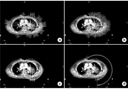
Fig. 1. Comparisons of the IMRT and VMAT with equally ((a); (c)) or partially ((b); (d)) beam arrange- ments. (a) IMRTEA; (b) IMRTPA; (c) VMATEA; (d) VMATPA.
고 성별은 남자 10명, 여자 2명으로 남자 환자가 많았다.
육안적 종양체적은 최저 0.4 cc에서 최고 44.3 cc로 다양하 였으며 평균 9.8 cc였고 계획용 표적체적은 최저 7.6 cc에서 최고 137.3 cc로 평균 39.7 cc였다. 병소의 위치는 우하엽 6 명으로 가장 많았고 우상엽 1명, 우중엽 1명, 좌상엽 3명, 좌하엽 1명이었고 환자의 특성은 아래와 같다(Table 1).
2. 빔 배치와 선량처방 최적화
세기조절방사선치료기법에서 360
o회전각도(IMRT
EA)는 0
o/
40
o/80
o/120
o/160
o/200
o/240
o/280
o/320
o로 40
o등간격으로 9개의 조사면을 이용하였고, 종양의 위치가 왼쪽에 있는 경우에 부분각도(IMRT
PA)는 335
o/0
o/25
o/50
o/75
o/100
o/125
o/150
o/175
o/ 200
o/225
o로 종양을 중심으로 11개의 조사면을 이용하였다.
종양이 오른쪽에 있는 경우는 왼쪽 폐의 선량이 적게 들어 가도록 320
o/345
o/10
o/35
o/60
o/85
o/110
o/135
o/160
o/185
o/210
o의 부 분각도로 배열하였다.
또한, 체적변조회전치료기법에서 360
o회전각도(VMAT
EA) 는 181
o∼179
o의 시계방향 1ARC와 179
o∼181
o의 반시계방 향의 1ARC로 구성된 총 2ARC로 치료계획을 하였고, 부분 각도(VMAT
PA)에서는 종양의 위치가 왼쪽 폐에 있는 경우 에는 320
o∼179
o의 시계방향 1ARC와 179
o∼320
o의 반시계 방향의 1ARC로 구성된 총 2ARC로 치료계획을 하였고, 오 른쪽 폐에 있는 경우에는 200
o∼50
o의 시계방향 1ARC와 50
o∼200
o의 반시계방향의 1ARC로 구성된 총 2ARC로 치 료계획을 하여 반대측 폐의 조사선량이 최소가 되게 하였 다.
이들의 빔 배열에 대한 구성은 Fig. 1에 표시하였다.
IMRT와 VMAT은 동일한 제약조건(constraint)을 사용하였
으며, 이들 제약조건은 RTOG-0813,
9)RTOG-0915,
10)AAPM
TG-101
11)등의 선량부피그림표한계(dose volume histogram
limits; DVH limits)조건을 만족시키기 위하여, 최적화
(optimizations)과정을 200회로 고정하여 실시 하였다. 선량
계산은 비균질 영역에서 비교적 정확하다고 보고된 Aniso-
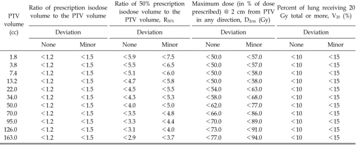
Table 2. Plan evaluation table for HDSvolume, D2cm, R50 in the RTOG9).
PTV volume
(cc)
Ratio of prescription isodose volume to the PTV volume
Ratio of 50% prescription isodose volume to the
PTV volume, R50%
Maximum dose (in % of dose prescribed) @ 2 cm from PTV in any direction, D2cm (Gy)
Percent of lung receiving 20 Gy total or more, V20 (%)
Deviation Deviation Deviation Deviation
None Minor None Minor None Minor None Minor
1.8 <1.2 <1.5 <5.9 <7.5 <50.0 <57.0 <10 <15
3.8 <1.2 <1.5 <5.5 <6.5 <50.0 <57.0 <10 <15
7.4 <1.2 <1.5 <5.1 <6.0 <50.0 <58.0 <10 <15
13.2 <1.2 <1.5 <4.7 <5.8 <50.0 <58.0 <10 <15
22.0 <1.2 <1.5 <4.5 <5.5 <54.0 <63.0 <10 <15
34.0 <1.2 <1.5 <4.3 <5.3 <58.0 <68.0 <10 <15
50.0 <1.2 <1.5 <4.0 <5.0 <62.0 <77.0 <10 <15
70.0 <1.2 <1.5 <3.5 <4.8 <66.0 <86.0 <10 <15
95.0 <1.2 <1.5 <3.3 <4.4 <70.0 <89.0 <10 <15
126.0 <1.2 <1.5 <3.1 <4.0 <73.0 <91.0 <10 <15
163.0 <1.2 <1.5 <2.9 <3.7 <77.0 <94.0 <10 <15
tropic Analytical Algorithm (AAA, Varian Medical Systems) 알고리즘을 사용하여
12)치료계획을 수행하였다.
3. 치료계획 평가 기준
계획용 표적체제(planning target volume; PTV)의 최대, 최 소 및 평균 방사선량(Gy)을 분석하였다. 또한 RTOG-0817/
0915에서 제안한 high dose spillage (HDS
location, HDS
volume)와 low dose spillage (D
2cm, R
50)을 분석하였다. HDS
location와
HDS
volume은 식 (1)와 (2)으로 표현할 수 있다.
HDS
location=
cumulative volume of all tissue outside the PTV receiving a dose>105% of prescription dose
(1)
volume of the PTV
HDS
volume=
volume of the prescription isodose volume of the PTV(2)
Low dose spillage의 2항목(D
2cm, R
50)은 계획용 표적체제 밖에서의 방사선량이 급격히 줄어들어 주위 주요장기에 선 량이 최소화 할 수 있도록 평가하는 항목이다. D
2cm은 계획 용 표적체제로부터 모든 방향으로 2cm을 확장하여, 계획용 표적체제를 뺀 용적의 최대 선량값으로 정의된다. R
50은 다 음과 같이 표현될 수 있다.
R
50=
volume of the 50% of the prescription isodose volume of the PTV(3)
RTOG에서는 HDS
location의 값을 15% 이하로 권고하고 있 으며, HDS
volume, D
2cm, R
50의 값은 Table 2에서 계획용 표적 체제에 따라서 각각 권고되고 있다.
각 치료계획의 계획용 표적체제의 방사선량의 품질을 평 가하기 위하여 선량일치지수(conformity index; CI)는 Van’t Riet et al.
13)이 제시한 아래 수식을 사용하였다.
CI=
TVRI× TVRI
TV VRI
(4)
여기서 TV
RI=target volume covered by 95% isodose, V
RI= volume of 95 % isodose, TV=target volume로 각각 정의 된다.
균질성지수(homogeneity index; HI)는 rDHI (radical dose homogeneity index)와 mDHI (moderate homogeneity index)로 구분하여 분석하였다. 이 지수들은 이전 연구
2)에서 정의 되었다.
결 과
총 200회의 치료계획 최적화 과정을 수행하였고, 계획용 표적체적은 최소 7.6 (cc)에서 최대 137.3 (cc)이며 평균 39.7 (cc)였다. 치료계획 평가 변수 비교에서 IMRT
EAIMRT
PAVMAT
EAVMAT
PA에서 표적체적선량의 분포는 65.37,
65.29, 64.94, 64.90를 나타내었으며, 선량일치지수는 0.74,
0.70, 0.68, 0.67를 나타내었으며, 균질성지수는 0.92, 0.86,
0.81, 0.87를 나타내었으며, 고선량 유출은 HDS
location에서
1.61, 2.05, 3.63, 4.93의 큰 차이를 나타내었으며, R
50은 0.74,
0.70, 0.68, 0.67를 나타내었으며, 모니터 단위는 5287, 4493,
3565, 303의 큰 차이를 나타내었다(Table 3).
Table 4. Dose difference analysis according to the equally angles and the partially angles for the beam arrangements.
Structure Parameter
IMRTEA IMRTPA Difference (%) VMATEA VMATPA Difference (%)
Mean±SD Mean±SD IMRTPAIMRT− IMRTEA ×100
PA Mean±SD Mean±SD IMRTPAIMRT− IMRTEA ×100
PA
Both MLD (Gy) 4.61±1.39 4.19±1.29 −10.04 4.69±1.51 4.55±1.43 −3.02
lung V5 (%) 20.35±5.66 17.21±5.57 −18.28 19.62±7.49 16.57±7.49 −18.36
V12.5 (cc) 407.49±224.81 321.68±224.81 −26.68 370.03±206.62 347.51±190.34 −6.48 V13.5 (cc) 375.44±214.44 302.32±162.60 −24.19 350.48±201.25 329.11±186.05 −6.49
V20 (%) 6.40±3.04 6.20±2.65 −3.14 7.18±3.35 7.05±3.24 −1.82
V25 (%) 4.39±2.26 4.51±2.09 2.69 4.80±2.49 5.34±2.68 10.04
Ipsilateral MLD (Gy) 7.40±2.42 7.26±2.36 −1.95 8.03±2.78 7.98±2.78 −0.69
lung V5 (%) 30.12±7.47 29.77±7.72 −1.18 31.61±7.81 31.54±7.92 −0.23
V10 (%) 23.18±7.44 21.98±8.05 −5.45 24.80±8.55 23.85±8.46 −3.99
V20 (%) 12.63±6.26 12.48±5.60 −1.24 14.51±7.17 14.25±6.90 −1.86
V30 (%) 7.24±3.76 7.29±3.58 0.67 8.34±4.50 8.64±4.67 3.47
Contralateral MLD (Gy) 1.75±0.77 1.15±0.50 −51.89 1.39±0.58 1.17±0.45 −18.70
lung V5 (%) 10.28±4.88 4.30±4.88 −139.34 7.50±8.30 3.93±4.63 −91.08
V10 (%) 5.40±2.50 0.11±0.27 −4645.17 0.33±0.72 0.17±0.36 −96.82
PRVSpinal cord Dmax (Gy) 13.36±0.85 10.37±2.56 −28.81 10.69±1.43 9.59±1.82 −11.46
OARTrachea Dmax (Gy) 11.78±10.31 9.08±8.02 −29.75 10.34±9.56 10.67±8.96 3.14
OARHeart Dmax (Gy) 23.53±12.39 18.85±13.63 −24.84 18.84±12.96 19.34±13.19 2.58
V5/10/20/25/30 (%): percentage volumes that received 5, 10, 20, 25 and 30 Gy, respectively, V12.5/13.5 (cc): absolute volumes that received 12.5 and 13.5 Gy, respectively, MLD: Mean Lung Dose.
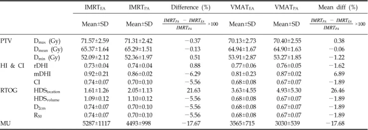
Table 3. Comparisons of PTV, HI, CI, RTOG plan evaluation’s parameter and MU between the equally angles and the partially angles for the beam arrangements.
IMRTEA IMRTPA Difference (%) VMATEA VMATPA Mean diff (%)
Mean±SD Mean±SD IMRTPA− IMRTEA ×100
IMRTPA Mean±SD Mean±SD IMRTPA− IMRTEA ×100 IMRTPA
PTV Dmax (Gy) 71.57±2.59 71.31±2.42 −0.37 70.13±2.73 70.40±2.55 0.38
Dmean (Gy) 65.37±1.64 65.29±1.51 −0.13 64.94±1.67 64.90±1.63 −0.06
Dmin (Gy) 52.09±2.12 52.36±1.97 0.51 53.91±2.87 53.27±1.85 −1.22
HI & CI rDHI 0.73±0.04 0.74±0.04 0.88 0.77±0.06 0.76±0.05 −1.62
mDHI 0.92±0.21 0.86±0.02 −6.29 0.81±0.23 0.87±0.02 6.89
CI 0.74±0.07 0.70±0.10 −5.56 0.68±0.08 0.67±0.07 −1.89
RTOG HDSlocation 1.61±1.26 2.05±1.13 21.63 3.63±4.55 4.93±5.30 26.46
HDSvolume 1.09±0.12 1.10±0.12 −5.56 0.68±0.08 0.67±0.07 −1.89
D2cm 0.74±0.07 0.70±0.10 −5.56 0.68±0.08 0.67±0.07 −1.89
R50 0.74±0.07 0.70±0.10 −5.56 0.68±0.08 0.67±0.07 −1.89
MU 5287±1117 4493±998 −17.67 3565±715 3030±539 −17.68
IMRTEA: Intensity-modulated radiotherapy arranged by equally beam angles, IMRTPA: Intensity-modulated radiotherapy arranged by partially beam angles, VMATEA: Volumetric-modulated arc therapy arranged by equally beam angles, VMATPA: Intensity-modulated radiotherapy arranged by partially beam angles, SD: standard deviation, rDHI: radical dose homogeneity index, mDHI: moderate dose homogeneity index, CI: conformity index, HDSlocation: location of the high dose spillage described by the RTOG, HDSvolume: volume of the high dose spillage described by the RTOG, D2cm: location of the low dose spillage described by the RTOG, R50: volume of the low dose spillage described by the RTOG, and MU: monitor units.
또한 치료계획 조사선량 평가 비교에서 IMRT
EAIMRT
PAVMAT
EAVMAT
PA에서 양쪽 폐를 고려한 경우에서 V
20(%) 은 6.40, 6.20, 7.18, 7.05를 나타내었으며, V
5(%)은 20.35,
17.21, 19.62, 16.57를 나타내었으며, 종양이 있는 부위의 폐
에 대해서는 V
20(%)은 12.63, 12.48, 14.51, 14.25를 나타내
었으며, V
5(%)은 30.12, 29.77, 31.61, 31.54를 나타내었으
며, 조양이 없는 반대편 폐에 대해서는 V
5(%)은 10.28, 4.30, 7.50, 3.93를 나타내었으며, 중요 장기 척추에 조사되 는 최대선량의 평균선량에 대해서는 13.36 Gy, 10.37 Gy, 10.69 Gy, 9.59 Gy를 나타내었다(Table 4).
고찰 및 결론
이중 양측 폐나 동측 폐, 척수, 기도, 심장에 분포하는 선 량지표는 큰 차이가 없었으며 반대측 폐의 V
10(%)이 부분 각 배열에서 5배이상의 큰 차이를 보여 방사선수술 이후 만성 합병증인 방사선폐렴과 드물게 발생하는 섬유화로 인 한 폐기능 저하 발생빈도를 감소시킨다.
최근 폐암의 정위적 체부 방사선치료(SBRT)는 RTOG와 EORTC 권고안에 따라 여러 가지 빔 배열 방법이 사용되 고 있다. SBRT에서 적합한 빔 방향은 표적체적에는 적절 한 선량분포를 얻고 손상위험장기는 최대한 피하며 단거리 빔 경로를 이용하고 치료기의 기계적인 제한점 없이 선택 하게 된다.
7)따라서 충분한 개수의 빔을 여러 방향에서 입 사되도록 하는 것이 좋으며 이전 연구에서 빔 갯수가 방향 보다 더 중요성을 가진다.
10)그러나 실제 환자 치료에서는 여러 가지 제약이 있고 SBRT에서 적절한 빔 배치에 대한 근거도 부족하며 다만 반대측 폐를 통과하는 빔 배치는 되 도록 제한하는 정도이다.
14,15)따라서 본 연구는 방사선치료를 시행한 환자의 영상을 이용하여 종양주위에 빔 배열을 균등하게 한 360
o회전각도 와 부분각도로 배열한 치료계획의 선량차이를 알아보기 위 해 세기조절방사선치료(Intensity-modulated radiation therapy, IMRT)와 체적변조회전치료(Volumetric-modulated arc ther- apy, VMAT)으로 분류하고 각각 선량분포를 비교하였다.
치료계획 평가 변수중 선량일치지수, 균질성지수, 고선량 유출, D
2cm, R
50은 빔 배열에 따라 크게 차이가 나지 않았다.
또한 손상위험장기인 척수, 기관지, 식도의 최고선량은 각 각의 방법에 따라 차이가 있었으나 빔 배열에 의한 차이보 다는 표적체적의 위치 차이에 의한 것으로 생각된다.
HDS
location에서는 360
o회전각도의 빔 균등배열에 비해 부분
각도 빔 배열이 빔의 집중성 때문에 21.63%와 26.46%의 큰 차이를 나타낸 것으로 생각된다.
모니터 단위는 부분각 빔 배열에서 작았으며 반대측 폐의 V
10(%)도 동일한 결과를 보였고 특히 세기조절방사선치료 에서 5배 이상의 큰 차이를 보였다.
본 연구에서 원발성 폐암의 정위적 체부 방사선치료시 빔 배열에 따른 선량분포의 차이를 분석하였고 결론적으로
부분 각 빔 배열에 따른 치료계획이 적은 모니터 단위로 환자의 치료에는 효율적이고 반대측 폐의 방사선량은 감소 시켜 부작용의 위험을 줄일 수 있다.
본 연구의 제한 점은 환자수가 적고 후향적 연구로 좀 더 많은 환자를 대상으로 전향적 연구를 시행하여 수치를 분석하는 것이 통계적인 의의를 나타낼 수 있을 것으로 생 각된다.
References
1. Robert D, Timmerman MD, Brain D, et al: Stereotactic Body Radiation Therapy in Multiple Organ Sites. Journal of Clinical Oncology 25:947-52 (2007)
2. Oh SA, Kang MK, Kim SK, Yea JW: Comparison of IMRT and VMAT techniques in spine stereotactic radiosurgery with in- ternational spine radiosurgery consortium consensus guidelines.
Prog Med Phys 24(3):145-153 (2013)
3. Lim DH, YI BY, Mirmiran A, et al: Optimal beam arrange- ment for stereotactic body radiation therapy delivery in lung tumors. Acta Oncologica 49:219-224 (2010)
4. Mara WR, Catherine MK, Kelly MPC, et al: Circumferen- tial or sectored beam arrangements for stereotactic body radia- tion therapy (SBRT) of primary lung tumors: Effect on target and normal-structure dose-volume metrics. Med Dos 38:407-412 (2013)
5. Robert T, Rebecca P, James G, et al: Stereotactic Body Radiation Therapy for Inoperable Early Stage Lung Cancer, JAMA 303(11):1070-1076 (201)
6. Nagata Y, Takayama K, Matsuo Y, et al: Clinical out- comes of a phase I/II study of 48 Gy of stereotactic body radio- therapy in 4 fractions for primary lung cancer using a stereotactic body frame. Int J Radiat Oncol Biol Phys 63(5):1427-1431 (2005) 7. Timmerman R, Galvin J, Michalski J, et al: Accreditation and quality assurance for Radiation Therapy Oncology Group:
multicenter clinical trials using stereotactic body radiation therapy in lung cancer. Acta Oncol 45(7):779-786 (2006)
8. Dirk De R, Corinne FF, Ursula N, et al: European Organi- zation for Research and Treatment of Cancer Recommenda- tions for Planning and Delivery of High-Dose, High-Precision Radiotherapy for Lung Cancer. 28(36):5301-5310 (2010) 9. RTOG 0813: Seamless Phase I/II Study of Stereotactic Lung
Radiotherapy (SBRT) for Early Stage, Centrally Located, Non-Small Cell Lung Cancer (NSCLC) in Medically Inoperable Patients. http://www.rtog.org/ClinicalTrials/ProtocolTable/StudyDetails.
aspx?study=0813.
10. RTOG 0915: A randomized phase II study comparing 2 ster- eotactic body radiation therapy (SBRT) schedules for medically inoperable patients with stage I peripheral non-small cell lung cancer. http://www.rtog.org/ClinicalTrials/ProtocolTable/StudyDetails.
aspx?study=0915.
11. Benedict SH, Yenice KM, Followill D, et al: Stereotactic body radiation therapy: The report of AAPM task group 101.
원발성 폐암에서 정위적 체부 방사선치료의 빔 배열에 따른 선량분포의 비교
영남대학교 의과대학 방사선종양학교실
예 지 원
원발성 폐암의 정위적 체부 방사선치료(Stereotatic Body Radiation Therapy; SBRT)시에, 종양주위의 빔 배열을 균등하게 한 360o회전각도(Equally angles; EA)와 종양주위의 빔 배열을 부분각도(Partially angles; PA)로 배열한 치료계획의 선량차 이를 확인하기 위하여, 세기조절방사선치료(Intensity-modulated radiation therapy, IMRT)와 체적변조회전치료(Volume- tric-modulated arc therapy, VMAT)의 종양의 선량, 동측 폐의 선량, 반대측 폐의 선량, 손상위험장기(Organ at risk, OAR)의 선량, 치료효율 등을 비교분석 하였다. 12명의 환자에서 각각 4종류(IMRTEA, IMRTPA, VMATEA, VMATPA)의 치료계획을 생 성하였으며, 처방선량은 총 선량 60 Gy, 4회 분할치료로 표적체적 95%에 대해 100% 선량이 포함되게 하였다. IMRT와 VMAT의 치료계획 평가에서 360o회전각도의 빔 균등배열과 부분각도 빔 배열에서 변수중 선량일치지수, 균질성지수, 고 선량 유출, D2cm, R50은 빔 배열에 따라 크게 차이가 나지 않았다. 또한 손상위험장기인 척수, 기관지, 식도의 최고선량은 각각의 방법에 따라 차이를 나타내었다. 특히 HDSlocation에서 21.63%와 26.46%의 큰 차이를 나타내었다.
중심단어: 선량분포, 전체각, 부분각, 정위적 체부 방사선치료 Med Phys 37:4078-101 (2010)
12. Oh SA, Kang MK, Yea JW, Kim SH, Kim KH, Kim SK:
Comparison of intensity modulated radiation therapy dose cal- culations with a PBC and AAA algorithms in the lung cancer.
Korea J Med Phys 23:48-53 (2012)
13. Feuvret L, Noel G, Mazeron JJ, Bey P: Conformity index:
a review. Int J Radiat Oncol Biol Phys 64:333-342 (2006) 14. Ong CL, Verbakel WF, Cuijpers JP, et al: Stereotactic
radiotherapy for peripheral lung tumors: A comparison of volu- metric modulated arc therapy with 3 other delivery techniques Radiother Oncol 97:437-42 (2010)
15. Holt A, van Vliet-Vroegindeweij C, Mans A, et al: Volume- tric-Modulated Arc Therapy for Stereotactic body radiotherapy of lung tumors: A comparison with intensity-modulated radio- therapy techniques. Int J Radiat Oncol Biol Phys 81:1560-7 (2011)