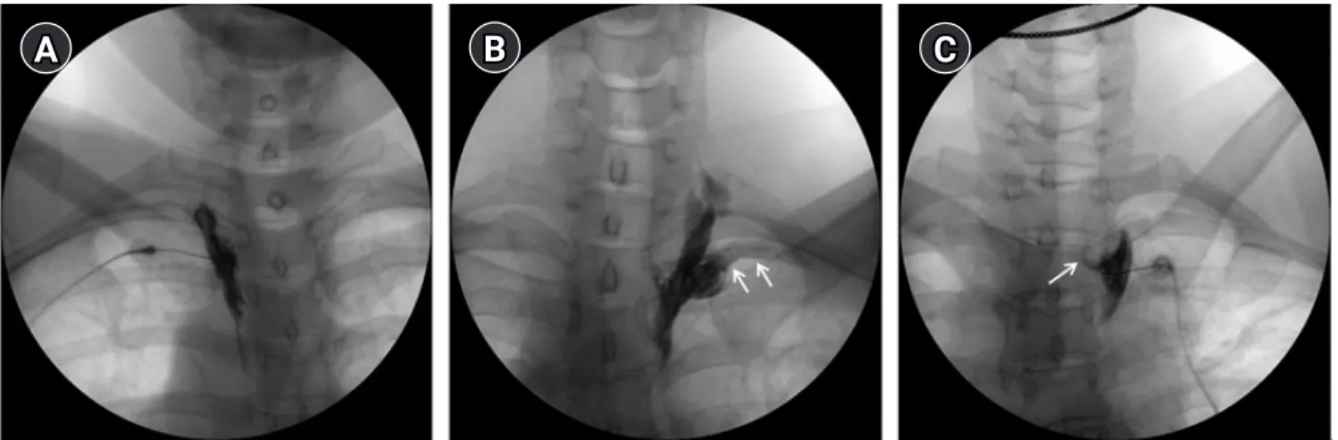
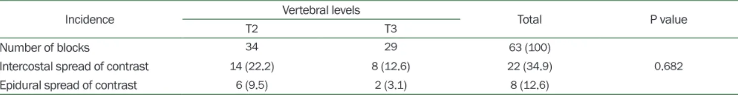
Incidence of inadvertent intercostal or epidural spread during thoracic sympathetic ganglion block
6
0
0
전체 글
(2)
(3)
(4)
(5)
(6)
수치
관련 문서
