Ⅰ. 서 론
환자의 무치악부에 기능적이고 심미적인 보철물 제작을 위 해서는 임플란트가 치조제의 정확한 위치에 식립되어야 한다.
지연형 임플란트 식립시 치조제가 흡수되어, 그 양이 부족한 경우가 많이 있다. 치조제의 흡수는 환자의 건강상태에 관계 없이 치아를 발거 후 발생하는 자연적인 현상이며, 치조제가 흡수됨에 따라 그 부피가 감소하고, 형태의 변형이 일어난다1,2). 발치 후 골소실은 잔존 치조골의 형성과 재형성 과정을 거치 며 발치 후 첫 6개월 내에 가속화 된다. 이러한 치조골의 소실 은 발치 후 첫 6개월에 치조골 높이의 40%, 폭의 60% 가량이 발
생한다. 치조골의 높이의 감소보다 치조골 폭의 감소가 많이 발생하며, 치조골 폭경에 있어서는 특히 협측 또는 안면측에 서의 감소가 두드러진다3). 이러한 발치와 주위 조직 형태의 소 실로 인해 이상적인 위치에 임플란트를 식립하기가 어려워지 며, 따라서 심미적인 치아수복 역시 힘들어진다4-6).
임플란트를 보다 안정적인 위치에 식립하고 기능적이고 심 미적인 보철물 수복을 위해서는 치아소실부 치조제의 흡수를 최소화 하는 술식이 요구되며, 현재까지 많은 시도들이 있어 왔다7-9). 1980년대 초반 Garver와 Wowern 등은 치아 치근을 발치 와에 남겨두는 방식으로 치조제의 붕괴를 방지하는 술식을 사 용하였으며, 1980년대 중반 치근 형태의 hydroxylapatite(HA)를 발치와에 삽입하는 술식이 시행된 후10,11) 최근까지 autografts, allografts, alloplasts, xenografts, barrier membrane 등 다양한 재료를 사용한 많은 치조제 보존술이 시행되고 있다12-15). 이와 같은 연 구들에서 발치후 치조제 흡수방지에 있어 치조제 보존술의 효 용성을 말하고 있다.
저자 등은 발치와에 bovine bone mineral particle과 absorbable collagen sponge를 삽입하는 술식을 사용하였으며, 이 술식은
임플란트 식립을 위한 치조제 보존술에 관한 연구
김종원∙전하룡∙홍종락
성균관대학교 의과대학 삼성서울병원 구강악안면외과
Abstract (J. Kor. Oral Maxillofac. Surg. 2006;32:430-435)
홍 종 락
135-710 서울 강남구 일원동50
성균관의대 삼성서울병원 구강악안면외과 Jong-Rak Hong
Dept. of OMFS, Samsung Medical Center, Sungkyunkwan Univ. School of Medicine,
#50, Ilwon-dong, Gangnam-gu, Seoul, 135-710, Korea Tel: +82-2-3410-2420 Fax: +82-2-3410-0038 E-mail: [email protected]
THE STUDY ON RIDGE PRESERVATION FOR IMPLANT SITE DEVELOPMENT
Jong-Won Kim, Ha-Ryong Jeon, Jong-Rak Hong Department of Oral and Maxillofacial Surgery, Samsung Medical Center,
Sungkyunkwan University School of Medicine, Seoul, Rep. of Korea
Purpose
The aim of this study was to investigate healed bovine bone particles (Bio-Oss�) and absorbable collagen sponge (CollaPlug�) applied extraction socket site at 4-6 months’post-extraction.
Material and methods
From August, 2004 to October, 2005, 17 sockets in 5 adult patients were selected out of the patients whose received ridge preservation using bovine bone particles and absorbable collagen sponges at Dept. of oral and maxillofacial surgery in Samsung Medical Center. There were 5 male patients, ages 30 to 58 years. Immediate postoperation and 4-6 months after operation study models were compared to evaluate the ridge dimension by measur- ing vertical height and horizontal width of alveolar ridge.
Results
The measurements at 4-6 months revealed, in the ridge dimension, a loss of vertical height of 0.91±0.40mm and horizontal width of 1.25±
0.58mm. There was no adverse reaction.
Conclusion
This study suggests that treatment of extraction sockets with graft materials and collagen sponges is valuable in preserving alveolar bone in extrac- tion sockets and preventing alveolar ridges defects.
Key words: Ridge preservation, Alveolar ridge width, Alveolar ridge height
allograft material 상방에 collagen sponge를 적용함으로써 일차 봉 합 없이도 graft material의 소실을 방지할 수 있으며, barrier mem- brane을 사용하는 것에 비해 적은 비용이 든다는 장점이 있다.
본 연구의 목적은 발치와 동시에 기술한 바와 같은 치조제 보존술을 시행 후 치조제의 수직적, 수평적 변화 양상을 관찰 하고자 하였다.
Ⅱ. 연구대상 및 방법 1. 연구대상
본 연구는 2004년 8월부터 2005년 10월까지 삼성서울병원 구 강악안면외과에서 발치 후 임플란트 식립을 위해 치조제 보존 술을 시행한 환자 중 4-6개월간 추적조사가 가능한 5명의 환자 를 대상으로 하였다. 5명은 모두 남환으로 30세에서 58세까지 48년 10개월의 평균연령을 보였다. 총 17개의 구치부 발치와를 대상으로 하였으며, 상악 11개, 하악 6개의 발치와를 연구 하였 다. 발치와 형태 변이관찰의 용이성을 위해 잔존 치조골 양이 1/2이상 남은 발치와를 대상으로 하였고, 심각한 치조골 위축 으로 임플란트 식립을 위해서 온레이, 인레이 형태의 추가적 인 골이식이 필요하거나, 현존하는 치주질환이 매우 심한 환 자는 대상에서 제외 하였다.
2. 재료와 술식 (Fig. 1, 2)
발거가 필요한 치아를 조심스럽게 발치한 후 발치와에 남아 있는 염증조직을 제거 후 하방부터 Bovine cancellous bone min- eral particle(Bio-Oss�, Geistlich Pharma AG, Wolhusen, Switzerland)
을 삽입하고 그 상부에 Absorbable collagen sponge(Collaplug�, Integra LifeSciences Corp., Plainsboro, USA)를 적용한 후 sponge가 빠져나가지 않게 4-0 vicryl을 사용하여 봉합을 시행하였다. 일 차봉합을 시도하지는 않았다.
Fig. 1. Materials used in ridge preservation. On average, 0.25g of Bio-Oss� particulated cancellous bone graft material is used to graft a single extraction site. CollaPlug� absorbable collagen dressing is used to isolate the grafted site from oral cavity. To prevent coronal displacement of the CollaPlug dressing, 4-0 vicryl suture is used on FS-2 needle.
step 1 step 2 step 3
Fig. 2. Diagrammatic representation of ridge preservation procedure using Bovine cancellous bone mineral particle and absorbable collagen sponge.
Step 1- atraumatic extraction of teeth. Care must be taken to preserve the socket walls and minimize microtrauma to the bone.
Step 2- following curettage to remove soft tissue remnants, Bio-Oss� is loosely packed into the sockets and Collaplug�is applied over the graft material.
Step 3- interrupted suture is placed. no attempt to achieve primary closure.
Collagen sponge를 발치와 상방에 적용함으로써 일차봉합 없 이도 삽입한 bovine cancellous bone particle의 소실을 방지할 수 있어 피판의 조작 없이도 술식을 시행할 수 있었다.
3. 연구방법 (Fig. 3)
연구방법은 5명의 환자에서 발치후 치조제 보존술을 시행하 고 술후 2-4주 내, 4-6개월 후 채득한 인상에서 얻은 study model 에서 alveolar ridge의 vertical, horizontal dimension을 측정하였다.
Acrylic stent를 reference tool로 사용하였으며, digital caliper를 이 용하여 계측하였다. 또한 각각의 계측치에 대한 통계적 유의 성을 검증하기 위해 paired t-test를 사용하여 통계적 분석을 시 행하였고, p<0.05에서 통계적으로 유의하다고 평가하였다.
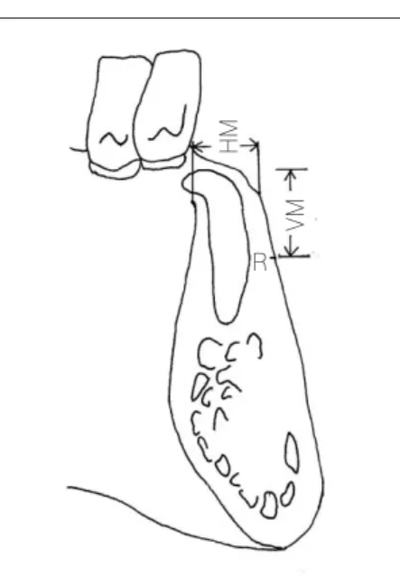
4. 계측항목 (Fig. 4)
A. Vertical dimension of alveolar ridge
술후 2-4주와 술후 4-6개월에 채득한 study model에서 acrylic stent를 장착하고 acrylic stent에서 15mm 하방에 reference point 정한 후 ridge의 crest에서 reference point까지의 거리를 측정하 였다.
B. Horizontal dimension of alveolar ridge
술후 2-4주와 술후 4-6개월에 채득한 study model에서 acrylic stent를 장착하고 reference line을 정한 후 ridge의 bucco-lingual width를 측정하였다.
Ⅲ. 연구결과
임플란트 식립을 위해 시행한 치조제 보존술 후 치조제의 수 직적 높이와 수평적 폭의 변화에 관한 연구 결과는 다음과 같다.
1. Vertical dimension (Table 1)
수술 2-4주 후 vertical height가 7.96±1.47mm, 수술 4-6개월 후 7.05±1.63mm로 수술 2-4주 후에 비해 술후 4-6개월에 치조제 의 높이가 평균 0.91±0.40mm 감소하였다.
이러한 치조제 높이의 감소는 통계적으로 유의성이 있음을 보였다 (p-value<0.01) .
2. Horizontal dimension (Table 2)
수술 2-4주 후 horizontal width가 10.77±1.53mm, 수술 4-6개월 후 9.52±1.81mm로 수술 2-4주 후에 비해 술후 4-6개월에 치조 제의 폭이 평균 1.25±0.58mm 감소하였다.
이러한 치조제 폭의 감소는 통계적으로 유의성이 있음을 보 였다 (p-value<0.01) .
치조제 보존술 시행 후 수직적 높이의 감소량은 평균 0.91±
0.40mm, 수평적 폭의 감소량은 평균 1.25±0.58mm으로 술 후
폭의 변화가 높이의 변화보다 큰 것으로 나타났다.
상악 치조제와 하악 치조제를 비교시 치조제의 높이와 폭이 모두 상악에서 하악보다 감소량이 크게 나타났다 (Table 3) .
Fig. 4.Measurement of vertical & horizontal dimension using digital caliper.
HM: horizontal measurement VM: vertical measurement R: reference point Fig. 3.Measurement of ridge dimension.
Immediate postoperation and 4-6 months after operation study models were measured to evaluate the ridge dimen- sional change using digital caliper
HM VM
R
Ⅳ. 고 찰
발치 후 발치부 치조제의 흡수는 자연적인 현상으로 2005년 Lindhe16)등은 12마리 mongrel dog의 발치된 하악 소구치부를 관 찰하였을 때 치조제가 흡수됨을 보고하였다. 발치창의 치유를 조직학적으로 연구시 파골세포를 볼 수 있었으며, 발치 후 치 조제의 흡수는 치조정 부위의 이러한 파골세포의 작용때문이 라 하였다. 발치 후 치조제의 흡수량에 대한 1997년 Lekovic17)의 연구에서 vertical dimension이 평균 0.88±0.26mm, horizontal dimension이 평균 4.43±0.52mm 감소했다고 하였다. 1년 후 연 구18)에 의하면 vertical dimension이 1.50±0.21mm, horizontal dimension이 4.59±0.23mm 감소했다 하였다. 2003년 Iasella19)등 의 연구는 발치 후 발치부 치조제의 흡수는 폭이 2.6±2.3mm, 높이가 협측에서는 0.9±1.6mm, 설측에서는 0.4±0.1mm라 발 표 하였다. 이들의 연구 결과 발치 후 치조제의 흡수는 설측에 서보다 순측 또는 협측에서 그 양이 많다 하였다. 또 하악보다 는 상악에서 발치 시 치조제의 흡수량이 크다 하였다. 본 연구 에서는 발치 후 아무런 처치를 하지 않은 대조군을 설정하지 않았으며, 앞선 연구들 에서 보고하고 있는 발치 후 치조골의 흡수양상을 참고하였다.
무치악부 임플란트 식립을 위해서는 일정량의 치조제의 폭
과 높이가 유지되어야 한다. 그러나 발치부의 치조제는 흡수 되어 임플란트 식립을 위해 인레이 또는 온레이 이식 등의 추 가적인 골이식을 필요로 하는 경우가 많다. 선학들은 발치 후 치조제의 흡수를 최소화 하기 위해 발치시 최소한의 외력을 가해야 한다 하였으며, 발치창에 골전도성을 지닌 물질 삽입 을 추천하기도 하였다. 이렇게 발치 후 발치창에 이식제를 삽 입하여 치조제 흡수를 최소화 하기위한 술식이 치조제 보존술 이라 할 수 있다. 치조제 보존술을 시행함에 따라 치조제 흡수 를 방지하는 기전에 대해 1992년 Hart20)등은 삽입된 이식체와 골사이에 elastic modulus차이로 bone remodeling이 활성화되고 이로 인해 골밀도가 증가하기 때문이라 하였다. 1996년 Rowe21) 등은 치조제 보존술 시행시 사용한 occlusive membrane이 발치 창으로의 구강상피침투를 방지해 bone fill을 증가시키기 때문 이라 하였다. 또한 graft material의 osteoclast inhibition, osteoblast stimulation 작용으로 골밀도를 향상시킨다 하였다.
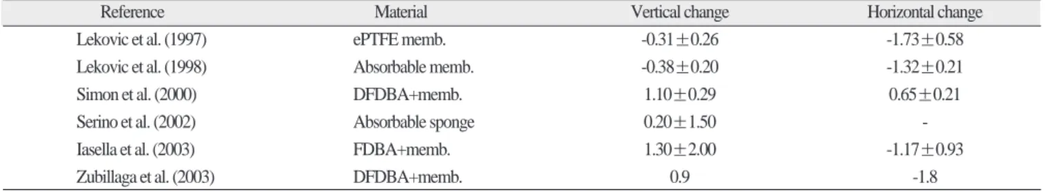
1980년대부터 현재까지 발치 후 임플란트 식립을 위한 치조 제 보존술에 대한 많은 연구들이 있어왔다. osteoconductive material, barrier membrane 등의 다양한 재료를 사용한 치조제 보 존술이 시행되었으며 그 결과 치조제의 높이와 폭의 변화에 대해 Lekovic, Simon, Serino, Iasella, Zubillaga 등은 발치 후 아무 런 처치를 하지 않은 군보다 발치와에 이식재료를 사용하여
maxilla mandible
change of VH 0.96±0.39 0.80±0.41
change of HW 1.49±0.51 0.81±0.42
VH: vertical height, HW: horizontal width
Table 2.Result (horizontal width). (mm)
Mean SD
T1 10.77 1.53
T2 9.52 1.81
T2-T1 1.25 0.58
T1: Immediate post-op, T2: Post-op. 4-6months, T2-T1: Change of hori- zontal width
Table 1.Result (vertical height). (mm)
Mean SD
T1 7.96 1.47
T2 7.06 1.63
T2-T1 0.91 0.40
T1: Immediate post-op, T2: Post-op. 4-6months, T2-T1: Change of verti- cal height
Table 4.Comparison of ridge preservation. (mm)
Reference Material Vertical change Horizontal change
Lekovic et al. (1997) ePTFE memb. -0.31±0.26 -1.73±0.58
Lekovic et al. (1998) Absorbable memb. -0.38±0.20 -1.32±0.21
Simon et al. (2000) DFDBA+memb. 1.10±0.29 0.65±0.21
Serino et al. (2002) Absorbable sponge 0.20±1.50 -
Iasella et al. (2003) FDBA+memb. 1.30±2.00 -1.17±0.93
Zubillaga et al. (2003) DFDBA+memb. 0.9 -1.8
Table 3.Dimensional changes in maxilla and mandible. (mm)
치조제 보존술을 시행한 군에서 치조제의 흡수가 적다고 하였 다. 이들의 연구는 각기 다른 재료를 사용 하였으며 이식재의 종류와 무관하게 치조제 보존술의 효용성을 보여 주고 있으 며, 치조제 보존술을 시행한 후에도 치조제 높이의 변화에 비 해 폭의 변화가 큰 것으로 나타났다(Table 4). 또한 하악 보다는 상악에서 치조제의 변화량이 컸음을 말하고 있다. 이와 유사 한 치조제 변화 양상을 이 번 연구를 통해서도 알 수 있었다. 이 러한 연구들에서 발치 후 자연적 창상치유를 기대한 대조군에 서 보다 치조제 보존술을 시행한 군에서 치조제의 흡수가 적 음을 나타냈으며, Artis22,23)등의 연구에 의하면 치조제 보존술을 시행한 발치부에서 골의 양과 골의 성숙도는 발치와의 깊은 곳일수록 높다 하였다.
본 연구는 bovine cancellous bone mineral particle(Bio-Oss�)과 absorbable collagen sponge(Collaplug�)를 사용하여 발치 후 치조 제의 흡수를 줄이고자 하였다. 이 술식 역시 널리 사용되고 있 으며, 치조제 보존술 시행시 무리한 피판의 조작으로 일차봉 합을 시도하지 않고도 graft material을 유지할 수 있으며, barrier membrane을 사용하는 것에 비해 적은 비용이 드는 술식이라 할 수 있다.
1997년 Berglundh와 Lindhe24)는 5마리 beagle dog으로 골 결손 부위에서 bovine cancellous bone mineral particle의 효과를 연구했 다. Beagle dog의 소구치 부위에 인위적인 골 결손부를 형성하 고 bovine cancellous bone을 삽입한 부위와 아무런 처치를 하지 않은 부위의 치유과정을 3개월, 6개월째 관찰 하였다. Bovine cancellous bone 삽입 후 6개월까지 시간이 경과함에 따라 bovine cancellous bone mineral의 양은 감소 하였으며, bone to implant contact(%)는 실험군과 대조군에서 비슷한 양을 보였다. 조직학 적으로 관찰시 bovine cancellous bone mineral particle 주위로 새로 운 골이 형성되는 것을 관찰할 수 있었다고 하였다. 이 연구는 bovine cancellous bone mineral particle이 골 전도성을 지닌 물질로 써 골 결손부에서 새로운 골을 형성하는 골격역할을 하며 이 식된 bovine bone은 시간이 지날수록 새로운 골로 대체된다는 결론을 내렸다. 1997년 Howell25)등은 24개월간의 human clinical trial을 통한 absorbable collagen sponge을 사용한 ridge preservation 시행결과 이 물질이 발치 후 골 결손부위에 안정적인 것이라 말하고 있다. Berglundh 및 Lindeh와 Howell 등의 연구에서 치조 제 보존술 시행시 사용한 bovine cancellous bone mineral particle, absorbable collagen sponge는 검증된 재료라 할 수 있다.
Bovine cancellous bone mineral particle과 absorbable collagen sponge를 이용해 치조제보존술을 시행한 본 연구를 통해 술 후 4-6개월에 임플란트 식립을 위한 적절한 치조제의 폭과 높이 가 유지되었음을 알 수 있었다.
Ⅴ. 결 론
치조제 보존술 시행 후 4-6개월동안 통계적 유의성이 있는 치조제의 폭과 높이의 감소를 보였으며, 치조제의 수직적 높 이의 감소에 비해 수평적 폭의 감소량이 더 큰 것으로 나타났
다. 하악의 치조제 감소보다는 상악에서의 감소량이 많았다.
비록 치조제 보존술 시행시 치조제의 폭과 높이의 변화가 통 계적으로는 유의하였으나 임상적으로 임플란트를 식립할 만 한 치조제는 유지되었고 특기할 합병증이나 후유증은 없었다.
참고문헌
1. Sobolik CF: Alveolar bone resorption. J Prosthet Dent 1960;10:612- 619.
2. Devlin H, Ferguson MJ: Alveolar ridge resorption and mandibular atrophy. A review of the role of local and systemic factors. Br Dent J 1991;170:101-104.
3. Pietrokovski J, Massler M: Alveolar ridge resorption following tooth extraction. J Prosthet Dent 1997;77:596-600.
4. Mecall RA, Rosenfeld AL: Influence of residual ridge resorption pat- terns on implant fixture placement and tooth position. Int J Periodontics Restorative Dent 1991;11:8-23.
5. Razavi R, Zena RB, Khan Z, Gould AR: Anatomic site evaluation of edentulous maxillae for dental implant placement. J Prosthodont 1995;4:90-94.
6. Eufinger H, Konig S, Eufinger A: The role of alveolar ridge width in dental implantology. Clin Oral Invest 1997;1:169-177.
7. Uhler IV: Oral surgery and the geriatric patient. Oral and Maxillofacial Surgery. Philadelphia, Pa: WB Saunders Co; 1975:976.
8. Garver DG, Fenster RK: Vital root retention in humans; a final report.
J Prosthet Dent 1980;43:368-373.
9. Von Wowern N, Winther S: Submergence of roots for alveolar ridge preservation. A failure(4-year follow-up study). Int J Oral Surg 1981;10:247-250.
10. Quinn JH, Kent JN: Alveolar ridge maintenance with solid nonporous hydroxylapatite root implants. Oral Surg Oral Med Oral Pathol 1984;58:511-521.
11. Kentros GA, Filler SJ, Rothstein SS: Six month evaluation of particu- late Durapatite in extraction sockets for the preservation of the alveo- lar ridge. Implantologist 1985;3:53-62.
12. Nemcovsky CE, Serfaty V: Alveolar ridge preservation following extraction of maxillary anterior teeth. Report on 23 consecutive cases.
J Periodontol 1996;67:390-395.
13. Gross J: Ridge preservation using HTR synthetic bone following tooth extraction. Gen Dent 1995;43:364-367.
14. Becker W, Becker BE, Caffesse R: A comparison of demineralized freeze-dried bone and cell occlusive membranes. J Periodontol 1996;67:821-825.
15. Schepers EJ, Ducheyne P, Barbier L, Schepers S: Bioactive glass of narrow size range: a new material for the repair of bone defects.
Implant Dent 1993;2:151-156.
16. Araujo MG, Lindhe J: Dimensional ridge alterations following tooth extraction. An experimental study in the dog. J Clin Periodontol 2005;32:212-218.
17. Lekovic V, Kenney EB, Weinlaender M: A bone regenerative approach to alveolar ridge maintenance following tooth extraction.
Report of 10 cases. J Periodontol 1997;68:563-570.
18. Lekovic V, Camargo P, Klokkevold P, Weinlaender M: Preservation of alveolar bone in extraction sockets using bioabsorbable mem- branes. J Periodontol 1998;69:1044-1049.
19. John MI, Henry G, Richard LM: Ridge preservation with freeze-dried bone allograft and a collagen membrane compared to extraction alone for implant site development: A clinical and histologic study in humans. J Periodontol 2003;74:990-999.
20. Hart RT, Hennebel VV, Thongpreda N, Van Buskirk WC, Anderson RC: Modeling the biomechanics of the mandible: a three-dimensional finite element study. J Biomech 1992;25:261-286.
21. Rowe DJ, Leung WW, Del Carlo DL: Osteoclast inhibition by factors from cells associated with regenerative tissue. J Periodontol 1996;
67:414-421.
22. Artiz Z, Tal H, Dayan D: Porous bovine bone mineral in healing of human extraction sockets. Part 1:Histomorphometric evaluations at 9 months. J Periodontol 2000;71:1015-1023.
23. Artiz Z, Tal H, Dayan D: Porous bovine bone mineral in healing of human extraction sockets: 2. Histochemical observations at 9 months.
J Periodontol 2001;72:152-159.
24. Berglundh T, Lindhe J: Healing around implants placed in bone
defects treated with Bio-Oss. An experimental study in the dog. Clin Oral Implants Res 1997;8(2):117-124.
25. Howell TH, Fiorellini J, Jones A: A feasibility study evaluating rhBMP-2/absorbable collagen sponge device for local alveolar ridge preservation or augmentation. Int J Periodontics Restorative Dent 1997;17:124-139.