R E S E A R C H Open Access
Molecular diagnosis of hereditary spherocytosis by multi-gene target sequencing in Korea: matching with osmotic fragility test and presence of spherocyte
Hyoung Soo Choi 1 † , Qute Choi 2 † , Jung-Ah Kim 3 , Kyong Ok Im 4 , Si Nae Park 4 , Yoomi Park 5 , Hee Young Shin 4,6 , Hyoung Jin Kang 4,6 , Hoon Kook 7 , Seon Young Kim 8 , Soo-Jeong Kim 9 , Inho Kim 10 , Ji Yoon Kim 11 , Hawk Kim 12 , Kyung Duk Park 4,6 , Kyung Bae Park 13 , Meerim Park 14 , Sang Kyu Park 15 , Eun Sil Park 16 , Jeong-A Park 17 ,
Jun Eun Park 18 , Ji Kyoung Park 19 , Hee Jo Baek 7 , Jeong Ho Seo 20 , Ye Jee Shim 21 , Hyo Seop Ahn 6 , Keon Hee Yoo 22 , Hoi Soo Yoon 23 , Young-Woong Won 24 , Kun Soo Lee 11 , Kwang Chul Lee 25 , Mee Jeong Lee 26 , Sun Ah. Lee 27 , Jun Ah Lee 28 , Jae Min Lee 29 , Jae Hee Lee 30 , Ji Won Lee 22 , Young Tak Lim 20 , Hyun Joo Jung 18 , Hee Won Chueh 31 , Eun Jin Choi 32 , Hye Lim Jung 33 , Ju Han Kim 5* † , Dong Soon Lee 3* † and The Hereditary Hemolytic Anemia Working Party of the Korean Society of Hematology 34
Abstract
Background: Current diagnostic tests for hereditary spherocytosis (HS) focus on the detection of hemolysis or indirectly assessing defects of membrane protein, whereas direct methods to detect protein defects are complicated and difficult to implement. In the present study, we investigated the patterns of genetic variation associated with HS among patients clinically diagnosed with HS.
Methods: Multi-gene targeted sequencing of 43 genes (17 RBC membrane protein-encoding genes, 20 RBC enzyme- encoding genes, and six additional genes for the differential diagnosis) was performed using the Illumina HiSeq platform.
Results: Among 59 patients with HS, 50 (84.7%) had one or more significant variants in a RBC membrane protein- encoding genes. A total of 54 significant variants including 46 novel mutations were detected in six RBC membrane protein-encoding genes, with the highest number of variants found in SPTB (n = 28), and followed by ANK1 (n = 19), SLC4A1 (n = 3), SPTA1 (n = 2), EPB41 (n = 1), and EPB42 (n = 1). Concurrent mutations of genes encoding RBC enzymes (ALDOB, GAPDH, and GSR) were detected in three patients. UGT1A1 mutations were present in 24 patients (40.7%). Positive rate of osmotic fragility test was 86.8% among patients harboring HS-related gene mutations.
(Continued on next page)
© The Author(s). 2019 Open Access This article is distributed under the terms of the Creative Commons Attribution 4.0 International License (http://creativecommons.org/licenses/by/4.0/), which permits unrestricted use, distribution, and reproduction in any medium, provided you give appropriate credit to the original author(s) and the source, provide a link to the Creative Commons license, and indicate if changes were made. The Creative Commons Public Domain Dedication waiver (http://creativecommons.org/publicdomain/zero/1.0/) applies to the data made available in this article, unless otherwise stated.
* Correspondence: [email protected]; [email protected]
†
Hyoung Soo Choi, Qute Choi, Ju Han Kim and Dong Soon Lee contributed equally to this work.
5
Division of Biomedical Informatics, Seoul National University Biomedical Informatics (SNUBI), Seoul National University College of Medicine, 101, Daehak-ro, Jongno-gu, Seoul 03080, Republic of Korea
3
Department of Laboratory Medicine, Seoul National University College of
Medicine, 101, Daehak-ro, Jongno-gu, Seoul 03080, Republic of Korea
Full list of author information is available at the end of the article
(Continued from previous page)
Conclusions: This constitutes the first large-scaled genetic study of Korean patients with HS. We demonstrated that multi-gene target sequencing is sensitive and feasible that can be used as a powerful tool for diagnosing HS. Considering the discrepancies of clinical and molecular diagnoses of HS, our findings suggest that molecular genetic analysis is required for accurate diagnosis of HS.
Keywords: Hereditary spherocytosis, RBC membrane disorder, Molecular diagnosis
Background
Hereditary spherocytosis (HS) is the most common cause of hereditary hemolytic anemia (HHA) character- ized by the presence of spherocytes in peripheral blood smear (PBS) [1, 2]. HS occurs in 1 in 2000 Caucasians, with less common frequency in Asians [1, 3, 4]. The crude incidence of HS in Korea was reported as 1 in every 5000 births [5]. Approximately 75% cases of HS are inherited as autosomal dominant (AD) mutations, whereas the remaining cases involve autosomal recessive (AR) or de-novo mutations [ 1].
HS is caused by a deficiency in or dysfunction of mem- brane proteins, including spectrin, ankyrin 1, band 3, and protein 4.2, associated with the RBC cytoskeleton [3, 4, 6].
Defective membrane proteins disrupt the vertical linkage between the RBC membrane cytoskeleton and the phospholipid bilayer, causing RBCs to lose its biconcave characteristics and become spherical in shape [3, 4, 6].
This abnormal RBC morphology leads to osmotically fra- gile cells that are selectively trapped and destroyed in the spleen [3, 4, 6]. A major clinical manifestation of HS is hemolytic anemia, which exhibits a wide range of clinical manifestations from asymptomatic to life-threatening anemia requiring regular RBC transfusions [1, 2]. Other clinical symptoms include splenomegaly, jaundice, and gallstones, depending on disease severity [1, 2].
We have been operating the Korean Hereditary Hemolytic Anemia Working Party (KHHAWP) of the Ko- rean Society of Hematology for 7 years since 2010, which name has been changed to RBC Disorder Working Party since November 2016. From 2007 to 2011, 195 patients (121 males and 74 females) diagnosed with HHA from 25 institutions were registered [7]. The KHHAWP presented standard operating procedure (SOP) for the diagnosis of HHA (Fig. 1) [5], which is similar to ICSH (International Council for Standardization in Haematology) guideline [8]
except for excluding acid glycerol lysis time test as a screening test. Instead of gel electrophoresis analysis of erythrocyte membranes, the KHHAWP adopted mass spectrometry method as a confirmatory test, which is per- formed in one central laboratory in Korea.
The diagnosis of HS is based upon a combination of positive family history, clinical features and presence of spherocytes in PBS, which are detectable in 97% of patients [9]. When the diagnosis of HS is equivocal,
additional laboratory tests are recommended such as os- motic fragility test (OFT), autohemolysis test, flow cy- tometry [OFT and eosin-5-maleimide (EMA) binding test] for screening test, and protein analysis using gel electrophoresis or mass spectrometry can be additionally tested [10 – 16]. However, none of the current diagnostic test can detect all patients with HS.
Considering the limitations of existing diagnostic tests, development of a simple and direct method to measure RBC membrane protein abnormalities to confirm HS is re- quired. Analysis of RBC membrane protein-encoding genes is expected that it can be used complementarily with the conventional confirmatory tests [1, 11]. Multi-gene target sequencing for RBC membrane protein-encoding genes is feasible and reliable diagnostic method to detect mutations in patients affected by various disorders of the RBC mem- brane. Particularly, gene testing is important in young chil- dren with congenital anemia, transfusion-dependent patients, and in families with variable clinical expression or complex inheritance patterns [17 – 19].
In the present study, we investigated the genetic vari- ation of RBC membrane protein-encoding genes using multi-gene target sequencing, comparing with clinical features. A total of 43 genes was included; 17 RBC mem- brane protein-encoding genes and 20 RBC enzyme- encoding genes, in context with six additional candidate genes for the purpose of differential diagnoses [thalas- semia, congenital dyserythropoietic anemia (CDA), paroxysmal nocturnal hemoglobinuria (PNH), and Gil- bert syndrome].
Methods Patients
A total of 59 patients with HS including 31 males and 28 females with a median age of 7 years (range: 1–81 years), were registered between July 2013 and July 2014 from the pediatrics and internal medicine departments of 25 institutions in Korea. HS was diagnosed according to the SOP recommended by the KHHAWP of the Ko- rean Society of Hematology (Fig. 1) [5].
Along with clinical data including age, sex, symptoms
and family history, we collected the results of laboratory
tests including CBC with RBC index, reticulocyte count,
total and direct bilirubin concentration, lactate dehydro-
genase (LDH), iron, total iron-binding capacity (TIBC),
ferritin, PBS, and OFT by reviewing medical records (Table 1). Blood samples were collected from each patient after obtaining their written consent.
Targeted sequencing
To gain insight into the genetic variations, we per- formed targeted sequencing for 43 gene panel (Add- itional file 1: Table S1). gDNA shearing to generate the standard library and the hybridization step targeting only exonic regions were performed by Celemics Inc.
(Seoul, Korea). The final quality was assessed using the Agilent 2200 TapeStation System (Santa Clara, CA, USA). We sequenced a total target length of 259-kb regions using the paired-end 150-bp rapid-run sequencing mode on an Illumina HiSeq 2500 platform.
The mean sequencing depth for the targeted regions (259-kb) was 231-fold (n = 59). Because a matched control sample was not included in this study, we ap- plied a stringent variant selection pipeline to prioritize the high-confidence set of somatic mutations.
Variant calling
The filtration process was performed as follows. Variants within non-exonic regions were removed. Variants that do not have enough depth were also filtered out to re- move false positives. Common variants on 1000 genome projects with more than 5% of allele frequency were fil- tered out. CADD score shows predictive pathogenicity of variants. It considers diverse annotations from allelic diversity to functionality, in order to estimate pathogenic variants. In this study, CADD scores below 10 were cut-off for filtration. After these filters, in-house variants were also removed to make filtered variant lists. Valid- ation of variant call was performed by target gene sequencing of involved genes.
Simulation of the effect of mutated genes on protein structure
To predict how gene mutation affect protein structure, we visualized three-dimensional (3-D) spatial protein struc- ture following acquisition of their structural information (http://www.proteinmodelportal.org) (Additional file 1:
Fig. 1 Standard operating procedure for the diagnosis of hereditary hemolytic anemia (HHA) by HHA Working Party of Korean Society of
Hematology [5]
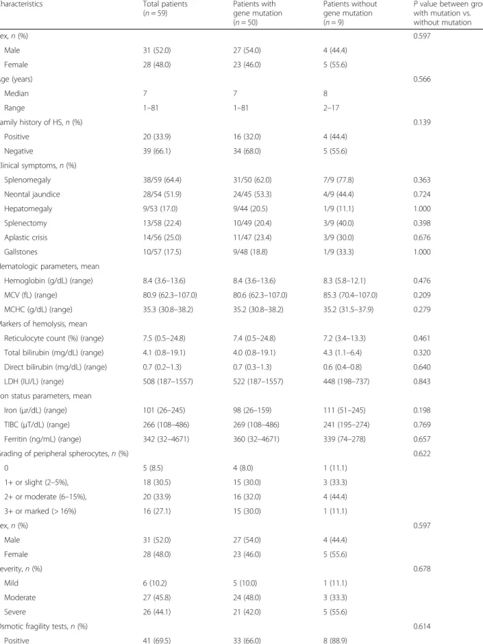
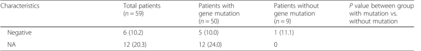
Table 1 Clinical characteristics of patients with HS in Korea
Characteristics Total patients
(n = 59)
Patients with gene mutation (n = 50)
Patients without gene mutation (n = 9)
P value between group with mutation vs.
without mutation
Sex, n (%) 0.597
Male 31 (52.0) 27 (54.0) 4 (44.4)
Female 28 (48.0) 23 (46.0) 5 (55.6)
Age (years) 0.566
Median 7 7 8
Range 1 –81 1 –81 2 –17
Family history of HS, n (%) 0.139
Positive 20 (33.9) 16 (32.0) 4 (44.4)
Negative 39 (66.1) 34 (68.0) 5 (55.6)
Clinical symptoms, n (%)
Splenomegaly 38/59 (64.4) 31/50 (62.0) 7/9 (77.8) 0.363
Neontal jaundice 28/54 (51.9) 24/45 (53.3) 4/9 (44.4) 0.724
Hepatomegaly 9/53 (17.0) 9/44 (20.5) 1/9 (11.1) 1.000
Splenectomy 13/58 (22.4) 10/49 (20.4) 3/9 (40.0) 0.398
Aplastic crisis 14/56 (25.0) 11/47 (23.4) 3/9 (30.0) 0.676
Gallstones 10/57 (17.5) 9/48 (18.8) 1/9 (33.3) 1.000
Hematologic parameters, mean
Hemoglobin (g/dL) (range) 8.4 (3.6 –13.6) 8.4 (3.6 –13.6) 8.3 (5.8 –12.1) 0.476
MCV (fL) (range) 80.9 (62.3 –107.0) 80.6 (62.3 –107.0) 85.3 (70.4 –107.0) 0.209
MCHC (g/dL) (range) 35.3 (30.8 –38.2) 35.2 (30.8 –38.2) 35.2 (31.5 –37.9) 0.279
Markers of hemolysis, mean
Reticulocyte count (%) (range) 7.5 (0.5 –24.8) 7.4 (0.5 –24.8) 7.2 (3.4 –13.3) 0.461 Total bilirubin (mg/dL) (range) 4.1 (0.8 –19.1) 4.0 (0.8 –19.1) 4.3 (1.1 –6.4) 0.320 Direct bilirubin (mg/dL) (range) 0.7 (0.2 –1.3) 0.7 (0.3 –1.3) 0.6 (0.4 –0.8) 0.640
LDH (IU/L) (range) 508 (187 –1557) 522 (187 –1557) 448 (198 –737) 0.843
Iron status parameters, mean
Iron ( μr/dL) (range) 101 (26 –245) 98 (26 –159) 111 (51 –245) 0.198
TIBC ( μT/dL) (range) 266 (108 –486) 269 (108 –486) 241 (195 –274) 0.769
Ferritin (ng/mL) (range) 342 (32 –4671) 360 (32 –4671) 339 (74 –278) 0.657
Grading of peripheral spherocytes, n (%) 0.622
0 5 (8.5) 4 (8.0) 1 (11.1)
1+ or slight (2 –5%), 18 (30.5) 15 (30.0) 3 (33.3)
2+ or moderate (6 –15%), 20 (33.9) 16 (32.0) 4 (44.4)
3+ or marked (> 16%) 16 (27.1) 15 (30.0) 1 (11.1)
Sex, n (%) 0.597
Male 31 (52.0) 27 (54.0) 4 (44.4)
Female 28 (48.0) 23 (46.0) 5 (55.6)
Severity, n (%) 0.678
Mild 6 (10.2) 5 (10.0) 1 (11.1)
Moderate 27 (45.8) 24 (48.0) 3 (33.3)
Severe 26 (44.1) 21 (42.0) 5 (55.6)
Osmotic fragility tests, n (%) 0.614
Positive 41 (69.5) 33 (66.0) 8 (88.9)
Table S2). We used PyMOL (http://www.pymol.org) to visualize 3-D representations of the protein, modified pro- tein structures based on genetic mutation profiles from next-generation sequencing (NGS) results.
Statistical analyses
Stata/SE (v.14; StataCorp, College Station, TX, USA) was used for data analyses. Statistical differences in terms of continuous clinical characteristic variables were esti- mated by two sample t test. The significance of differ- ences in categorical variables between groups was determined by the Pearson χ2 test or Fisher’s exact test.
The level of significance was set at P < 0.05.
Results
Clinical characteristics
Among 59 patients with HS, 20 (33.9%) had a family his- tory of HS, whereas symptoms of splenomegaly, neo- natal jaundice, and hepatomegaly were exhibited in 38 of 59 (64.4%), 28 of 54 (51.9%), and 10 of 59 (16.7%) pa- tients, respectively. Mean values for laboratory tests were as follows: hemoglobin concentration 8.4 g/dL (3.6–13.6 g/dL); corpuscular volume 80.9 fL (62.3–107.0 fL); cor- puscular hemoglobin concentration 35.3 g/dL (30.8–
38.2 g/dL); reticulocyte count indicating hemolysis 7.5%
(0.5–24.8%); total bilirubin/direct bilirubin 4.1/0.7 mg/dL (0.8–19.1/0.2–1.3 mg/dL); LDH 508 IU/L (187–1557 IU/
L); parameters representing iron profile, including iron 101 μg/dL (26–245 μg/dL), TIBC 266 μg/dL (108–
486 μg/dL); and ferritin concentration, 342 ng/mL (32–
4671 ng/mL). PBS was rated for spherocytes on a four-point scale [20] from 0, 1+ or slight (2–5%), 2+ or moderate (6–15%), and 3+ or marked (> 16%) and the number of smears returning 0, 1+ or slight, 2+ or mod- erate and 3+ or marked were 5 (8.5%), 18 (30.5%), 20 (33.9%), and 16 (27.1%) patients, respectively. According to HS-severity criteria [11], severe, moderate, and mild cases were 26 (44.1%), 27 (45.8%), and 6 (10.2%) pa- tients, respectively (Table 1).
Variants profile of RBC membrane protein-encoding genes
Among 17 RBC membrane protein-encoding genes examined, significant disease-related mutations were ob- served in six: SPTB (spectrin, beta), ANK1 (ankyrin 1), SLC4A1 (solute carrier family 4, member 1), SPTA1
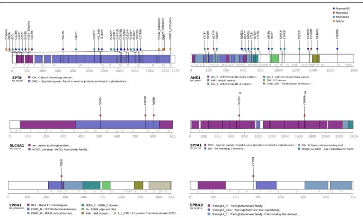
(spectrin, alpha 1), EPB41 (erythrocyte membrane pro- tein band 4.1), and EPB42 (erythrocyte membrane pro- tein band 4.2) (Fig. 2). A total of 54 significant mutations were observed, of which eight were previously reported as pathogenic in patients with HS and 46 vari- ants were novel mutations (Additional file 1: Table S3).
The highest number of mutations were found in SPTB (n = 28), and followed by ANK1 (n = 19), SLC4A1 (n = 3), SPTA1 (n = 2), EPB41 (n = 1), and EPB42 (n = 1). Ac- cording to the American College of Medical Genetics and Genomics guidelines [21], 12 were pathogenic muta- tions (including eight previously reported variants), 29 were likely pathogenic mutations, and 13 were classified as having uncertain significance. All the variants have been confirmed by Sanger sequencing using 35 primer sets (Additional file 1: Table S4).
Variant characteristics in patients with HS
Among 59 patients with HS, 50 (84.7%) had at least one mutation in a RBC membrane protein-encoding gene (Fig. 3). Twenty eight patients carried mutations in the SPTB gene, and 20 patients had mutations in the ANK1 gene. Forty patients (67.8%) carried a single mutation, and 10 patients (16.9%) carried two mutations. Among 40 patients with a single mutation, the most frequently mutated genes were SPTB and ANK1, which were mu- tated in 21 and 17 patients, respectively. The SCL4A1 mutation was found in two patients. Among the 10 pa- tients harboring two mutations, one carried two muta- tions in a single gene (ANK1), and three patients carried mutations in both SPTB and SPTA1. Combinations of mutations in SPTB and ANK1, SPTB and EPB41, and SPTB and EPB42 were detected in one patient each. In addition, combination with RBC enzyme-encoding gene mutations were found in three patients [SLC4A1 and GAPDH (glyceraldehyde-3-phosphate dehydrogenase), ANK1 and GSR (glutathione reductase), SPTB and ALDOB (aldolase B)] (Additional file 1: Table S5).
Nine patients carried no mutation on the RBC mem- brane protein- or enzyme-encoding genes. Coexisting mu- tations of UGT1A1 (UDP glycosyltransferase 1 family, polypeptide A1) gene were detected in 24 of 59 HS pa- tients (40.7%), with UGT1A1 mutations combined with other gene mutations in 20 patients and without other gene mutation in four patients (Table 2, Additional file 1:
Table S6). Total bilirubin level or presence of neonatal Table 1 Clinical characteristics of patients with HS in Korea (Continued)
Characteristics Total patients
(n = 59)
Patients with gene mutation (n = 50)
Patients without gene mutation (n = 9)
P value between group with mutation vs.
without mutation
Negative 6 (10.2) 5 (10.0) 1 (11.1)
NA 12 (20.3) 12 (24.0) 0
Abbreviation: HS hereditary spherocytosis, NA not assessable
Fig. 2 Characteristics of significant variants for RBC membrane protein-encoding genes; SPTB, ANK1, SLC4A1, SPTA1, EPB41, EPB42. Abbreviations: SPTB, spectrin, beta; ANK1, ankyrin 1; SLC4A1, solute carrier family 4, member 1; SPTA1, spectrin, alpha 1; EPB41, erythrocyte membrane protein band 4.1; EPB42, erythrocyte membrane protein band 4.2
Fig. 3 Number of patients with RBC membrane protein-encoding gene mutations. Abbreviations: SPTB, spectrin, beta; SPTA1, spectrin, alpha 1; EPB41, erythrocyte membrane protein band 4.1; EPB42, erythrocyte membrane protein band 4.2; ALDOB, aldolase B; ANK1, ankyrin 1; GSR, glutathione reductase;
SLC4A1, solute carrier family 4, member 1; GAPDH, glyceraldehyde-3-phosphate dehydrogenase
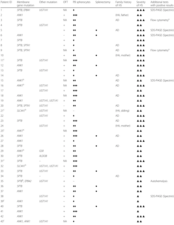
Table 2 Gene mutations, laboratory tests and clinical characteristics Patient ID Membrane
gene mutation
Other mutation OFT PB spherocytes Splenectomy Family history of HS
Severity of HS
Additional tests with positive results
1 SPTB, EPB41 UGT1A1 NA ♦ ▲▲▲ SDS-PAGE (Spectrin)
2 ANK1 + ♦♦♦ (HA, father) ▲▲
3 SPTB NA ♦♦ AD ▲▲▲ Flow cytometry
a4 SPTB UGT1A1 + ♦♦ ▲▲
5 + ♦♦ ● AD ▲▲▲ SDS-PAGE (Spectrin)
6 ANK1 – ♦♦ ● ▲▲▲ SDS-PAGE (Spectrin)
7 SPTB + ♦ ▲▲▲
8 SPTB, SPTA1 + ♦ AD ▲▲▲
9 SPTB, SPTA1 NA ♦ AD ▲▲▲ Flow cytometry
a10 + ♦♦ ● (HA, mother) ▲▲▲
11
cSPTB UGT1A1 NA ♦♦♦ ▲▲▲
12 ANK1 + ♦♦ ● ▲▲▲
13 SPTB UGT1A1 + ♦ ▲▲
14 + ♦ ● AD ▲▲▲
15 ANK1
bNA ♦♦ AD ▲▲ SDS-PAGE (Spectrin)
16 ANK1
bUGT1A1 NA ♦♦♦ AD ▲▲▲
17 UGT1A1 + ♦♦♦ ▲▲
18 ANK1 NA ♦♦♦ AD ▲▲▲
19 ANK1 UGT1A1, UGT1A1 + ♦♦ ▲▲
20 SPTB, SPTA1 UGT1A1 – ♦♦ AD ▲▲▲
21
cSLC4A1
bUGT1A1 NA – (HA, sibling) ▲▲
22 UGT1A1 + ♦ AD ▲▲▲
23 SPTB + ♦♦♦ AD ▲▲▲
24 UGT1A1 + ♦♦ (HA, mother) ▲▲▲
25
cANK1
bNA ♦♦♦ ▲▲
26 ANK1 + ♦♦♦ ● AD ▲▲
27 ANK1 + ♦ ▲▲▲
28 SPTB + ♦♦ ● AD ▲▲
29 ANK1
bGSR + ♦♦ ▲▲
30 SPTB ALDOB + ♦♦♦ ▲▲
31
cSPTB – NA ♦♦♦ ▲▲▲
32 SLC4A1
bUGT1A1, UGT1A1 + ♦♦♦ ▲▲
33 SPTB UGT1A1 + ♦♦ ● ▲▲▲
34 SPTB – – ♦ AD ▲▲
35 SPTB
b, EPB42 UGT1A1 + – ▲▲ Autohemolysis
36 SPTB + ♦♦ ● ▲▲
37 ANK1 + ♦♦ ● ▲▲
38 UGT1A1 + ♦ ▲ SDS-PAGE (Spectrin)
39
cANK1 UGT1A1 – ♦ ▲
40 SPTB + ♦♦ ● ▲▲▲
41 ANK1 + ♦♦♦ ▲
42 ANK1 + ♦♦ ▲▲▲
43
cANK1, ANK1 UGT1A1 NA ♦ ▲▲
jaundice did not differ significantly from those without UGT1A1 mutations.
Genotype and phenotype correlations in patients with HS Comparisons of laboratory findings and clinical charac- teristics showed no significant differences in hematologic parameters, hemolysis markers, iron status parameters, sex, family history of HS, number of splenectomized pa- tients, and disease severity according to the gene muta- tion type and number of mutation or presence of UGT1A1 mutation (Table 1, Additional file 1: Table S6).
Among 59 patients with HS, nine patients (15.3%) without mutation associated with RBC membrane pro- tein-encoding genes showed similar baseline character- istics in most aspects as compared with those with mutations (Table 1). Median age of patients without mutation was 8 years, and the proportion of family his- tory, clinical symptoms, grading of peripheral sphero- cytes, and OFT results did not differ significantly from those with mutation.
Intercorrelations between gene mutations and laboratory findings: OFT, the presence of spherocytes in PBS, and gene mutations
The results of genetic test were matched with routine diagnostic tests for HS including OFT and the presence
of spherocytes in PBS (Table 3, Fig. 4). Among 59 pa- tients with clinical HS, results of NaCl induced OFT (room temperature and/or 24 h incubated) was avail- able in 47 patients and 41 of them (87.2%) showed posi- tive results (Additional file 1: Figure S2). Thirty three of 47 patients (70.2%) showed positivity in both OFT and gene test, while one patients (2.1%) showed negative re- sults in both OFT and gene test. In six out of 47 pa- tients (12.7%) with negative OFT, five carried mutations in RBC membrane protein-encoding genes. Among 38 patients harboring HS-related gene mutations, 33 showed positive OFT (86.8%).
Spherocytes in PBS were present in 54 of 59 patients (91.5%). Among five patients without spherocytes in PBS, four carried mutations in RBC membrane protein- encoding genes (Additional file 1: Table S7). One of 59 patients who had anemia and family history of HS showed negative results on all three tests.
Discussion
Using multi-gene target sequencing, 50 of 59 patients (84.7%) of clinically diagnosed HS proved to be molecu- lar HS and three patients harbored coexisting gene mu- tations of RBC enzymes (ALDOB, GAPDH, and GSR) in this study. Mutations of six kinds of RBC membrane Table 2 Gene mutations, laboratory tests and clinical characteristics (Continued)
Patient ID Membrane gene mutation
Other mutation OFT PB spherocytes Splenectomy Family history of HS
Severity of HS
Additional tests with positive results
44 SPTB,ANK1 UGT1A1 + ♦ ▲▲
45 ANK1 UGT1A1 + ♦♦♦ ▲▲
46 SPTB UGT1A1 + ♦♦♦ ▲▲
47 SPTB + ♦ (HA, sibling) ▲▲▲
48
cSPTB
bNA ♦ ▲▲▲
49
cSPTB UGT1A1 + – ▲▲
50 ANK1 + – AD ▲▲
51 SPTB UGT1A1 + ♦ AD ▲▲
52 + ♦♦ ▲▲
53 ANK1 + ♦ ▲
54 – – ● AD ▲▲▲
55 SPTB – ♦♦♦ AD ▲
56 SLC4A1 UGT1A1, GAPDH + ♦ ▲
57 SPTB + ♦♦♦ AD ▲▲▲
58 SPTB UGT1A1 + ♦♦ AD ▲▲
59 SPTB + ♦ ▲▲▲
a
Flow cytometry (OFT and EMA binding test),
bPreviously reported variants (see Additional file 1: Table S3),
cEight patients who did not meet the diagnostic criteria of HS without genetic testing
PB spherocytes [20] ♦, 1+; ♦♦, 2+; ♦♦♦, 3+, Severity of HS [ 8] ▲, mild; ▲▲, moderate; ▲▲▲, severe
Abbreviations: AD autosomal dominant, ALDOB aldolase B, ANK1 ankyrin 1, EPB41 erythrocyte membrane protein band 4.1, EPB42 erythrocyte membrane protein
band 4.2, GAPDH glyceraldehyde-3-phosphate dehydrogenase, GSR glutathione reductase, HA hemolytic anemia, SLC4A1 solute carrier family 4, member 1, SPTA1
spectrin, alpha 1, SPTB spectrin, beta, UGT1A1, UDP glycosyltransferase 1 family, polypeptide A1, OFT osmotic fragility test, NA not assessable
protein-encoding genes (total 54 variants) were de- tected in order of SPTB, ANK1, SLC4A1, SPTA1, EPB41, and EPB42.
To find whether there is an ethnic difference in HS related variants, we reviewed the literatures on the re- ports of HS related mutations in comparison with the results of the present study, although the methods are different among reported mutations of HS. Table 4 shows summary of comparison among previous reports by NGS [22 – 24]. With regards to the frequency of mu- tated gene, the SPTA1 mutation was the most common followed by the SPTB mutation in the reports from the United States [22, 23]. Meanwhile, a study in Netherland revealed that the ANK1 mutation was the most common
mutation followed by the SPTA1 mutation [ 24]. In the present study, SPTB mutations was the most common mutation, followed by ANK1 mutations. Particularly note- worthy, SPTA1 mutations was rarely detected, compared to that of the United States. Briefly, mutation frequency by NGS study in Korean was different from those of Cauca- sian. Korean patients with HS showed higher frequency of ANK1 mutation. Consistent with our study, another study in Korea reported that 25 patients with HS carried one heterozygous mutation of ANK1 (n = 13) or SPTB (n = 12) but none carried mutations in SPTA1, SLC4A1, or EPB42 by Sanger sequencing [25]. Previous molecular testing demonstrated that mutations in the ANK1, SPTB, SLC4A1, SPTA1, and EPB42 genes account for 60, 10, 15, Table 3 Comparison of OFT, PBS and gene test results in patients with HS
RBC membrane protein-encoding genes
No. of patients with mutation (%) No. of patients without mutation (%) OFT
(n = 47)
Positive 33 (70.2) 8 (17.0)
Negative 5 (10.6) 1 (2.1)
PBS (n = 59)
Positive 46 (78.0) 8 (13.6)
Negative 4 (6.8) 1 (1.7)
Abbreviation: OFT osmotic fragility test, PBS peripheral blood cell smear
Fig. 4 A diagram showing the number of patients with positive results of gene mutation, osmotic fragility test, and peripheral blood (PB)
spherocytes in 58 of 59 patients with HS. One of 59 patients who had anemia and family history of HS showed negative result on all three tests
10, and 5% cases of HS, respectively, in the United States and Europe [26, 27].
Ethnic differences in RBC membrane protein defects were also reported in previous studies according to sodium dodecyl sulfate polyacrylamide gel electrophoresis (SDS- PAGE) analyses (Table 5) [9, 16, 28–32]. A Korean study in 2000 [28] reported that protein 4.2 defects were detected at a higher frequency than those of band 3 in the United States and Europe. That study also reported that most de- fects were found in ankyrin 1 according to SDS-PAGE ana- lysis, whereas most mutations were detected in the SPTB followed by ANK1, according to our NGS results. Addition- ally, protein defects were not observed was nine out of 27 patients (33.3%) [28]. Meanwhile, single defects in band 3 and spectrin constitute the primary variants reported in Italy [9, 16], and a combined defect in spectrin/ankyrin is frequently detected in patients in the United States and Spain [6, 29, 30]. Regarding to the incidence of HS, an inci- dence of Japan is highest among Asian countries, and the defect in the 4.2 protein in Japan is more frequent as com- pared to the United States and Europe [31, 32]. Those dif- ferent profiles of HS among countries might be due to complexity associated with SDS-PAGE methods and lack of objectiveness in the interpretation of the results. The
interpretation of SDS-PAGE is based on the comparison with normal healthy control. For that reason, the standardization is not possible and the comparison of SDS-PAGE results cannot give a meaningful conclusion. By contrast, nucleotide sequence analysis gives us straightfor- ward results, and the interpretation of results is objective.
Inherited pattern of HS differs depending on the gene.
In most HS patients, inheritance is AD and each of HS patients has a unique mutation [11 ]. However, SPTA1 or EPB42 mutation is inherited with AR pattern. Rarely, double dominant HS due to defects in SLC4A1 or SPTB are reported [33], which results in fetal death or severe transfusion-dependent hemolytic anemia presenting in the neonatal period. SPTB and SPTA1 mutations can be AD or de novo, whereas ANK1mutation can be AD, AR, or de novo. SLC4A1 mutation is AD and EPB42 is AR.
Inherited pattern is not clearly revealed in EPB41. Of note, all the significant variants in RBC membrane protein-encoding genes are heterozygous. Hence, muta- tions of genes inherited in AR pattern such as EPB41 and EPB42 gene possibly cannot be a direct cause of HS, requiring additional mutation to cause hemolytic pheno- type. In the present study, two patients harboring EPB41 and EPB42 mutations also carried another mutation in Table 4 NGS results of RBC membrane protein-encoding genes in patients with HS
RBC membrane-encoding gene USA 1 [22] USA 2 [23] Netherlands [24] Korea (this study)
No. of patients with mutation (%) 10/20
a(50.0) 16 /19
b(84.2) 52 /66 (78.9) 50/59 (84.7)
No. of total mutations 13 21 73 57
No. of different variants 11 15 53 54
ANK1 1 3 14 19
SPTA 6 5 25 2
SPTB 4 4 8 28
SCL4A1 0 3 4 3
EBP41 NA 0 1 1
EBP42 NA 0 1 1
a
including 2 patients suspected having hereditary elliptocytosis
b
including patients with diagnosed as HHA Abbreviation; NA not assessable
Table 5 Literature review on SDS-PAGE results of RBC membrane protein abnormalities in patients with HS (%) RBC membrane protein Italy2[16]
(n = 87)
Italy1[9]
(n = 300)
USA2[6]
(n = 55)
USA1[29]
(n = 166)
Spain[30]
(n = 62)
Japan2*[31]
(n = 60)
Japan1[32]
(n = 47)
Korea[28]
(n = 27)
Band 3 23 (26) 158 (53) 10 (18) 38 (23) 0 (20) 15 (32) 3 (11)
Spectrin only 36 (41) 98 (33) 7 (13) 0 19 (31) 0 8 (15) 2 (7)
Ankyrin only 0 13 (4)
†0 0 4(6) (7) 1 (2) 8 (30)
Spectrin/ankyrin 16 (18) 6 (11) 100 (60) 34 (55) 0 1 (2) 1 (4)
Other combination – – – – – – 15 (34) –
4.2 protein 6 (7) 2 (1) 0 3 (2) 0 (45) 3 (6) 4 (15)
Undetected 6 (7) 29 (10) 32 (58) 25 (15) 5 (8) (28) 4 (9) 9 (33)
*Only % without the number of the patients was presented in this study
†
Including both Ankyrin only and Spectrin/ankyrin
the SPTB gene (EPB41 and SPTB, EPB42 and SPTB in each patient).
Interestingly, concurrent mutations of genes encod- ing RBC enzymes (ALDOB, GAPDH, and GSR) were detected along with heterozygous mutations of RBC membrane protein-encoding genes in three patients.
Further analysis of enzyme activities in these patients is necessary for validation. Of the 59 patients with HS ex- amined in this study, 24 (40.7%) had significant UGT1A1 variants. It was reported that a polymorphism of UGT1A1 gene promoter homozygous insertion of TA pairs (genotype UGT1A1*28/*28) might results in a decrease in bilirubin glucuronidation activity, leading to hyperbilirubinemia and late complication of patients with HS, such as development gallstones [34, 35]. In contrast, there are debates on the late impact of geno- type of UGT1A1 [ 36]. However, a polymorphism of UGT1A1 gene promoter was not included in this study.
Based on the results of the present study showing high frequency of UGT1A1 variant with low enzymatic activity, we infer that genotyping of UGT1A1 poly- morphism might help to predict the development of gallstones in HS.
The laboratory diagnosis of HS routinely relies on the presence of spherocytes in PBS, OFT, and more recently EMA binding test [10, 11, 37, 38]. Yet, there is no single test that can confirm HS. We have matched the results of genetic test with those of routine diagnos- tic tests (Table 3). Among 50 patients harboring muta- tions of encoding RBC membrane protein, 86.8%
showed positive OFT, while 70.2% of clinical HS showed positive OFT. On the contrary, eight patients (17.0%) with positive OFT result revealed no mutation of membrane genes, and five (10.6%) with negative OFT proved to harbor membrane gene mutation. Re- garding to spherocytes, four of 50 patients (8%) harbor- ing membrane gene mutation did not show spherocytes in PBS. We retrospectively reviewed PBS to determine the presence of spherocytes in those four patients who did not show spherocytes in PBS but with RBC mem- brane protein-encoding gene mutations. However, we could not detect additional spherocytes. Conclusively, OFT and spherocytes in PBS can be used in conjunc- tion with genetic test for the -diagnosis of HS, giving higher sensitivity and specificity.
With regards to the genotype-phenotype relationship, we could not find any correlation between the genetic test results and clinical characteristics including disease severity, mean hemoglobin concentrations, splenomeg- aly, gallstones, aplastic crisis and bilirubin levels ac- cording to mutations of four genes (SPTB, ANK1, SPTA1, and SLC4A1), except EPB41 and EPB42, which were found in only one patient each, However, one study reported that anemia was most severe in HS
patients with mutations on the ANK1 spectrin-binding domain and splenectomy was more frequently per- formed in patients with ANK1 mutations than in those with SPTB mutations [ 25]. In addition, the other re- ported that hemoglobin concentration was slightly lower in patients with spectrin deficiency than with band 3 deficiency [39].
Other NGS study on RBC membrane diseases re- ported similar results (86.3%, 44 of 51 patients) [24].
This finding suggested a close correlation between clin- ical diagnosis and gene mutations. In the present study, molecular test could detect additional HS which could be missed without molecular test (Fig. 4). Furthermore, molecular test would be an effective method for neo- nates or transfused individuals, since the result of OFT and spherocytes in PBS can be unreliable, especially when the patients are transfused [11]. Collectively, our results suggest that mutation analyses will complement with other conventional tests for accurate diagnosis of HS. We consider the molecular test needs to be inte- grated to the diagnostic criteria of HS.
The limitation of this study is that we did not per- form the analysis on RBC membrane protein as a valid- ation. Instead, we simulated 3-D spatial structure of protein encoding mutated genes, predicting the effects of gene mutations in silico. Although exact changes in protein structure cannot be predicted based on 3-D spatial structure, large-scale modification of the protein due to frame shift or nonsense mutations can be visual- ized and subsequent functional changes can be ex- pected from structure analysis. Further family study or functional studies using knockout mice needs to be conducted to validate the significance of variants. An- other limitation is that we could not match the results of EMA binding test with genetic results, since our study was done retrospectively. Nine patients who did not harbor gene mutation of RBC membrane protein (Additional file 1: Table S8), satisfied the diagnostic cri- teria of HS suggested in the guideline [11]. Though they satisfied those criteria, there are two possibilities that they have other forms of hemolytic anemia or other membrane gene mutations that is not included in our multi-gene panel (e.g. channel defects such as KCNN4 as found in hereditary stomatocytosis) [40].
When we target the most frequent mutations only,
composition of gene panel with genes over 10% fre-
quency (SPTB and ANK1) will cover 94% (47 of 50 pa-
tients) of the diagnosis of HS. This could provide a
cheaper and more convenient method than current
strategies for diagnosis of HS. Regarding to the diag-
nostic guidelines suggested by international working
parties, we suggest that genetic test should be con-
ducted at least in patients without clues of laboratory
tests in spite of clinically suspected HS.
Conclusions
This constitutes the first large-scaled genetic study of Korean patients with HS. We detected 54 significant HS-related mutations, including 46 novel mutations in RBC membrane protein-encoding genes. We demon- strated that multi-gene target sequencing is sensitive and feasible that can be used as a powerful tool for diag- nosing HS. Considering the discrepancies between clin- ical and molecular diagnoses, use of molecular genetics analysis provides an effective method for improving the accuracy of HS diagnosis.
Additional file
Additional file 1: Figure S1. Significant variants diagrams for UGT1A1 gene. Figure S2. Results of NaCl induced OFT. Table S1. Multi-gene panel for targeted sequencing. Table S2. List of protein simulation templates. Table S3.
List of significant variants detected in RBC membrane protein-encoding genes. Table S4. Primer sets for all significant variants in RBC membrane protein-encoding genes. Table S5. List of significant variants detected in RBC enzyme-encoding genes among patients with HS. Table S6. List of UGT1A1 gene variants in patients with HS in Korea. Table S7. Clinical characteristics of patients with HS without peripheral blood spherocytes. Table S8. Patients without RBC membrane-encoding gene mutation. (DOCX 114 kb)
Abbreviations
AD: Autosomal dominant; ALDOB: Aldolase B; ANK1: Ankyrin 1;
AR: Autosomal recessive; CDA: Congenital dyserythropoietic anemia;
EMA: Eosin-5-maleimide; EPB42: Erythrocyte membrane protein band 4.2;
GAPDH: Glyceraldehyde-3-phosphate dehydrogenase; GSR: Glutathione reductase; HHA: Hereditary hemolytic anemia; HS: Hereditary spherocytosis;
ICSH: International Council for Standardization in Haematology;
IRB: Institutional Review Board; KHHAWP: The Korean Hereditary Hemolytic Anemia Working Party; LDH: Lactate dehydrogenase; NA: Not assessable;
NGS: Next-generation sequencing; OFT: Osmotic fragility test; PBS: Peripheral blood smear; PNH: Paroxysmal nocturnal hemoglobinuria; SLC4A1: Solute carrier family 4, member 1; SNP: Single nucleotide polymorphism;
SOP: Standard operating procedure; SPTA1: Spectrin, alpha 1; SPTB: Spectrin, beta; TIBC: Total iron-binding capacity
Acknowledgments
The authors thank the participating patients and their families. We also thank Dr.
YM Park and the Division of Statistics at the Medical Research Collaborating Center, Seoul National University Bundang Hospital for assistance with statistical analysis.
Funding
Support was provided by: the National Research Foundation of Korea (NRF) grant funded by the Korea government(MSIT) (NRF-2017R1A2A1A17069780) http://www.nrf.re.kr/.
Availability of data and materials
The datasets used and/or analysed during the current study are available from the corresponding author on reasonable request.
Authors ’ contributions
HLJ, JHK and DSL designed the study. HSC, QC, HYS, HJK, HK, SJK, IK, JAK, HK, KDP, KBP, MP, SKP, ESP, JAP, JEP, JKP, HJB, JHS, YJS, HSA, KHY, HSY, KSL, KCL, MJL, SAL, JML, JHL, JAL, JWL, YWW, YTL, HWC, EJC, HLJ and DSL collected study samples and data. QC, JAK, KOI, SNP, YP, JHK, and DSL processed blood samples, performed mutation analysis and analyzed the study data. CHS, QC, and DSL wrote the manuscript. HLJ, JHK and DSL provided final review of the manuscript. All authors read and approved the final manuscript.
Ethics approval and consent to participate
This study was approved by the Institutional Review Board (IRB) of each participating institution (Seoul National University Hospital IRB No. 1308 –006-507).
Consent for publication
As details on individuals reported within the manuscript are entirely
unidentifiable, consent for publication in OJRD was not requested from parents.
Competing interests
The authors declare that they have no competing interests.
Publisher ’s Note
Springer Nature remains neutral with regard to jurisdictional claims in published maps and institutional affiliations.
Author details
1
Department of Pediatrics, Seoul National University Bundang Hospital, Seongnam, Republic of Korea.
2Department of Laboratory Medicine, Chungnam National University Hospital, Daejeon, Republic of Korea.
3
Department of Laboratory Medicine, Seoul National University College of Medicine, 101, Daehak-ro, Jongno-gu, Seoul 03080, Republic of Korea.
4
Cancer Research Institute, Seoul National University College of Medicine, Seoul, Republic of Korea.
5Division of Biomedical Informatics, Seoul National University Biomedical Informatics (SNUBI), Seoul National University College of Medicine, 101, Daehak-ro, Jongno-gu, Seoul 03080, Republic of Korea.
6
Department of Pediatrics, Seoul National University College of Medicine, Seoul, Republic of Korea.
7Department of Pediatrics, Chonnam National University Hwasun Hospital, Chonnam National University Medical School, Gwangju, Republic of Korea.
8Department of Laboratory Medicine, Chungnam National University School of Medicine, Daejeon, Republic of Korea.
9Division of Hematology, Department of Internal Medicine, Yonsei University College of Medicine, Severance Hospital, Seoul, Republic of Korea.
10
Department of Internal Medicine, Seoul National University College Medicine, Seoul, Republic of Korea.
11Department of Pediatrics, Kyungpook National University School of Medicine, Daegu, Republic of Korea.
12
Department of Hematology and Oncology, Ulsan University Hospital, University of Ulsan College of Medicine, Ulsan, Republic of Korea.
13
Department of Pediatrics, Soonchunhyang University Hospital Cheonan, Cheonan, Republic of Korea.
14Department of Pediatrics, Chungbuk National University College of Medicine, Cheongju, Republic of Korea.
15Department of Pediatrics, Ulsan University Hospital, Ulsan, Republic of Korea.
16
Department of Pediatrics, Gyeongsang National University College of Medicine, Jinju, Republic of Korea.
17Department of Pediatrics, Inje University College of Medicine, Busan, Republic of Korea.
18Department of Pediatrics, Ajou University School of Medicine, Suwon, Republic of Korea.
19Department of pediatrics, Inje University College of Medicine, Busan Paik Hospital, Busan, Republic of Korea.
20Department of Pediatrics, Pusan National University College of Medicine, Yangsan, Republic of Korea.
21Department of Pediatrics, Keimyung University School of Medicine and Dongsan Medical Center, Daegu, Republic of Korea.
22Department of Pediatrics, Sungkyunkwan University School of Medicine, Samsung Medical Center, Seoul, Republic of Korea.
23Department of Pediatrics, Kyung Hee University School of Medicine, Seoul, Republic of Korea.
24Department of Internal Medicine, Hanyang University Guri Hospital, Guri, Republic of Korea.
25Department of Pediatrics, Korea University College of Medicine, Seoul, Republic of Korea.
26Department of Pediatrics, University of Dankook College of Medicine, Cheonan, Republic of Korea.
27Department of Internal Medicine, Daegu Fatima Hospital, Daegu, Republic of Korea.
28Department of Pediatrics, Korea Cancer Center Hospital, Seoul, Republic of Korea.
29Department of Pediatrics, College of Medicine, Yeungnam University, Daegu, Republic of Korea.
30Department of Pediatrics, Chosun University School of Medicine, Gwangju, Republic of Korea.
31
![Fig. 1 Standard operating procedure for the diagnosis of hereditary hemolytic anemia (HHA) by HHA Working Party of Korean Society of Hematology [5]](https://thumb-ap.123doks.com/thumbv2/123dokinfo/4954706.299375/3.892.95.806.134.642/standard-operating-procedure-diagnosis-hereditary-hemolytic-working-hematology.webp)

![Table 5 Literature review on SDS-PAGE results of RBC membrane protein abnormalities in patients with HS (%) RBC membrane protein Italy2[16]](https://thumb-ap.123doks.com/thumbv2/123dokinfo/4954706.299375/10.892.86.811.147.356/literature-results-membrane-protein-abnormalities-patients-membrane-protein.webp)