pISSN 2233-8233 · eISSN 2233-8241 Clin Exp Reprod Med 2015;42(2):72-76
A case of 17 alpha-hydroxylase deficiency
Sung Mee Kim
1, Jeong Ho Rhee
21Saint Mary’s Women’s Hospital, Daegu; 2Department of Obstetrics and Gynecology, Keimyung University College of Medicine, Daegu, Korea
17α-hydroxylase and 17,20-lyase are enzymes encoded by the CYP17A1 gene and are required for the synthesis of sex steroids and cortisol. In 17α-hydroxylase deficiency, there are low blood levels of estrogens, androgens, and cortisol, and resultant compensatory increases in adreno- corticotrophic hormone that stimulate the production of 11-deoxycorticosterone and corticosterone. In turn, the excessive levels of mineralo- corticoids lead to volume expansion and hypertension. Females with 17α-hydroxylase deficiency are characterized by primary amenorrhea and delayed puberty, with accompanying hypertension. Affected males usually have female external genitalia, a blind vagina, and intra-ab- dominal testes. The treatment of this disorder is centered on glucocorticoid and sex steroid replacement. In patients with 17α-hydroxylase defi- ciency who are being raised as females, estrogen should be supplemented, while genetically female patients with a uterus should also receive progesterone supplementation. Here, we report a case of a 21-year-old female with 17α-hydroxylase deficiency who had received inadequate treatment for a prolonged period of time. We also include a brief review of the recent literature on this disorder.
Keywords: 17 alpha-Hydroxylase deficiency; Amenorrhea; Delayed puberty; Hypertension
Introduction
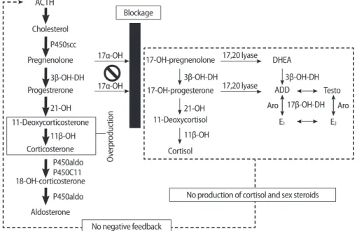
17α-hydroxylase and 17,20-lyase (P450c17) are enzymes that con- vert pregnenolone and progesterone to 17α-hydroxypregnenolone (17-OHPreg) and 17α-hydroxyprogesterone (17-OHP), which are pre- cursors of sex steroids and cortisol. In cases of P450c17 deficiency, androgens, estrogens, and cortisol cannot be produced. A compen- satory increase in adrenocorticotrophic hormone (ACTH), due to the failure of cortisol production, stimulates the production of 11-deoxy- corticosterone and corticosterone, which have powerful mineralo- corticoid activity. In turn, the excessive levels of these mineralocorti- coids lead to volume expansion, hypertension, and electrolyte imbal- ances such as hypokalemia (Figure 1).
Deficiency in sex steroids and excessive mineralocorticoid produc- tion cause characteristic symptoms and signs such as delayed puber-
ty with primary amenorrhea and hypertension. Congenital adrenal hyperplasia (CAH) is an autosomal recessive disorder due to a defect in any of several enzymes involved in steroidogenesis; different gen- otypes induce different phenotypes. 17α-hydroxylase/17,20-lyase deficiency (17OHD) is a rare type of CAH that causes cortisol and sex hormone deficiency and aldosterone excess [1]. Here, we report the case of a 21-year-old woman who had been inadequately treated for 17α-hydroxylase/17,20-lyase deficiency for a prolonged period of time.
Case report
An 18-year-old female was transferred for evaluation of low serum potassium levels (2.6 mmol/L) found incidentally during a preopera- tive workup for an appendectomy at a local medical center. The pa- tient had no past medical or family history associated with electro- lyte imbalance. The hypokalemia appeared to be corrected after po- tassium supplementation. Then, the patient was referred to the car- diology department because she had constant high blood pressure, with a maximal reading of 180/110 mm Hg. The patient was treated with an angiotension II receptor antagonist (telmisartan) and a calci- um channel blocker (amlodipine). However, her blood pressure was
Received: Apr 13, 2015 ∙ Revised: Jun 8, 2015 ∙ Accepted: Jun 10, 2015 Corresponding author: Jeong Ho Rhee
Department of Obstetrics and Gynecology, Keimyung University Dongsan Medical Center, 56 Dalsung-ro, Daegu 700-712, Korea
Tel: +82-53-250-7871, Fax:+82-53-250-7599, E-mail: [email protected] This is an Open Access article distributed under the terms of the Creative Commons Attribution Non-Commercial License (http://creativecommons.org/licenses/by-nc/3.0/) which permits unrestricted non-commercial use, distribution, and reproduction in any medium, provided the original work is properly cited.
still not well controlled and she was subsequently diagnosed with stage 1 hypertensive retinopathy.
Upon further investigation, it became apparent that the patient had had hypertension since the age of 15-year-old, when the patient visited her gynecologist. At that time, the patient was found to have primary amenorrhea and features of sexual infantilism. On physical examination, the patient was quite tall and slender, with a height of 166.2 cm and a body weight of 44 kg (body mass index, 15.9 kg/m
2), and was phenotypically female. The patient’s breast development
had only progressed to Tanner stages 1–2 and pubic hair was under- developed. Ultrasonography of the abdomen and pelvis demon- strated a hypoplastic uterine corpus, bilateral small multicystic ova- ries, and a very thin endometrium. Considering the patient’s history and clinical signs, hormone profiles and karyotype were checked.
The patient’s karyotype was 46,XX and hormone studies showed a normal to hypergonadotropic hypogonadal state with low testoster- one and dehydroepiandrosterone sulfate (DHEA-S) levels (Table 1).
Because there was a high suspicion of 17OHD, further evaluation was
Table 1. Serum hormone levels before and after medication
First visit (August 2008) Second visit (February 2013) After medication (October 2014) Reference range
FSH (mIU/mL) 8.29 12.58 - Female, 1.8–9.0
LH (mIU/mL) 14.58 11.52 - Female, 0.5–10.4
Estradiol (pg/mL) 14.97 31.05 - Female, 50–482
Testosterone (ng/mL) 0.01 0.01 - Female, 0.1–1.0
DHEA-S (μg/dL) 3.84 2.29 - Female, 35–430
ACTH (μg/dL) - 123.39 13.42 Morning, 10–60
Cortisol (μg/dL) - 2.44 - Morning, 9.41–26.06
Renin (ng/mL) - 0.01 2.70 Upright, 0.5–1.9
Aldosterone (pg/mL) - 114.66 18.37 Upright, 34–273
Progesterone (ng/mL) - 8.21 0.92 Female, 0.06–25
Sodium (mmol/L) - 144 142 132–146
Potassium (mmol/L) - 2.9 5.1 3.5–5.5
Chloride (mmol/L) - 105 103 95–109
FSH, follicle stimulating hormone; LH, luteinizing hormone; DHEA-S, dehydroepiandrosterone sulfate; ACTH, adrenocorticotrophic hormone.
Figure 1. Pathogenesis in 17 alpha-hydroxylase deficiency (17OHD). The blockage at the 17α-OH impairs cortisol and sex steroid synthesis. The low cortisol increases ACTH production. Accumulated deoxycorticosterone and corticosterone cause plasma volume expansion and hyperten- sion, and hypokalemia, which suppress renin and aldosterone. ACTH, adrenocorticotrophic hormone; 17α-OH, 17α-hydroxylase; 3β-OH-DH, 3β- hydroxysteroid dehydrogenase; 21-OH, 21-hydroxylase; 11β-OH, 11β- hydroxylase; 17β-OH-DH, 17β-hydroxysteroid dehydrogenase; ADD, androstenedione; Testo, testosterone; Aro, aromatase; E
1, estrone; E
2, estradiol.
ACTH
Pregnenolone
11-Deoxycorticosterone
Corticosterone Progestrerone
18-OH-corticosterone
Aldosterone Cholesterol P450scc
3β-OH-DH
11β-OH 21-OH
P450C11 P450aldo
P450aldo
Overproduction
No negative feedback
No production of cortisol and sex steroids Blockage
17α-OH
17α-OH
17,20 lyase
17,20 lyase 17-OH-pregnenolone
17-OH-progesterone
11-Deoxycortisol
Cortisol
3β-OH-DH 3β-OH-DH
17β-OH-DH
11β-OH 21-OH
DHEA
ADD
Aro Aro
E1 E2
Testo
recommended; however, the patient refused further investigation at this time. After a period of 4.5 years, the patient revisited the gyne- cology department with a complaint of persistent amenorrhea.
Upon physical examination, there was no interval change in the pa- tient’s sexual development. A repeat hormone profile showed ele- vated levels of follicle-stimulating hormone (FSH) and luteinizing hormone (LH) with low levels of estradiol, indicating hypergonado- tropic hypogonadism. Studies also demonstrated very low cortisol, high ACTH, and low levels of both testosterone and DHEA-S (Table 1).
With a clinical suspicion of an enzyme defect in steroidogenesis, par- ticularly 17OHD, an ACTH stimulation test and a CYP17A1 gene anal- ysis were performed. The ACTH stimulation test showed no increase in 17-OHP after intravenous administration of synthetic ACTH (Syn- acthen 0.25 mg, Dalim Biotech, Jecheon, Korea) when compared with the basal level (Table 2), suggesting an impairment in the path- way catalyzed by 17α-hydroxylase. Genetic analysis showed a com- pound heterozygous mutation in exon 6 of the CYP17A1 gene (c.985–987TAC>AA and c.1118A>T). After being diagnosed with 17α-hydroxylase deficiency, the patient stopped taking antihyper- tensive medication and started oral hydrocortisone (Rapison 30 mg daily, Kolon Pharmaceuticals, Daejeon, Korea), which is a cornerstone for the treatment for CAH. In addition, the patient commenced oral estradiol and cyclic medroxyprogesterone (Progynova 2 mg daily, Bayer, Lys-Lez-Lannoy, France and Provera 10 mg daily for 13 days each month, Pfizer, Tronto, Italy) in order to induce secondary sexual development and maintain cyclic uterine bleeding. After appropriate therapy, the patient’s blood pressure normalized without antihyper- tensive therapy, and five months later, follow up laboratory investi- gations revealed normal levels of ACTH (11.04 μg/dL), progesterone (0.04 ng/mL) and serum electrolytes. Physical examination showed stage 2 Tanner breast development with scanty pubic and axillary
hair production, and the uterus was normal in size and shape upon ultrasonographic examination.
Discussion
17α-hydroxylase deficiency is a rare form of CAH, with an estimated incidence of 1 in 50,000 to 100,000, and accounts for about 1% of all CAH cases [2,3]. The most common enzyme defect causing CAH is 21-hydroxylase deficiency (21-OHD), followed by 11β-hydroxylase deficiency (11β-OHD). Clinically, these enzyme deficiencies are char- acterized by hyperandrogenism or heterosexual precocious puberty in females; however, clinical features of 17α-hydroxylase deficiency differ from those of 21-OHD or 11β-OHD in that there is no hyperan- drogenism, but rather, sexual infantilism.
The enzyme P450c17 has both 17α-hydroxylase and 17,20-lyase activity. The gene encoding the enzyme P450c17 is CYP17A1, which is located at chromosome 10q24.3. Human 17α-hydroxylase defi- ciency involving a loss of only 17α-hydroxylase or 17,20-lyase has been observed [4,5], but in most affected patients, both enzymes are deficient [6]. Blockage of the pathway catalyzed by 17α-hydroxylase enhances the mineralocorticoid pathway, resulting in overproduc- tion of 11-DOC, corticosterone and aldosterone. This also causes a decline in 17-OHPreg, 17-OHP, 11-deoxycortisol, cortisol, DHEA, an- drostenedione, and testosterone [1]. High levels of 11-DOC induce sodium and fluid retention and loss of potassium and hydrogen, and consequently hypertension, because of its potent mineralocorticoid effect [7]. Affected males have ambiguous or female external genita- lia due to a deficiency in androgen synthesis. Female patients have normal genitalia but show immature sexual development and pri- Table 2. The results of ACTH stimulation test
Basal One hour after
ACTH stimulation Reference range
Pregnenolone (ng/mL) 3.9 5.9 0.1–1.0
Progesterone (ng/mL) 8.21 15.62 Female, 0.06–25 11-Deoxycorticosterone
(ng/mL)
0.39 3.88 Female, 0.03–0.33
Aldosterone (pg/mL) 42.14 91.47 Upright, 34–273 17-Hydroxypregnenolone
(ng/mL)
0.9 1.54 0.1–4.0
17-Hydroxyprogesterone (ng/mL)
1.32 1.25 0.18–4.69
Cortisol (μg/dL) 1.71 3.18 Morning, 9.41–26.06
DHEA (ng/mL) 0.11 0.15 Female, 0.12–19.0
Androstenedione (ng/mL) 0.2 <0.1 Female, 0.3–3.9 ACTH, adrenocorticotrophic hormone; DHEA, dehydroepiandrosterone.
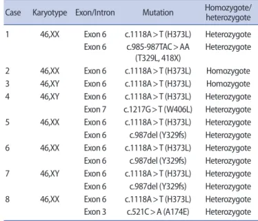
Table 3. CYP17A1 gene mutations reported among Koreans
Case Karyotype Exon/Intron Mutation Homozygote/heterozygote 1 46,XX Exon 6 c.1118A>T (H373L) Heterozygote
Exon 6 c.985-987TAC>AA (T329L, 418X)
Heterozygote
2 46,XX Exon 6 c.1118A>T (H373L) Homozygote 3 46,XY Exon 6 c.1118A>T (H373L) Homozygote 4 46,XY Exon 6 c.1118A>T (H373L) Heterozygote Exon 7 c.1217G>T (W406L) Heterozygote 5 46,XX Exon 6 c.1118A>T (H373L) Heterozygote Exon 6 c.987del (Y329fs) Heterozygote 6 46,XX Exon 6 c.1118A>T (H373L) Heterozygote Exon 6 c.987del (Y329fs) Heterozygote 7 46,XY Exon 6 c.1118A>T (H373L) Heterozygote Exon 6 c.987del (Y329fs) Heterozygote 8 46,XX Exon 6 c.1118A>T (H373L) Heterozygote Exon 3 c.521C>A (A174E) Heterozygote