ORIGINAL ARTICLE
J Cardiovasc Ultrasound 2016;24(2):144-152• *Dae-Hee Shin currently works in the Incheon St. Mary’s Hospital, College of Medicine, The Catholic University of Korea, Incheon, Korea.
• Received: March 31, 2016 • Revised: May 10, 2016 • Accepted: May 10, 2016
• Address for Correspondence: Seung Woo Park, Division of Cardiology, Department of Medicine, Heart Vascular Stroke Center, Samsung Medical Center, Sungkyunkwan University School of Medicine, 81 Irwon-ro, Gangnam-gu, Seoul 06351, Korea
Tel: +82-2-3410-3419, Fax: +82-2-3410-3849, E-mail: [email protected]
• This is an Open Access article distributed under the terms of the Creative Commons Attribution Non-Commercial License (http://creativecommons.org/licenses/by-nc/3.0) which permits unrestricted non-commercial use, distribution, and reproduction in any medium, provided the original work is properly cited.
Normal Echocardiographic Measurements in a Korean Population Study: Part II.
Doppler and Tissue Doppler Imaging
Jin-Oh Choi, MD
1, Mi-Seung Shin, MD
2, Mi-Jeong Kim, MD
3, Hae Ok Jung, MD
4,
Jeong Rang Park, MD
5, Il Suk Sohn, MD
6, Hyungseop Kim, MD
7, Seong-Mi Park, MD
8, Nam Jin Yoo, MD
9, Jung Hyun Choi, MD
10, Hyung-Kwan Kim, MD
11, Goo-Yeong Cho, MD
12, Mi-Rae Lee, MD
13, Jin-Sun Park, MD
14, Chi Young Shim, MD
15, Dae-Hee Kim, MD
16,
Dae-Hee Shin, MD
17*, Gil Ja Shin, MD
18, Sung Hee Shin, MD
19, Kye Hun Kim, MD
20, Jae-Hyeong Park, MD
21, Sang Yeub Lee, MD
22, Woo-Shik Kim, MD
23,
and Seung Woo Park, MD
11Division of Cardiology, Department of Medicine, Heart Vascular Stroke Center, Samsung Medical Center, Sungkyunkwan University School of Medicine, Seoul, Korea
2Division of Cardiology, Department of Internal Medicine, Gil Hospital, Gachon University of Medicine and Science, Incheon, Korea
3Division of Cardiology, Department of Internal Medicine, Incheon St. Mary’s Hospital, College of Medicine, The Catholic University of Korea, Incheon, Korea
4Department of Internal Medicine, Seoul St. Mary’s Hospital, College of Medicine, The Catholic University of Korea, Seoul, Korea
5Division of Cardiology, Department of Internal Medicine, Gyeongsang National University Hospital, Gyeongsang National University School of Medicine, Jinju, Korea
6Department of Cardiology, Kyung Hee University School of Medicine, Kyung Hee University Hospital at Gangdong, Seoul, Korea
7Division of Cardiology, Keimyung University Dongsan Medical Center, Daegu, Korea
8Division of Cardiology, Department of Internal Medicine, Korea University College of Medicine, Seoul, Korea
9Department of Internal Medicine, Wonkwang University Hospital, Institute of Wonkwang Medical Science, Iksan, Korea
10Division of Cardiology, Department of Internal Medicine, Pusan National University School of Medicine, Busan, Korea
11Division of Cardiology, Department of Internal Medicine, Cardiovascular Center, Seoul National University College of Medicine, Seoul, Korea
12Division of Cardiology, Department of Internal Medicine, Seoul National University and Cardiovascular Center, Seoul National University Bundang Hospital, Seongnam, Korea
13Division of Cardiology, Department of Medicine, Samsung Changwon Hospital, Sungkyunkwan University School of Medicine, Changwon, Korea
14Department of Cardiology, Ajou University School of Medicine, Suwon, Korea
15Division of Cardiology, Severance Cardiovascular Hospital, Yonsei University College of Medicine, Seoul, Korea
16Department of Cardiology, Asan Medical Center, University of Ulsan College of Medicine, Seoul, Korea
17Division of Cardiology, Gangneung Asan Hospital, University of Ulsan College of Medicine, Gangneung, Korea
18Division of Cardiology, Department of Internal Medicine, Ewha Womans University School of Medicine, Seoul, Korea
19Division of Cardiology, Department of Internal Medicine, Inha University College of Medicine, Incheon, Korea
20Department of Cardiology, Chonnam National University Hospital, Gwangju, Korea
21Division of Cardiology, Department of Internal Medicine, Chungnam National University Hospital, Chungnam National University School of Medicine, Daejeon, Korea
22Division of Cardiology, Department of Internal Medicine, Chungbuk National University School of Medicine, Cheongju, Korea
23Department of Internal Medicine, Cardiovascular Center, Kyung Hee University Medical Center, Seoul, Korea
Introduction
As Doppler and tissue Doppler images were able to provide important hemodynamic information in various cardiovascu- lar disorders noninvasively, echocardiography has been widely adopted as a noninvasive tool of choice for clinical and hemo- dynamic evaluation of patients with heart failure.
1)2)However, there have been only few studies which evaluated normal echo- cardiographic reference values of Doppler and tissue Doppler imaging (TDI) variables according to age and sex in a large number of normal subjects and no such data are available from Korean population yet.
3-6)Previous study about normal echocardiographic reference val- ues did not include variables from TDI and reference values spe- cific to sex were not provided.
7)In this regard, we sought to pro- vide normal reference values for variables from Doppler and TDI according to sex and age groups using the Normal echO- caRdiographic Measurements in a KoreAn popuLation (NOR- MAL) study.
8)Methods
Study populations
Inclusion and exclusion criteria of the NORMAL study were presented in the part I of the current study.
8)Briefly, this was a prospective nationwide multicenter (23 centers) study evaluat- ing normal Korean normal adult subjects (age; 20–79 years- old) who had no significant cardiac disorders or clinical illnesses that might affect cardiac structure and function, such as hyper- tension and diabetes. We also excluded subjects if a structural or functional abnormality on the cardiac valve or cardiac cham-
ber was evident during echocardiographic examination. All study patients agreed to provide their information for purposes of the research and the study protocol was approved by the In- stitutional Review Board of each institute. Written informed consent was waived.
Echocardiography
Echocardiographic images were acquired and measured at each institute. Like previous report, images were stored in digi- tal image communication in medicine format and transferred to the Echocardiographic Core Laboratory (ECL) in Samsung Medical Center.
8)Final measurements and analysis were per- formed in ECL with a dedicated software package (EchoPAC, GE Medical Systems, Horten, Norway). All echocardiographic measurements were performed according to the American So- ciety of Echocardiography guidelines.
1)Briefly, on the apical 4-chamber view, mitral inflow velocities were obtained using pulsed-wave (PW) Doppler imaging with a 1–3 mm sample volume placed between the mitral leaflet tips during diastole.
Early diastolic (E) velocity, late diastolic (A) velocity, E to A ra- tio (E/A), and mitral E wave deceleration time (DT). Continu- ous-wave Doppler imaging for the measurement of isovolumic relaxation time (IVRT) were performed by placing the sample volume in the left ventricular outflow tract (LVOT) to simulta- neously display the end of aortic ejection and the onset of mi- tral inflow. Tricuspid inflow velocity was obtained using PW Doppler with sample volume placed between the tips of tri- cuspid valve leaflet on apical 4-chamber view. Mitral annular velocities were obtained at the lateral and septal mitral annulus from the apical 4-chamber view using TDI. Tricuspid lateral an-
Background:Hemodynamic and functional evaluation with Doppler and tissue Doppler study as a part of comprehensive echocardiography is essential but normal reference values have never been reported from Korean normal population especially ac- cording to age and sex.
Methods:
Using Normal echOcaRdiographic Measurements in a KoreAn popuLation study subjects, we obtained normal ref- erence values for Doppler and tissue Doppler echocardiography including tricuspid annular velocities according to current guide- lines and compared values according to gender and age groups.
Results:
Mitral early diastolic (E) and late diastolic (A) velocity as well as E/A ratio were significantly higher in women com- pared to those in men. Conversely, mitral peak systolic and late diastolic annular velocity in both septal and lateral mitral annulus were significantly lower in women compared to those in men. However, there were no significant differences in both septal and lateral mitral early diastolic annular (e’) velocity between men and women. In both men and women, mitral E velocity and its de- celeration time as well as both E/A and E/e’ ratio considerably increased with age. There were no significant differences in tricus- pid inflow velocities and tricuspid lateral annular velocities between men and women except e’ velocity, which was significantly higher in women compared to that in men. However, changes in both tricuspid inflow and lateral annular velocities according to age were similar to those in mitral velocities.
Conclusion:
Since there were significant differences in Doppler and tissue Doppler echocardiographic variables between men and women and changes according to age were even more considerable in both gender groups, normal Doppler echocardiographic values should be differentially applied based on age and sex.
KEY WORDS:
Transthoracic echocardiography · Doppler · Tissue Doppler · Normal population · Reference value.
nular velocities were also obtained at lateral annulus of tricuspid valve from the apical 4-chamber view. Peak systolic (s’), early diastolic annular (e’) velocity and late diastolic annular (a’) ve- locity of each annulus were measured.
LVOT flow velocity was measured at apical long-axis view or anteriorly angulated 4-chamber view using PW Doppler with sample volume positioned on the left ventricular side of the aortic valve just proximal to the region of flow acceleration.
Right ventricular outflow tract (RVOT) flow velocity was also measured on parasternal short-axis view at aortic valve level plac- ing sample volume just below pulmonary valve. Peak velocity and velocity-time integral (VTI) of both ventricular outflow ve- locities were obtained.
Spectral Doppler signal of pulmonary venous flow was ob- tained in the apical 4-chamber view placing a 2–3 mm sample volume placed 0.5 cm into the pulmonary vein for optimal re- cording of the spectral waveforms. Pulmonary vein systolic (PVS) and diastolic (PVD) velocities as well as pulmonary vein rever- sal flow velocity during atrial contraction (PVAr) were measured.
Every conventional and tissue Doppler parameter was mea- sured in 3 consecutive beats, and averaged.
Statistical analysis
Mean ± SD and 95% confidence intervals (CIs) for continu- ous variables are presented. Independent t-test was used for the comparison of mean values between men and women and a one-way analysis of variance test was performed to evaluate whether mean values differed according to age groups. To eval- uate the intra- and interobserver variability, we randomly se-
lected 50 cases and calculated intraclass correlation coefficients (ICC). To determine intraobserver measurement variability, one researcher repeated measurements at least 2 weeks after the first measurements, and another researcher who did not have information about the measurement value repeated mea- surements to evaluate interobserver variability. We considered p values < 0.05 as statistically significant. All statistical analy- ses were performed using SPSS statistics version 21 (SPSS Inc., Chicago, IL, USA).
Results
Variables from mitral inflow and annular velocities
A total of 1003 normal subjects from 23 centers were evalu- ated in the current study. Demographic and clinical data are provided in previous report.
8)M-mode variables according to gender groups and according to age and gender groups are presented in Table 1 and Supplementary Table 1, respectively.
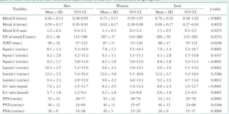
Mitral E and A velocity were significantly higher in women compared to men. Mitral E/A ratio was also greater in women compared to men. DT of mitral E velocity was longer in men compared to women. Mitral A velocity and DT of E velocity in- creased and mitral E velocity and E/A ratio decreased with age in both men and women. IVRT was slightly longer in men com- pared to women and increased with age. There were no signifi- cant differences in mitral septal and lateral e’ velocities between men and women. However, both septal and lateral s’ and a’
velocities were significantly higher in men compared to wom-
Table 1. Mitral inflow and annular velocities and pulmonary venous flow velocities according to gender
Variables Men Women Total
p value
Mean ± SD 95% CI Mean ± SD 95% CI Mean ± SD 95% CI
Mitral E (m/sec) 0.66 ± 0.14 0.38–0.95 0.73 ± 0.17 0.39–1.07 0.70 ± 0.16 0.38–1.02 < 0.0001 Mitral A (m/sec) 0.59 ± 0.17 0.26–0.91 0.62 ± 0.17 0.28–0.96 0.60 ± 0.17 0.27–0.94 0.0029
Mitral E/A ratio 01.2 ± 0.4 00.4–2.1 01.3 ± 0.5 00.2–2.4 01.3 ± 0.5 00.3–2.2 0.0255
DT of mitral E (msec) .212 ± 40 .133–290 .207 ± 37 .134–280 .209 ± 39 .133–285 0.0354
IVRT (msec) 0.90 ± 16 0.57–122 0.87 ± 17 0.53–120 0.88 ± 17 0.55–121 0.0108
Septal s’ (cm/sec) 08.1 ± 1.4 05.3–10.9 07.8 ± 1.3 05.1–10.4 07.9 ± 1.4 05.2–10.7 0.0001
Septal e’ (cm/sec) 09.2 ± 2.6 04.2–14.2 09.3 ± 3.1 03.3–15.3 09.3 ± 2.8 03.7–14.8 0.4347
Septal a’ (cm/sec) 09.2 ± 1.7 05.8–12.6 08.5 ± 1.8 05.0–12.0 08.8 ± 1.8 05.3–12.4 < 0.0001 Lateral s’ (cm/sec) 10.2 ± 2.5 05.3–15.0 09.6 ± 2.3 05.0–14.1 09.9 ± 2.4 05.1–14.6 0.0001 Lateral e’ (cm/sec) 12.3 ± 3.5 05.4–19.2 12.6 ± 3.8 05.1–20.0 12.4 ± 3.7 05.2–19.6 0.2386 Lateral a’ (cm/sec) 09.4 ± 2.3 04.9–13.9 09.0 ± 2.2 04.6–13.3 09.2 ± 2.3 04.7–13.6 0.0032 E/e’ ratio (septal) 07.6 ± 2.1 03.5–11.7 08.4 ± 2.5 03.4–13.4 08.0 ± 2.4 03.4–12.7 < 0.0001
E/e’ ratio (lateral) 05.7 ± 1.8 02.2–9.3 06.2 ± 1.8 02.6–9.8 06.0 ± 1.8 02.4–9.6 0.0001
PVS (cm/sec) 0.52 ± 12 0.28–77 0.55 ± 12 0.30–79 0.54 ± 12 0.29–78 0.0083
PVD (cm/sec) 0.46 ± 12 0.23–69 0.46 ± 11 0.25–67 0.46 ± 11 0.24–68 0.6368
PVAr (cm/sec) 0.26 ± 6 0.14–38 0.26 ± 5 0.15–36 0.26 ± 6 0.15–37 0.4066
E: early diastolic inflow velocity, A: late diastolic inflow velocity, DT: deceleration time, IVRT: isovolumic relaxation time, s’: systolic annular velocity, e’: early diastolic annular velocity, a’: late diastolic annular velocity, PVS: pulmonary venous systolic velocity, PVD: pulmonary venous diastolic velocity, PVAr: pulmo- nary venous reversal flow velocity during atrial contraction, CI: confidence interval
en. The s’ and e’ velocities decreased and a’ velocity increased with age in both sex groups and the changes according to ages were greater in women compared to men. E/e’ ratio calculated from the e’ values of both septal and lateral annulus were sig- nificantly higher in women compared to men. E/e’ ratio in- creased with age in both men and women.
Variables from pulmonary vein flow velocities
Measurement values of pulmonary vein flow Doppler vari- ables according to gender groups and according to age and gender groups are also presented in Table 1 and Supplementa- ry Table 1, respectively. PVS was significantly higher in wom- en compared to men. However, there were no significant dif- ferences in PVD and PVAr between men and women. PVS and PVAr increased with age in both gender groups. However, PVD decreased according to age in both men and women.
Variables from tricuspid inflow and annular velocities
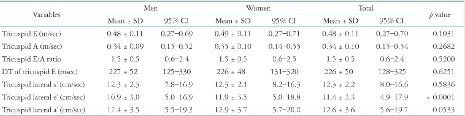
Measurement values of the tricuspid inflow and annular ve- locity according to gender groups and according to age and gen- der groups are presented in Table 2 and Supplementary Table 2, respectively. There were no significant differences in tricus- pid E and A velocities, E/A ratio and DT of tricuspid E veloci- ty between men and women. Like mitral inflow velocities, tri- cuspid E velocity decreased and A velocity increased with age in both men and women. Thus tricuspid E/A ratio decreased according to age. DT of tricuspid E velocity also increased ac- cording to age in both sex. Tricuspid e’ velocity was greater in
women compared to men. However, there were no significant difference in tricuspid lateral s’ and a’ velocity between men and women. However, tricuspid lateral s’ and e’ velocities de- creased and a’ velocity increased with age in both sexes.
Variables from left and right ventricular outflow flow velocities
Measurement values of LVOT and RVOT flow velocities ac- cording to gender groups and according to age and gender groups are presented in Table 3 and Supplementary Table 3, respectively. LVOT peak systolic flow velocity and LVOT VTI value were higher in women compared with men. However, there were no significant differences in RVOT peak flow ve- locity between men and women and RVOT VTI was slightly greater in women compared to men. LVOT peak flow veloci- ties and LVOT VTI increased with age in both men and wom- en. However, RVOT peak flow velocity showed decreasing trends according to age in both men and women. And RVOT VTI showed decreasing trends only in men and there were no significant changes according to age in women.
Intra- and interobserver variability
ICCs for both intra- and interobserver variability testing are presented in Supplementary Table 4. For both intraobserver variability, ICCs for echo variables were above 0.9, except that of the DT of mitral E velocity (ICC = 0.896, 95% CI = 0.812–
0.944), IVRT (ICC = 0.855, 95% CI = 0.684–0.929), and DT of tricuspid E velocity (ICC = 0.879, 95% CI = 0.779–0.936).
For interobserver variability, ICCs of echocardiographic vari-
Table 2. Measurement values of the tricuspid inflow and annular velocity according to gender
Variables Men Women Total
p value
Mean ± SD 95% CI Mean ± SD 95% CI Mean ± SD 95% CI
Tricuspid E (m/sec) 0.48 ± 0.11 0.27–0.69 0.49 ± 0.11 0.27–0.71 0.48 ± 0.11 0.27–0.70 0.1031 Tricuspid A (m/sec) 0.34 ± 0.09 0.15–0.52 0.35 ± 0.10 0.14–0.55 0.34 ± 0.10 0.15–0.54 0.2682
Tricuspid E/A ratio 01.5 ± 0.5 00.6–2.4 01.5 ± 0.5 00.6–2.5 01.5 ± 0.5 00.6–2.4 0.5200
DT of tricuspid E (msec) .227 ± 52 .125–330 .226 ± 48 .131–320 .226 ± 50 .128–325 0.6251
Tricuspid lateral s’ (cm/sec) 12.3 ± 2.3 07.8–16.9 12.3 ± 2.1 08.2–16.3 12.3 ± 2.2 08.0–16.6 0.5836 Tricuspid lateral e’ (cm/sec) 10.9 ± 3.0 05.0–16.9 11.9 ± 3.5 05.0–18.8 11.4 ± 3.3 04.9–17.9 < 0.0001 Tricuspid lateral a’ (cm/sec) 12.4 ± 3.5 05.5–19.3 12.9 ± 3.7 05.7–20.0 12.6 ± 3.6 05.6–19.7 0.0533 E: early diastolic inflow velocity, A: late diastolic inflow velocity, DT: deceleration time, s’: systolic annular velocity, e’: early diastolic annular velocity, a’: late diastolic annular velocity, CI: confidence interval
Table 3. Measurement values of LVOT and RVOT flow velocity according to gender
Variables Men Women Total
p value
Mean ± SD 95% CI Mean ± SD 95% CI Mean ± SD 95% CI
LVOT peak velocity (cm/sec) 0.96 ± 15 0.66–126 0.99 ± 16 0.67–131 0.97 ± 16 0.66–129 0.0119
LVOT VTI (cm) 20.0 ± 3.3 13.5–26.6 21.5 ± 3.7 14.2–28.8 20.8 ± 3.6 13.7–27.9 < 0.0001
RVOT peak velocity (cm/sec) 0.76 ± 15 0.47–104 0.76 ± 13 0.51–101 0.76 ± 14 0.49–102 0.5891
RVOT VTI (cm) 17.2 ± 3.1 11.2–23.2 17.7 ± 3.0 11.8–23.6 17.4 ± 3.0 11.5–23.4 0.0070
LVOT: left ventricular outflow tract, VTI: velocity-time integral, RVOT: right ventricular outflow tract, CI: confidence interval
ables were above 0.8 except for IVRT (ICC = 0.898, 95% CI = 0.815–0.944), and DT of tricuspid E velocity (ICC = 0.832, 95% CI = 0.698–0.910).
Discussion
This study provided normal reference measurement values of Doppler and TDI variables for comprehensive echocardio- graphic evaluation according to age and gender using data from the NORMAL study. Briefly, there were statistically signifi- cant differences between men and women in most of the Dop- pler and TDI variables. But the differences according to gender did not seem to be clinically important. Interestingly, changes of the variables according to the age were more considerable compared with those according to gender.
As the prevalence and incidence of heart failure with pre- served ejection fraction increased with age, evaluation of dia- stolic function and filling pressure in the elderly subject is more important.
9)Although current echocardiographic guideline suggested normal value of Doppler and TDI variables accord- ing to the age, diagnostic algorithm for the grading of diastolic dysfunction did not consider normal reference values of these variables according to age for routine echocardiographic evalu- ation.
10)Actually if we apply the diagnostic scheme to elderly women of current study subjects more than half of them would be classified as having diastolic dysfunction grade I or II. There- fore, consideration of the age factor, when evaluating diastolic function especially in elderly female subject is needed to avoid unnecessary sophisticated cardiac evaluation for asymptomatic normal subjects.
In this regard, mitral inflow and annular velocities, which are representative variables for diastolic functions and filling pres- sure, changed according to ages suggesting trends of more di- astolic dysfunction and increased filling pressure in the elderly subjects. That is to say, A velocity and E/e’ ratio increased whereas E velocity, E/A ratio, and e’ velocity decreased with ages in both men and women. These results were well consis- tent with previous studies from European and Japanese popu- lation and confirms that age reference values should be taken into account when evaluating diastolic function or filling pres- sures according to those variables.
3)4)Interestingly, those chang- es seemed to be more significant in women compared to men and these trends were very similar to the results from the stud- ies from Japan, which evaluated normal echocardiographic values for Japanese subjects.
4)As suggested in the previous stud- ies, these findings might partially explain why elderly women have relatively higher incidence of heart failure with preserved ejection fraction and higher filling pressure and why there were higher cardiovascular mortality in elderly female. However, in other studies evaluating European populations which evaluat- ed the reference values according to age groups of 20–40, 40–
60, and more than 60 years old, those trends of more signifi- cant changes according to age especially in the elderly female was not noted.
5)11)Although mean values of both septal and lateral e’ velocities were not significantly different between men and women, val- ues according to the age groups were significantly different be- tween the gender groups. And there were considerable differ- ences in septal and lateral mitral annular velocities and every TDI variables measured from mitral and tricuspid lateral an- nulus were greater compared with the values from septal an- nulus, which was consistent with previous reports.
12)The abso- lute value as well as relative value of each annular velocity might be useful for differentiating normal subjects from those with constrictive pericarditis or restrictive cardiomyopathy.
13)Lower velocities of s’ and e’ and higher value of a’ were observed espe- cially female older subjects. This again might explain why women are susceptible for heart failure with preserved ejection fraction.
Although clinical implication of pulmonary venous and tri- cuspid inflow velocities are often regarded as less important compared with those of mitral valves, variables from those flow velocities might be useful for differential diagnosis of con- strictive pericarditis from restrictive cardiomyopathy and esti- mating filling pressures of both ventricles.
14)For pulmonary venous flow variables, PVS and PVAr increased and PVD de- creased according to age in both men and women, which find- ings were consistent with previous reports in that healthy old- er subjects had higher PVS, PVAr, and lower PVD compared with younger subjects.
15)And these results were also consistent with the prior study from healthy Korean population.
7)How- ever, higher PVS value in men compared to women in our study was not consistently observed in the previous studies.
4)11)Likewise, no significant differences were found between men and women for the tricuspid inflow velocities and its DT. In- terestingly, the tricuspid inflow velocities and its DT showed significant differences according to age group, which were sim- ilar to those of mitral inflows. And the mean values of the vari- ables were very similar to the previous study for Korean popu- lation.
7)There are several limitations to be acknowledged for this study. First, we included only normal Korean subjects in the NORMAL study and there might be considerable differences in clinical and demographic characteristics as we discussed in the prior reports.
8)Thus our data might not be applicable to other populations. Second, we did not evaluate myocardial ve- locity using TDI or speckle tracking techniques. Lastly, as ac- knowledged in the previous report, patients with significant hypertension and diabetes were excluded based on past medi- cal histories obtained from the study subjects, and results of blood sampling and/or other clinical tests were not obtained.
8)Therefore, patients with subclinical hypertension or coronary artery disease might be included in the current study. However, their effects on the variables of the current study are unlikely to be significant.
In conclusion, we provided normal values for Doppler and
TDI variables for comprehensive echocardiography including
right-sided heart from the NORMAL study. As there were sig- nificant changes among different age groups, normal reference values according to age should be used for Doppler and TDI variables.
• Acknowledgements
NORMAL study was supported by a research fund from Korean Society of Echocardiography.
References