Research Articles Open Access
8
Isolation and Identification of Antagonistic Bacteria for Biological Control of Large Patch Disease of Zoysiagrass Caused by
Rhizoctonia solani AG2-2 (IV)
Chi Hyun Song1, Md Rezuanul Islam1, Taehyun Chang2, and Yong Se Lee3*
1Department of Biotechnology, Daegu University, Gyeongsan 712-714, Korea
2Plant Resources and Environment Major, Kyungpook National University, Sangju 742-711, Korea
3Division of Life and Environmental Science, Daegu University, Gyeongsan 712-714, Korea
ABSTRACT. The objective of this study was to identify bacterial antagonists of R. solani AG2-2 (IV) on zoysiagrass and to evaluate their antifungal activity in vitro and in vivo to select an antagonistic isolate. Antagonistic isolates that inhibit large patch disease caused by R. solani AG2-2 (IV) in zoysiagrass were selected from several soils, and their antagonistic activities were investigated in vitro and in vivo. Of 216 bacterial isolates, 67 inhibited several plant pathogenic fungi. The isolates that inhibited stem-segment colonization by R. solani AG2-2 (IV) in zoysiagrass were tested in a growth chamber. Eleven isolates were active as plant growth promoting isolates. Among them, five plant growth promoting isolates and their concentration dependent efficiency on zoysiagrass following inoculation with R.
solani AG2-2 (IV) was evaluated. Isolate H33 was one of the potential antagonistic isolates, and it was further tested against various plant pathogens. H33 not only suppressed the disease caused by R. solani AG2-2 (IV) on zoysiagrass but also promoted leaf weight and leaf height of zoysiagrass under growth chamber and greenhouse conditions. The H33 isolate, which belongs to Streptomyces arenae, was identified through physiological, biochemical, and 16S rDNA studies. Further studies will investigate the cultural characterization of S. arenae H33 and isolation and identification of antifungal substance produced by S. arenae H33.
Key words: Biological control, Large patch, Zoysiagrass, Rhizoctonia solani, Streptomyces arenae
Introduction
Soil-borne pathogenic microorganisms affecting plant health are the main and constant threat to turfgrass management on golf courses worldwide. Of several turfgrass diseases, large patch disease caused by Rhizoctonia solani Kühn is the most destructive disease of zoysiagrass. Over the past decade, many golf course green keepers have relied on chemicals as a relatively dependable method of protecting turfgrass against soil-borne pathogens (Compant et al., 2005; Chang and Lee, 2010). However, increasing the use of chemicals causes several adverse affects on the environment and on human health. Consequently, there has been an increased restriction on a variety of chemicals (Gerhardson, 2002). Therefore, efforts have been directed towards developing new alternatives to chemical disease control.
Many studies have reported on the natural activity of some fungi and bacteria against fungal pathogens, and this is
considered as a very appealing alternative to the use of chemical fungicides (Gerhardson, 2002; Welbaum et al., 2004).
Despite the long list of currently available antibiotics on the market, antifungal antibiotics are a very small but significant group of chemicals that plays an important role in the control of fungal diseases. The development of new anti- fungal agents, preferably naturally occurring with novel mechanisms of action, is an urgent need.
Disease-suppressing soils have been described for several soil-borne plant pathogens, including Gaeumannomyces graminis var. tritici (Cook and Rovira, 1976), Fusarium oxysporum (Alabouvette, 1986), Plasmodiophora brassicae (Worku and Gerhardson, 1996), and Rhizoctonia solani (Garbeva et al., 2004). Although soil abiotic factors are believed to play a role, there is also evidence that microbial activity contributes to disease suppression. Thus, soils are regarded as sources of natural, effective, and valuable antagonists for biological control. Isolates with antagonistic activity towards plant pathogens have been studied among the most abundant soil and plant-associated bacteria in a study involving agricultural sites in Germany (Berg et al., 2002, 2006). Different mechanisms contribute to suppressing soil-borne plant pathogens, including parasitism, production
*Corresponding author; Tel: +82-53-850-6763 E-mail : [email protected]
Received : March 14, 2012, Revised: March 29, 2012, Accepted:
April 05, 2012.
of antifungal compounds, nutrients and colonization sites, and inducing systemic resistance against pathogens in plants (Fravel, 1988; Han et al., 2000; Lugtenberg et al., 2001;
Berg et al., 2005; Dahiya, 2005).
Bacterial biocontrol agents have several advantages over fungal agents, including ease of propagation, immediate fungicide compatibility, and compatibility with current liquid delivery systems. Actinomycetes are sources of several secondary metabolites, antibiotics, and lytic enzymes that affect fungal growth (Okami and Hotta, 1988; Nolan and Cross, 1998,). The species belonging to the genus Strepto- myces constitute 50% of the total population of soil actino- mycetes and 75-80% of the commercially and medicinally useful antibiotics that have been derived from this genus (Mellouli et al., 2003).
Therefore, the objective of this study was to identify bacterial antagonists of R. solani AG2-2 (IV) on zoysiagrass and to evaluate their antifungal activity in vitro and in vivo to select an antagonistic isolate.
Materials and Methods
Selection of antagonistic isolates
Soil samples (80-100 g) were collected from several agriculture crop fields (peppers, vineyards, peach orchards) in Gyeongsan of Gyeongbuk and stored at 4oC in sterile plastic bags until use. Samples (5 g) were diluted with 45 mL of sterile and distilled water and thoroughly dispersed by shaking at 150 rpm for 30 min at 27oC and then diluted 103- 105 folds. A diluted aliquot (100µl) of each soil sample was spread on a triptic soy agar (TSA) culture medium plate and incubated at 27oC overnight. The single colony was subcul- tured on TSA and stored at 4oC until dual culture screening of the isolates.
Screening of antagonistic isolates
The isolates were tested for an in vitro mycelial inhibition effect against R. solani on TSA medium using the dual plate culture technique (Anith et al., 2003). Antagonistic isolates were examined for a mycelial inhibition effect against two strains of R. solani which are isolate collected from large patch diseased plants in Daegu country club and R. solani AG2-2 (IV) (KACC No. 40152) isolate obtained from the Korean Agricultural Culture Collection (KACC). The extent of antagonistic activity against 19 selected isolates was measured on TSA medium (Table 1). A minimum amount of sample from the single colony was streaked 4 cm wide on a TSA plate and incubated for 48 h at 27oC. A mycelial disk (5 mm i.d.) from the margin region of R. solani was placed on the opposite middle of the plate and incubated for 48 h at 27oC. The distance of the fungal colony towards and away from the bacterial colony was measured, and the inhibition
rate (%) was calculated. Isolates that achieved more than 50% inhibition against the tested pathogen were stored in tryptic soy broth (TSB) suspended with 20% glycerol at −70oC for the in vitro and in vivo experiments.
Growth chamber evaluation
Seed germination efficiencies of 19 isolates were as follows: Zoysiagrass seeds (5 g) were submerged with 100 mL of sterile water (check), 100 mL mycelial suspension (106 cfu/mL) of R. solani AG2-2 (IV) (control) in a 250 mL Erlenmeyer flask. One mL of 19 isolates cell suspension (108 cfu/mL), that were cultured in TSB for 48 hours at 27oC, added in control flask. All samples were incubated at 27oC for 1 h on an oscillatory shaker (100 rpm), and 100 seeds from each treatment were sown in plastic pots filled with soil and sand (1 : 1, by volume) medium.
Pots were incubated at 27±2oC, 85% relative humidity, Table 1. Antagonistic effect of various microorganisms on mycelial growth of Rhizoctonia solani AG2-2 (IV).
Isolate
Inhibition rate (%) of mycelial growth R. solani AG2-2 (IV)
(KACC 40152)
R. solani (Locally isolated)
A10 69.8gh 67.8bca
A17 68.8h 65.3de
B4 76.3e 60.5gh
C9 83.3bc 67.3bcd
C10 64.7j 54.8ij
C47 71.0fg 68.5b
E2 59.3l 55.3ij
E7 62.9k 62.9f
E13 82.1c 55.6i
E19 79.8d 62.6fg
E26 66.2i 53.1j
F8 72.4f 59.4h
F17 75.5e 59.7h
F30 71.0fg 61.4fgh
F32 76.7e 60.5gh
F33 78.7d 56.4i
H6 88.4a 79.1a
H33 83.9b 63.4ef
H81 75.3e 65.8cd
Inhibition rate (IR) was determined as follows; IR (%) = 100− (a/b×
100) where, a = mycelial growth (mm) toward the antagonistic isolate and b = mycelial growth (mm) opposite direction to the antagonistic isolate.
aThe same letters in a column denote no significant difference at P ≤ 0.05 by Duncan’s multiple range test.
and maintained on a 18 h photoperiod until sampling.
The seedling viable capacity of the 19 isolates was deter- mined as follows surface sterilized zoysiagrass seeds (100 seeds/pot) were sown in plastic pots, which were filled with soil and sand mixed media, incubated at 27±2oC, 85%
relative humidity, and maintained on a 18 h photoperiod until germination. After germination, the pots were divided into three groups. Group 1 treatment (check) was supplied with tap water, Group 2 treatment (control) was inoculated with a mycelial suspension of R. solani AG2-2 (IV) (106 mfu/mL), and the Group 3 treatment (treated) was inoculated with R. solani AG2-2 (IV) and drenched with a 30 mL cell suspension (108 cfu/mL) of antagonistic microorganisms.
After a week incubation, the seedling viable efficiencies of the antagonistic microorganisms were evaluated as the per- centage of seedling viability per pot compared to the control.
The efficiency of the antagonistic microorganisms on stem-segment colonization was determined by the following method. Zoysiagrass seedlings (45 days after sowing) were inoculated with a mycelial suspension (106 mfu/mL) of R.
solani AG2-2 (IV) and a 24 h old culture (108 cfu/mL) of antagonistic microorganisms (30 mL/pot). The seedlings were kept in a growth chamber under conditions of 27±2oC, 85% relative humidity, and a 18 h photoperiod. After a 3- week incubation, stem-segment colonization was measured using the following formula, and disease reduction was calculated in comparison to the control.
Stem-segment colonization (%) = (0×A + 2×B + 3×C + 5×D + 8×E + 10×F)/(total number of stems × highest score (10))× 100.
Using the stem-segment colonizing scale, where A (0):
healthy stem; B (2): normal with little blight; C (3): blight
< 10%; D (5): blight = 11-50%; E (8): blight = 51-90%; F (10): blight = 91-100%.
Greenhouse evaluation
Zoysiagrass seeds (5 g) were submerged in 100 mL of sterile water for germination, and incubated at 27oC for 1 h.
Approximately 100 seeds were sown in plastic pots (Ø 11.5× 10.5 cm), which were filled with soil and sand (1:1, by volume) mixed media, incubated at 27±2oC, 85% relative humidity, and maintained under a 18 h photoperiod until germination. The plants were grown in a greenhouse at 18- 28oC, respectively, and mowed with scissors to 2.5-3.0 cm until they were 80 days old and adult plants. Water soluble fertilizer (10-5-6: J Agro Co., Daegu, South Korea) was applied weekly at 2 g/L after the beginning of mowing at 21 days. Cell suspensions of 11 bacterial isolates were applied to the grass under greenhouse conditions. The plants were inoculated with R. solani AG2-2 (IV) after 7 days of bacterial isolate application. The control was used the same amount of TSB media. Three weeks after the inoculation,
the efficiency of the isolates was measured based on leaf weight and leaf height.
Antagonistic effect of isolate H33
The extent of the antagonistic activity against KACC - plant pathogens of isolate H33 was examined on TSA medium. Isolates grown in TSB for 132 h at 27oC were streaked in 4 cm long inoculates on one side of a plate. A mycelial disk (5 mm in diameter) of a tested pathogen was placed 3.5 cm from the streaks and incubated at 27oC for 3-5 days. The distance (mm) of the fungal colony towards (a) and away (b) from the bacterial colony was measured. The percentage growth inhibition rate (IR %) was calculated as follows:
IR (%) = 100− (a/b×100).
Identification of isolate H33
The Gram reaction was performed for the antagonistic microorganisms according to standard methods. Biochemical identification was assessed in accordance with the proced- ures described in Bergey’s Manual of General Bacteriology and the Analytical Profile Index System (API 50 CHB, BioMerieux, France). Various compositions of ISP (Inter- national Streptomyces Project) media were prepared to grow the antagonistic actinomycetes isolate, including TSA, LBA, and PDA. After streaking plates with the isolate, plates were incubated for 14 days at 27oC. The growth conditions, material color released, and mycelial color were evaluated by comparison with the ISCC-NBS color centric system.
The relationship between the colors released and melanin production of the isolate was evaluated. The morphological appearances of the antagonistic microorganisms were ob- served using binocular microscopy (ZEISS, Axiolab-990308) and scanning electron microscopy (SEM; Hitachi, S-4300) at 7 kV. Cells were prefixed with glutaraldehyde (2.5%
solution in, 0.1 M phosphate butter, pH 7.0) and then immersed into 2.0% OsO4 at 4oC for 16 h for SEM. The fixed cells were dehydrated using an ethanol series and dried in a freeze dryer. DNA was extracted from selected isolates and Sequencing of the 16S rRNA using universal primers (forward-27F, reverse-1492R) and a BLAST search of the National Center for Biotechnology Information (NCBI) database (http:www.ncbi.nlm.) were used for identification of the isolate. A phylogenetic tree was constructed with the neighbor-joining method described by Tamura et al. (2007).
Results and Discussion
Screening of antagonistic isolates
The effects of antagonistic activity against R. solani AG2-2 (IV) was tested (Fig. 1). A total of 216 isolates were screened
from agricultural soil through the dual culture method.
Among them, 19 isolates inhibited mycelial growth of R.
solani AG2-2 (IV) and R. solani to varying degrees (Table 1). Significant differences (P < 0.05) were observed for inhibition of mycelial growth among the isolates. Fifteen effectively inhibited > 70% of R. solani AG2-2 (IV) mycelial growth.
Identification of isolate H33
The isolate H33 grows on a wide range of agar media and shows the typical tracts of Streptomyces (Locci, 1989). The colonies were slow growing and contained aerial and substrate mycelia of different colors (Table 2). Additionally, all colonies possessed an earthy odor. When acid-fastness and Gram-stain methods were performed, the H33 isolate was aerobic and Gram positive (Fig. 2B), and the plate
culture showed that the isolate had aerial mycelia with a sporangium (Fig. 2A). It was also mesophilic and catalase positive. Moreover, the spores appeared round under light and SEM (Locci, 1989). Based on microscopic, macroscopic and biochemical analyses, the isolate was likely similar to Streptomyces spp. (Fig. 2C, D).
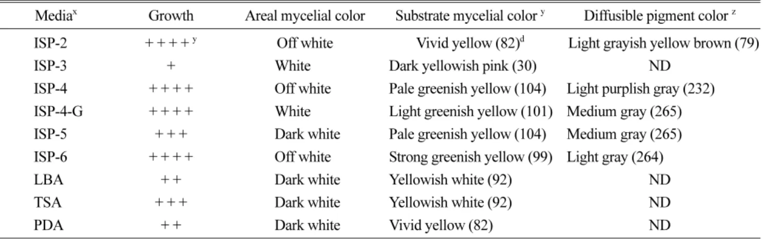
Generally, ISP (International Streptomyces Project) media is a suitable growth media for Streptomyces sp. In this study, several media were applied to observe the growing status and ISP2, ISP4, ISP4 + glucose, and ISP6 were the most effective for growing isolate H33 (data not shown). Pinkish red pigment diffused into the surrounding ISP medium (Table 2). The H33 isolate produced a maximum number of spores in the above mentioned media. The H33 isolate did
Table 2. Growth and pigment production of isolate H33 on various media.
Mediax Growth Areal mycelial color Substrate mycelial color y Diffusible pigment color z ISP-2 + + + + y Off white Vivid yellow (82)d Light grayish yellow brown (79)
ISP-3 + White Dark yellowish pink (30) ND
ISP-4 + + + + Off white Pale greenish yellow (104) Light purplish gray (232) ISP-4-G + + + + White Light greenish yellow (101) Medium gray (265) ISP-5 + + + Dark white Pale greenish yellow (104) Medium gray (265) ISP-6 + + + + Off white Strong greenish yellow (99) Light gray (264)
LBA + + Dark white Yellowish white (92) ND
TSA + + + Dark white Yellowish white (92) ND
PDA + + Dark white Vivid yellow (82) ND
aIsolate H33 was incubated on various media at 27oC for 5 days.
b+, poor; ++, moderate; +++, good; ++++, very good mycelial growth on various solid media
cISCC-NBS color centroid system was used to identify the released mycelial color
dISCC-NBC color code
ND: did not release any pigment on growing media.
Fig. 1. Efficacy of antagonistic isolates on mycelial growth of Rhizoctonia solani AG2-2 (IV) in the dual culture on TSA. Each plate number represents an antagonistic isolate, respectively.
Fig. 2. Morphological and microscopic appearance of isolate H33. A: ISP 4 culture plate, B: Gram reaction (magnification,
×400), C: Mycelial spore chains of isolate H33 through scanning electron microscopy (SEM), D: Spores of isolate H33 under SEM.
not produce any spores on ISP3 or PDA media during a 14- day incubation period. Based on the growing conditions and pigment production on different ISP media, the isolate H33 seemed to be a Streptomyces sp. (Loci, 1989). Similar to many fungi, which also have an immobile life style, Streptomyces spp. produce spores to aid their dispersal.
Spore chains are produced through fragmentation of aerial hyphae that form on the substrate mycelium. The shape of the spore chains, which may be straight, branched, spiral, or wavy, is an important taxonomic character (Goodfellow, 1988; Loci, 1989). Streptomyces species have DNA with a high proportion of guanine and cytosine, and at least some species possess a single linear chromosome (Goodfellow, 1988).
16S rDNA gene sequence analyses were performed for further confirmatory identification. Amplification and se- quencing reactions were conducted as described previously (Heyrman and Swings, 2001). BLAST matching was per-
formed to screen for similar sequences as isolate H33, which had a 99% homology with the reference strain Streptomyces arenae.
Growth chamber evaluation
The 19 isolates selected from the dual culture method were tested for effect on Zoysiagrass seed germination, seedling viability, and suppression of large patch disease under growth chamber conditions (Table 3). Highly significant differences (P < 0.05) were observed. Eleven isolates sup- pressed (> 40%) the disease incidence of stem-segment colonization after inoculation with R. solani AG2-2 (IV) in zoysiagrass. Five isolates promoted > 70% seed emergence and increased seedling viability (> 40%) of five isolates when compared to the controls at 11.86% and 12.33%.
Greenhouse evaluation
Based on the growth chamber evaluation, 11 isolates were
Table 3. Efficacy of the isolates for seed germination, seedling viability, and large patch disease suppression on zoysiagrass (Zoysia japonica ‘Zenith’) under growth chamber conditions.
Isolate Seed germination (%) Seedling viability (%) Disease suppression
Stem-segment colonization (%)x Disease Control value (%)
A10 37.1±3.0yef 40.2±2.2b 35.4±1.0hi 50.0±1.5
A17 41.3±4.7d 35.6±0.9cd 45.5±2.8cd 35.7±4.0
B4 36.2±1.3f 41.3±4.2b 42.7±3.1def 39.8±4.4
C9 54.6±4.6a 49.1±3.1a 23.8±0.7k 66.4±1.0
C10 39.3±3.6de 28.5±2.0ef 56.6±6.1b 20.1±8.6
C47 40.2±1.4d 48.9±3.7a 41.9±1.2def 40.8±1.7
E2 15.5±3.4j 22.2±1.9hi 37.5±0.9gh 47.0±1.3
E7 26.1±2.8h 26.1±3.3fg 32.7±3.5ij 53.8±4.9
E13 35.4±5.1f 23.4±2.8gh 40.0±3.6efg 43.6±5.1
E19 39.3±1.5de 34.4±4.5d 38.9±0.5fgh 45.1±0.8
E26 30.6±7.1g 25.7±7.8fgh 43.2±6.6de 38.9±9.4
F8 22.8±6.8i 19.3±4.3i 56.7±4.2b 19.9±5.9
F17 30.4±5.1g 22.5±3.1ghi 44.7±2.9cd 36.9±4.1
F30 27.4±4.2h 26.2±2.4fg 47.0±2.1c 33.7±3.0
F32 30.0±4.8g 26.1±3.5fg 37.5±2.6gh 47.0±3.7
F33 26.3±0.1h 23.0±3.4gh 43.2±2.6de 39.0±3.7
H6 54.7±5.5a 52.4±4.0a 30.8±3.9j 56.5±5.4
H33 48.3±8.4b 32.2±2.8de 33.5±2.0ij 52.7±2.8
H81 45.0±6.5c 39.2±2.4bc 32.7±2.0ij 53.8±2.8
Control 11.9±2.1k 12.3±1.1j 70.8±1.0a -
Stem segment colonizing scale, A(0), healthy stem; B(2), growth is normal but little blight; C(3), blight < 10%; D(5), blight = 11-50%; E(8), blight = 51-90%; F(10), blight = 91-100%.
x Stem-segment colonization (%) = [{(0×A + 2×B + 3×C + 5×D + 8×E + 10×F)/Total number of stems × highest score (10)} × 100]. Disease reduction (%) = {(Control− Treated / Control) × 100}
y Figures are averages and standard deviations of five replications. The same letters in a column denote no significant difference at P ≤ 0.05 by Duncan’s multiple range test.
tested for their ability to suppress large patch disease incidence and promote zoysiagrass growth under greenhouse conditions (Table 4). Disease incidence was reduced in all isolates (Table 4, Fig. 3). Tested 5 isolates were significantly promoted leaf weight and leaf height of zoysiagrass (Table 5).
Nutritional and environmental conditions could affect the antagonistic effects of the selected isolate under greenhouse.
Therefore, it is desirable that in vitro tests to select pro- spective isolates for biological control should be conducted with several target pathogen isolates prior to field tests. It is
probable that the antagonists could be used to control plant diseases caused by fungal pathogens. However, several researchers have reported no correlation between in vitro inhibition tests and field performance of biocontrol agents (Fravel, 2005). Thus, further field trials using the prospective antagonists are needed.
Antagonistic effect of isolate H33
The dual culture antagonistic assays revealed that isolate H33 showed varying degrees of mycelial inhibition on six turfgrass pathogens and 11 plant pathogenic fungi (Table 5).
Isolate H33 showed antagonistic activity with a broad anti- fungal spectrum in vitro.
We confirmed the antifungal effects of EtOAc extract of isolate H33 through light and SEM observations. Mycelial deformation, abnormal hyphal swelling, and cell lysis of R.
solani AG2-2 (IV) and R. solani AG4 were observed after treatment with EtOAc extract, and decolorization of Fusarium oxysporum was observed (data not shown). My- celial lysis and abnormal hyphae were found in the presence of the antifungal substance.
The SEM observations confirmed that the H33 isolate produced lytic enzymes, which could degrade the cell walls of the fungal pathogen (data not shown), R. solani AG2-2 (IV). A significant clear zone level (35 mm i.d.) was observed with strong chitinolytic activity when the cell-free culture supernatant of isolate H33 was placed near a paper disc.
Thus, the biocontrol mechanism of an EtOAc extract of isolate H33 was associated with the activity of the antibiotic Table 4. Efficacy of isolates for growth and disease severity on zoysiagrass in a greenhouse.
Isolate Grass leaf weight (gm/pot) Grass leaf height (cm/pot) Disease severity (0-10 scale)x
A10 2.1±0.38f y 16.3±3.23d 06.3±0.87c
B4 2.8±0.81b 17.8±2.94abc 07.2±0.35bc
C9 2.7±0.38c 18.1±2.90a 07.1±0.39bc
C47 2.7±0.59c 17.3±2.77abcd 07.2±0.50bc
E7 2.4±0.48e 16.9±3.48bcd 06.4±0.85c
E13 2.7±0.70c 18.1±3.26ab 07.3±0.45bc
E19 2.4±0.62e 17.5±3.87abc 06.9±0.46c
F32 2.4±0.51e 17.6±2.96abc 06.7±0.43c
H6 2.7±0.74c 18.3±3.71a 08.2±0.20b
H33 2.9±0.67a 17.9±2.40ab 07.4±0.83bc
H81 2.5±0.79d 16.6±3.52cd 07.3±0.71bc
Control 0.7±0.24g 09.3±2.16e 12.6±0.55a
x Disease severity was measured according to 0 to 10 scaling, 0% = no symptom of damage followed by 10 (1 scaling), 20, 30, 40, 50, 60, 70, 80, 90 and 100%.
It was inoculated with R. solani AG2-2 (IV) 7 days after isolate application.
Disease severity rate was applied 30 days after the isolates were applied.
yThe same letters in a column denote no significant difference at P ≤ 0.05 by Duncan,s multiple range test.
Fig. 3. Efficiency of isolate H33 for inhibiting stem-segment colonization by Rhizoctonia solani AG2-2 (IV) on zoysiagrass (Zoysia japonica ‘Zenith’) in a greenhouse. Control: Not inoculated, RS+H33: R. solani AG2-2 (IV) and isolate H33 were inoculated, RS: Inoculated with R. solani AG2-2 (IV).
Inoculation with 5 mycelial disc (i.d. 5 mm) of R. solani AG2-2 (IV), and the cell suspension of isolate H33 was drenched on the treated grass pot at the same time.
by producing a high level of chitinase. Elad et al. (1982) and Singh et al. (1999) reported that efficient microbial biocontrol agents such as Streptomyces spp. excrete extracellular lytic enzymes that are responsible for their antagonistic ability.
Chitinases play a role in hyphal swelling and lysis of fungal pathogen cell walls, resulting in inhibited fungal hyphae growth (Jung et al., 2003).
Isolate H33 seemed to have a comparatively broad antagonistic spectrum and is a promising biocontrol agent for soil borne diseases of plants. If several studies including safety, stability, identification of the active compound, and fermentation method to enhance antagonistic activity are completed successfully in the future, this antagonistic acti- nomycetes strain will be useful for developing an agent to control several fungal diseases, particularly stem-segment colonization of zoysiagrass caused by R. solani AG2-2 (IV).
Acknowledgment
This research was supported by Daegu University research fund in 2010.
References
Alabouvette, C. 1986. Fusarium wilt-suppressive soils from the Chateaurenard region: review of a 10-year study. Agronomie 6:273-284.
Anith, K.N., N.V. Radhakrishnam, and T.P. Manomohandas. 2003.
Table 5. Efficacy of antagonistic isolates on leaf weight and leaf height of zoysiagrass (Zoysia japonica ‘Zenith’).
Isolate 103 cfu/mL 105 cfu/mL 107 cfu/mL
Az B A B A B
C9 1.87±0.68 3.15±0.61 2.54±0.38 3.20±0.21 2.79±0.31 3.69±0.35
E19 1.95±0.44 2.73±0.68 2.49±0.38 3.18±0.85 2.63±0.69 3.72±0.89
Leaf H6 2.28±0.38 2.75±0.14 2.49±0.49 3.48±0.54 2.76±0.60 3.86±0.45
weight H33 2.30±0.58 2.94±0.52 2.16±0.69 4.05±1.04 2.74±0.49 4.38±1.18
H81 1.80±0.25 3.95±0.35 2.35±0.58 3.74±0.58 1.97±0.38 4.28±0.61
C. 0.68±0.25 1.72±0.26
N. 1.10±0.43 1.69±0.14
C9 15.0 ± 2.0 18.0 ± 1.6 17.5±2.3 22.1±1.7 19.3±1.9 24.8±1.2
E19 14.4 ± 1.8 17.6 ± 2.0 16.8±2.5 22.2±0.5 19.2±3.2 22.8±1.0
Leaf H6 15.4 ± 3.6 17.3 ± 3.6 17.0±2.9 19.3±3.5 18.9±2.7 23.5±1.9
height H33 15.3 ± 2.7 18.0 ± 0.5 16.6±2.4 20.2±0.2 18.0±1.8 23.3±1.0
H81 13.9 ± 3.5 18.8 ± 1.2 14.9±2.9 22.6±0.7 16.3±3.3 17.5±5.5
C. - - 7.8±1.7 8.9±1.8 - -
N. - - 9.2±3.3 10.1±1.4 - -
zA: Inoculation of Rhizoctonia solani AG2-2 (IV) 7 days before isolate application; B: Isolate was inoculated with R. solani G2-2 (IV) 7 days after isolate application. Control used the same amount of TSB media without microbes accordingly.
Table 6. Efficacy of isolate H33 on mycelial growth of plant pathogenic fungi on TSA media.
Plant pathogenic fungi Inhibition rate (%) Rhizoctonia solani AG2-2 a 76.9±8.34bcde R. solani AG2-2(IV) KACC 40132 73.1±2.1cdef R. solani AG2-2(IV) KACC 40152 82.7±2.0ab R. solani AG2-2(IIIB) KACC 40151 69.2±2.9fg R. solani AG4-4 KACC 40141 62.4±3.9h
R. cerealis KACC 40154 86.9±7.1a
Botrytis cinerea KACC 40573 88.9±3.6a Chatomium globosum KACC 40308 68.3±3.6fgh Collectotrichum circinans KACC 40641 79.3±3.3bc C. gloeosporioides KACC 40690 72.3±4.5def Corynespora cassiicola KACC 40964 79.3±3.5bc Fusarium oxysporum KACC 40052 68.3±4.5fgh Glomerella cingulata KACC 40299 65.2±3.6gh Penicillium ailli KACC 41337 89.3±4.6a Phytophthora cactorum KACC 40166 78.3±5.3bcd Pythium graminicola KACC 40155 52.6±3.5i Trichoderma harzianum KACC 40791 71.3±2.9efg The H33 isolate was streaked (35 mm) on one side of a test plate and then incubated for 48 h at 27oC. A mycelial disk (5 mm in diameter) of tested fungi was placed 35 mm from the antagonistic isolate and incu- bated at 27oC.
a isolate from Technogreen Co. Ltd., Korea.
Screening of antagonistic bacteria for biological control of nursery wilt of black pepper (Piper nigrum). Microbiological Research 15:91-97.
Berg, G., N. Roskot, A. Steidle, L. Eber, A. Zock, and K. Smalla.
2002. Plant-dependent genotypic and phenotypic diversity of antagonistic rhizobacteria isolated from different Verticillium host plants. Applied and Environmental Microbiology 68:3328- 3338.
Berg, G., A. Krechel, M. Ditz, R. Sikora, A. Ulrich, and J. Hall- mann. 2005. Endophytic and ectophytic potato-associated bac- terial communities differ in structure and antagonistic function against plant pathogenic fungi. FEMS Microbiol. Eco. 51:215- 229.
Berg, G., K. Opelt, C. Zachow, J. Lottmann, M. Gotz, R. Costa, and K. Smalla. 2006. The rhizosphere effect on bacteria antagonistic towards the pathogenic fungus Verticillium differs depending on plant species and site. FEMS Microbiol. Eco. 56:250-261.
Chang, T.H. and Y.S. Lee. 2010. Evaluation of occurrence of yel- low patch caused by Rhizoctonia cerealis of cool season turf- grass cultivars and species. Kor. Turfgrass Sci. 24(1):24-30. (in Korean)
Compant, S., B. Duffy, J. Nowak, C. Clement, and E. Barka. 2005.
Use of plant growth-promoting bacteria for biocontrol of plant diseases: principles, mechanisms of action and future pros- pects. App. Environ. Microbiol. 71: 4951-4959.
Cook, R.J. and A.D. Rovira. 1976. The role of bacteria in the bio- logical control of Gaeumannomyces graminis by suppressive soils. Soil Biology & Biochemistry 8:269-274.
Dahiya, N. 2005. Production of an antifungal chitinase from Enter- obacter sp. NRG4 and its application in protoplast production.
World J. Microbiol. Biotechnol. 21:8-9.
Elad, Y., I. Chet, and Y. Henis, 1982. Degradation of plant patho- genic fungi by Trichoderma harzianum. Canadian journal of Microbiology 28:719-725.
Fravel, D.R. 1988. Role of antibiosis in the biocontrol of plant dis- eases. Annual Review of Phytopathology 26:75-91.
Fravel, D.R. 2005. Commercialization and implementation of bio- control, Annual Review of Phytopathology 43:337-359.
Garbeva, P., J.A. Veen, and J.D. Elsas. 2004. Assessment of the diversity, and antagonism towards Rhizoctonia solani AG3, of Pseudomonas species in soil from different agricultural regimes.
FEMS Microbiol Eco. 47:51-64.
Gerhardson, B. 2002. Biological substitutes for pesticides. Trends in Biotechnol. 20:338-343.
Goodfellow, M., S.T. Williams, and M. Mordarski. 1988. Actino-
mycetes in Biotechnology. pp. 130-175, Academic Press, Lon- don.
Han, D.Y., D.L. Coplin, W.D. Bauer, and H.A. Hoitink. 2000. A rapid bioassay for screening rhizosphere microorganisms for their ability to induce systemic resistance, Phytopathology 90:
327-332.
Heyrman, J. and J. Swings. 2001. 16S rDNA sequence analysis of bacterial isolates from biodeteriorated mural paintings in the servilia tomb (Necropolis of Carmona, Seville, Spain) system.
App. Microbiology 24:417-422.
Jung, W.Y., K.N. An, Y.L. Jin, R.D. Park, K.T. Lim, and K.Y. Kim.
2003. Bioloical control of damping-off caused by Rhizoctonia solani using chitinase-producing Paenibacillus illinoiseniss KJ-424, Soil biology Biotechemistry 35:1261-1264.
Lugtenberg, B.J.J., L. Dekkers, and G.V. Bloemberg. 2001. Molec- ular determinants of rhizosphere colonization by Pseudomo- nas. Annual Review of Phytopatholgy 39:461-490.
Locci, R. 1989. Streptomyces and related genera, In: Bergey’'s manual of systematic bacteriology, pp. 2451-2508, fourth. ed.
Williams & Wilkins Company, Baltimore.
Mellouli, L., R.B. Mehdi, S. Sioud, M. Salem, and S. Bejar. 2003.
Isolation, purification and partial characterization of antibacte- rial activities produced by a newly isolated Streptomyces sp.
US24 strain. Res. Microbiol. 154:345-352.
Nolan, R. and T. Cross. 1998. Isolation and screening of actino- mycetes, in: Goodfellow, M., Williams, S. T., Mordarski, M.
(Eds). Actinomycetes in biotechnology. Academic Press, Lon- don, pp. 33-67.
Okami, Y. and T. Hott. 1988. Search and discovery of new antibi- otics. in: Goodfellow, M., Williams, S. T., Mordarski, M. (Eds), Actinomycetes in biotechnology. Academic Press, Inc., New York, pp. 33-67.
Singh, P., Y.C. Shin, C.S. Park, and Y.R. Chung. 1999. Biological control of Fusarium wilt of cucumber by chitinolytic bacteria.
Phytopathology 89: 92-99.
Tamura, M., Tsushida, T., Shinohara, K., 2007. Isolation of an isoflavone-metabolizing, Clostridium-like bacterium, strain TM-40, from human faces. Physiology microbial chemistry, Anaerobe 13: 32-35.
Welbaum, G., A.V. Sturz, Z. Dong, and J. Nowak. 2004. Fertilizing soil microorganisms to improve productivity of agroecosys- tems, Critical Reviews in Plant Sciences 23:175-193.
Worku, Y. and B. Gerhardson, 1996. Suppressiveness to clubroot, pea root and Fusarium wilt in Swedish soils. Journal of Phyto- pathology 144:143-146.
들잔디 갈색퍼짐병의 생물학적 방제를 위한 길항 세균의 분리와 동정
송치헌1·Md Rezuanul Islam1·장태현2·이용세3*
대구대학교 생물공학과1, 경북대학교 식물자원환경전공2, 대구대학교 생명환경학부3
요 약: 한국 들잔디에 발생하는 Rhizoctonia solani에 의한 라지패치를 생물학적으로 방제하기위해 일반토양에서
길항미생물을 분리하여 in vitro와 in vivo에서 길항효과 및 병 발생억제효과를 검정하였다. 토양에서 분리한 216 개 균주 중 15개 균주가 R. solani AG2-2 (IV)의 균사생장을 70%이상 억제하였으며, 온실실험 결과 11개 균주는 잔디의 생장을 촉진시켰으며, 병 발생 억제효과가 있었다. 분리한 길항미생물 중 H33 균주는 in vitro 및 in vivo 에서 R. solani AG2-2 (IV)에 대한 길항효과가 다른 균주에 비해 높았으며, 공시한 17개 식물병원성 진균에 대해 길항효과가 높아 생물방제균으로 선발하였다. H33 균주를 ISP 배지에 배양한 후 배양적 특성 및 형태를 관찰한 결과 Streptomyces sp.로 동정되었으며, 16S rDNA를 분석한 결과 Streptomyces arenae와 99% 상동성을 보였다.
주요어: 생물학적방제, 갈색퍼짐병, 들잔디, Rhizoctonia solani, Streptomyces arenae