Intrinsic and Extrinsic Regulation of Innate Immune Receptors
Eunshil Jeong and Joo Young Lee
School of Life Science, Gwangju Institute of Science and Technology, Gwangju, Korea.
Received: March 4, 2011
Corresponding author: Dr. Joo Young Lee, School of Life Science, Gwangju Institute of Science and Technology, Oryong-dong 1, Buk-gu, Gwangju 500-712, Korea.
Tel: 82-62-715-2505, Fax: 82-62-715-2484 E-mail: [email protected]
∙ The authors have no financial conflicts of interest.
© Copyright:
Yonsei University College of Medicine 2011 This is an Open Access article distributed under the terms of the Creative Commons Attribution Non- Commercial License (http://creativecommons.org/
licenses/by-nc/3.0) which permits unrestricted non- commercial use, distribution, and reproduction in any medium, provided the original work is properly cited.
Pattern recognition receptors (PRRs) in innate immune cells play a pivotal role in the first line of host defense system. PRRs recognize pathogen-associated molecu- lar patterns (PAMPs) or danger-associated molecular patterns (DAMPs) to initiate and regulate innate and adaptive immune responses. PRRs include Toll-like recep- tors (TLRs), RIG-I-like receptors (RLRs) and NOD-like receptors (NLRs), which have their own features in ligand recognition and cellular location. Activated PRRs deliver signals to adaptor molecules (MyD88, TRIF, MAL/TIRAP, TRAM, IPS-1) which act as important messengers to activate downstream kinases (IKK complex, MAPKs, TBK1, RIP-1) and transcription factors (NF-κB, AP-1, IRF3), which produce effecter molecules including cytokines, chemokines, inflammatory en- zymes, and type I interferones. Since excessive PRR activation is closely linked to the development of chronic inflammatory diseases, the role of intrinsic and extrin- sic regulators in the prevention of over- or unnecessary activation of PRRs has been widely studied. Intracellular regulators include MyD88s, SOCS1, TOLLIP, A20, and CYLD. Extrinsic regulators have also been identified with their molecu- lar targets in PRR signaling pathways. TLR dimerization has been suggested as an inhibitory target for small molecules such as curcumin, cinnamaldehyde, and sul- foraphane. TBK1 kinase can be a target for certain flavonoids such as EGCG, luteolin, quercetin, chrysin, and eriodictyol to regulate TRIF-dependent TLR path- ways. This review focuses on the features of PRR signaling pathways and the ther- apeutic targets of intrinsic and extrinsic regulators in order to provide beneficial strategies for controlling the activity of PRRs and the related inflammatory diseas- es and immune disorders.
Key Words: Pattern recognition receptor, toll-like receptor, dimerization, TBK1, therapeutic target
INTRODUCTION
Innate and adaptive immunity is required to eliminate pathogens as host defense system. Pattern recognition receptors (PRRs), which are germ-line encoded recep- tors, play a critical role in initiating and regulating innate and adaptive immune re- sponses by recognizing pathogen-associated molecular patterns (PAMPs) or danger- associated molecular patterns (DAMPs).1-3 PRRs are quite ubiquitously expressed in a variety of cells including monocytes, dendritic cells, neutrophils, and epithelial
and B cells; however, they are also present in non-immune cells, such as epithelial cells, endothelial cells, and fibro- blasts. TLR1, TLR2, TLR4, TLR5, TLR6, and TLR11 are expressed on the cell surface; TLR3, TLR7, TLR8, and TLR9 are expressed in intracellular vesicles such as endo- somes, lysosomes, and the endoplasmic reticulum.15 Epi- thelial TLR4 is expressed in phagosomes with a unique cel- lular expression profile.
Of the thirteen TLRs, TLR4 was characterized first.13 TLR4 recognizes lipopolysaccharide (LPS) in the outer membrane of Gram-negative bacteria, with the assistance of co-receptors such as CD14 and MD2.16,17 LPS binds first to LPS binding protein (LBP) and membrane-bound GPI (glycosylphosphatidylinositol)-anchored CD14, and is then transferred to the TLR4 and MD2 (myeloid differentiation proten-2) complexes.18,19 In the MD2 complex, LPS binds to a large hydrophobic pocket, through non-covalent interac- tions such as hydrogen bonding and hydrophobic and hydro- philic interactions, which results in the dimerization of the two TLR4/MD2 complexes. In addition, TLR4 recognizes mannan from Cadida albicans, glycoinositolphospholipids from Trypanosoma,20 and the envelope proteins from mouse mammary tumor virus (MMTV) and respiratory syncytial virus (RSV).21,22 TLR4 is also activated by endogenous mol- ecules, including heat-shock proteins (HSP60, HSP70, and HSP gp96),23-25 fibrinogen,26 oligosaccharides of hyaluronic acid,27 extracellular domain A of fibronectin,28 heparan sul- fate,29 myeloid-related proteins (Mrp8 and Mrp14),30 oxi- dized LDL,31 saturated fatty acid32 and amyloid-β.33 Further- more, human TLR4 senses chemical elements such as nickel (Ni2+), conferring immunostimulatory activity to Ni2+.34 Non- conserved histidine residues in human TLR4 provide binding pockets for nickel and trigger an immune response and con- tact hypersensitivity.
TLR2 recognizes a variety of PAMPs derived from mi- crobial organisms, including bacteria, fungi, virus, yeasts, and parasites.14 TLR2 detects peptidoglycan, lipoprotein, and lipoteichoic acid from Gram-positive bacteria, lipoara- binomannan from mycobacteria,35 glycosylphosphatidylino- sitol from Trypanosoma cruzi,36 a phenol-soluble modulin from Staphylococcus epidermises,37 hemagglutinin protein from the measles virus,38 and polysaccharides (known as zymosan) from Saccharamysec cerevisiae.39,40 TLR2 di- merizes with either TLR1 or TLR6. A study of macrophages from TLR1- or TLR6-deficient mice revealed that triacyl li- popeptide, from Gram-negative bacteria, is the ligand for the TLR1/TLR2 complex, and that diacyl lipopeptide, from cells. The best studied and characterized PRRs are Toll-like
receptors (TLRs).4,5 TLRs are a family of type I transmem- brane receptors with an extracellular domain that contains leucine-rich-repeat motifs, a transmembrane domain, and a conserved cytoplasmic domain known as the toll/interleu- kin-1 receptor homology domain.6 Another family of PRRs is the RIG-I-like receptors (RLRs), which include retinoic acid-inducible gene I (RIG-I), melanoma differentiation-as- sociated gene-5 (MDA-5), and laboratory of genetics and physiology 2 (LGP2).7 RLRs are located in the cytoplasm and recognize RNA species that are generated by invading viruses producing type I IFNs and cytokines.8 The nucleo- tide binding and oligomerization domain (NOD)-like re- ceptors (NLRs) are also cytoplasmic PRRs. NOD1 and NOD2 belong to the NLR family, and recognize bacterial components in order to protect the host from bacterial in- fection.
It is now well established that dysregulation of TLRs re- sults in an increase of uncontrolled inflammation and meta- bolic syndromes, which contributes to the development and progression of chronic diseases, such as atherosclerosis, rheumatoid arthritis, asthma, and cancer.9-11 In this report, we intend to provide a review of what TLRs, RLRs, NODs, and their stimulators or inhibitors are, and show how the intracel- lular signaling pathways are composed. This information contributes to the development of therapeutic intervention strategies for chronic inflammatory diseases and immune disorders, through the manipulation of PRR activation in a beneficial way.
PATTERN RECOGNITION RECEPTORS
Toll-like receptors
Toll protein, which plays an important role in antifungal de- fense, was first identified in Drosophila melanogaster (fruit-fly).12 Subsequently, the human homologue of Toll protein was discovered, and this analogue is referred to as the Toll-like receptor.13 So far, at least thirteen members of the TLR family have been identified and characterized in the mammalian system. TLR1 to TLR9 are conserved in both humans and mice. TLR10 is expressed in human, while TLR11 to TLR13 are present in mice.14 A study with mice deficient of TLRs 1-9, identified each TLR ligand, leaving the ligands for TLR10, TLR12, and TLR13 un- known. TLRs are expressed mainly in various immune cells, including monocytes, macrophages, dendritic cells
RIG-I-like receptors
RLRs are the primary sensor molecules for detecting viral RNA in the cytoplasm.7,66 Three RLRs have been identi- fied: RIG-I (also known as DDX58), MDA5 (also known as Helicard), and LGP2. RIG-I and MDA5 contain both a caspase recruitment domain (CARD) and a RNA helicase domain.67
Activation of RIG-I generates type I IFNs in response to both viral infection and synthetic RNA introduced into the cytoplasm.68 RIG-I is essential for the recognition of ss- RNA viruses, such as paramyxoviruses, the influenza virus, and VSV (vesicular stomatitis virus). Therefore, RIG-I defi- ciency disrupts immune responses to specific ssRNA virus- es resulting in the increased susceptibility of mice exposed to RNA viruses.69 Host cells contain an abundance of their own RNA, but host RNA, unlike viral RNA, fails to be rec- ognized by RIG-I. RIG-I binds to the 5’-triphosphate moi- ety, the signature of which is exposed in the process of viral entry or replication. This specificity explains the strict dis- crimination between self and non-self RNA by RIG-I; be- cause most endogenous RNAs lose their 5’-triphosphate group during maturation, and thereby escape detection by RIG-I. Short dsRNA (<1 kb) also behaves as a RIG-I ligand in a sequence- and 5’-triphosphate-independent manner.70 In- deed, short segments of reovirus, a segmented dsRNA virus, and short polyI:C can activate RIG-I-mediated signaling.71 Infection by DNA viruses is also detected by RIG-I, through generation of dsRNA by polymerase III.72 RIG-I is coupled with signaling pathways that activate NF-κB, MAPKs, and IRFs, which result in production of type I IFNs with Inter- feron beta promoter stimulator-1 (IPS-1) as an adaptor. IPS- 1 has an N-terminal CARD-like domain, sharing homology with RIG-I. The IPS-1 C-terminal domain contains a trans- membrane segment that targets mitochondria.73-76 IPS-1-defi- cient mice, exposed to RNA viruses, fail to activate NF-κB and IRF3, with the loss of type I IFN induction illustrating the critical role of IPS-1 in antiviral defense.77,78 However, in pDCs, IPS-1 deficiency did not affect type I IFN produc- tion, indicating that TLRs contribute more than RLRs to vi- ral recognition by pDCs. In other cell types, such as macro- phages and fibroblasts, RLRs play central roles in viral recognition. The C-terminal domain (CTD) was identified as the RNA recognition domain of RIG-I. Structural analy- sis revealed that CTD forms a cleft-like surface, with posi- tively charged amino acids that specifically interact with A- form dsRNA.34 However, it remains to be understood how CTD specifically recognizes the 5’-triphosphate group in mycoplasma, is the ligand for the TLR2/TLR6 complex.41,42
In addition, TLR2 forms a complex with non-TLR mole- cules such as CD36 and dectin-1. CD36, a member of the scavenger receptor type B family, has a role as a co-receptor for diacylglyceride recognition by the TLR2/TLR6 com- plex.43 Dectin-1, a C-type lectin receptor, recognizes β-glucan from fungal cell wall components, together with TLR2 triggering inflammatory responses.44 TLR2 is also activated by non-microbial molecules including HSP70 and HSP gp96,24,25 hyaluronan,45 and saturated fatty acids.46 In addition, TLR2 recognizes carboxyalkylpyrroles which are the end products of lipid oxidation.47 The wide responsive- ness of TLR2 and TLR4 to danger signals, such as sub- stances released from tissue injury and environmental toxi- cants, reinforces the theory that TLRs are strongly implicated in the development of chronic inflammatory diseases.
TLR5 recognizes flagellin, which is a monomeric con- stituent of bacterial flagella and an important structural pro- tein for motile bacteria.48 TLR5 is mainly expressed on the luminar surface of epithelial cells in the mucosal tissues and respiratory tract.49,50
TLR11 recognizes profilins from the protozoan parasite Toxoplasma gondii51 and uropathogenic E. Coli.52 TLR11 is expressed on epithelial cells in the mouse bladder. TLR11- deficient mice have displayed an increased susceptibility to uropathogenic bacteria.52
TLR3, TLR7, TLR8, and TLR9 sense oligonucleotides derived from microbes and host cells. TLR3 recognizes double-stranded RNA (dsRNA) from the West Nile virus,53 RSV,54 and encephalomycarditis virus (EMCV)55; recogni- tion results in the synthesis of type I interferons, such as IFNα and IFNβ which are important aspects of the antiviral response.56 TLR3 is expressed in myeloid dendritic cells, macrophages, B cells and NK cells; but not in plasmacytoid dendritic cells (pDCs).57 TLR7 and TLR8 detect viral and non-viral single-stranded RNA (ssRNA), and activate IRF3 and IRF7, leading to production of interferons and cyto- kines58,59; they also recognize imiquimod and its deriva- tives. TLR7 is highly expressed in pDCs, but TLR8 is mainly present in myeloid dendritic cells and macrophages.
TLR9 recognizes DNA from the murine cytomegalovirus (MCMV)60,61 and Herpes simplex virus 1/2 (HSV1/
HSV2),62,63 and unmethylated CpG motifs from bacteria and viruses, which induce inflammatory cytokines and type I IFNs.64 CpG DNA is a potent inducer of Type I IFNs in plasmacytoid dendritic cells, and is utilized as a vaccine ad- juvant against viral infection.65
the recognition of dsRNA.82 However, the concave surface of MDA5 CTD adopts a relatively open structure, suggest- ing that access by dsRNA may be difficult. The affinity be- tween MDA5 CTD and dsRNA was so low that recognition of dsRNA by MDA5 is likely to require additional adaptor molecules.
NOD-like receptors
NOD-like receptors (NLRs) like RLRs, recognize intracel- lular PAMPs.83 NLRs include NOD1 and NOD2, which are differentiated by their ligand specificity. A ligand of NOD1 is dipeptide γ-D-glutamyl-meso-diaminopimelic acid (iE- DAP),84 which is derived from most Gram-negative and cer- tain Gram-positive bacteria. NOD2 recognizes muramyl di- peptide (MDP), which is a component of peptidoglycan.85,86 When NOD1 and NOD2 are activated by ligands, NF-κB, MAP kinase p38, ERK, and JNK are activated through an signaling cascade, resulting in the production of cytokines.87,88 In order to activate MAP kinase, CARD9, a CARD-contain- ing adaptor protein, acts as a downstream component of NOD2.89 The NF-κB and MAP kinase pathways cooperate, viral dsRNA. The recognition of an RNA ligand by CTD, in-
duces a conformational change in RIG-I, which allows the N-terminal CARD to interact with the mitochondrial adaptor molecule IPS-1.77 The formation of a RIG-I/IPS-1 complex on the mitochondria triggers the assembly of downstream proteins to initiate signal transduction. TRAF3/6, caspase 8/10, RIP1, and Fas-associated death domain (FADD) have been demonstrated to be involved in RIG-I signaling.79
MDA5 is responsible for the detection of Picornaviridae, including the Encephalomyocarditis virus and Mengo vi- rus.80 Since Picornaviridae is known to generate long dou- ble-stranded replication intermediates in infected cells,81 the double-stranded RNA structure has been predicted to be a ligand for MDA5. A relatively long poly I:C (>1 kb) is se- lectively recognized by MDA5, whereas a shorter poly I:C generated by enzyme digestion (<1 kb) is detected by RIG- I. Therefore, the dsRNAs appearing in virus-infected cells are recognized differentially by RIG-I and MDA5 depend- ing on their length. Structural analysis of MDA5 CTD in solution and crystal has indicated that its global fold is simi- lar to that of RIG-I CTD, suggesting that it plays a role in
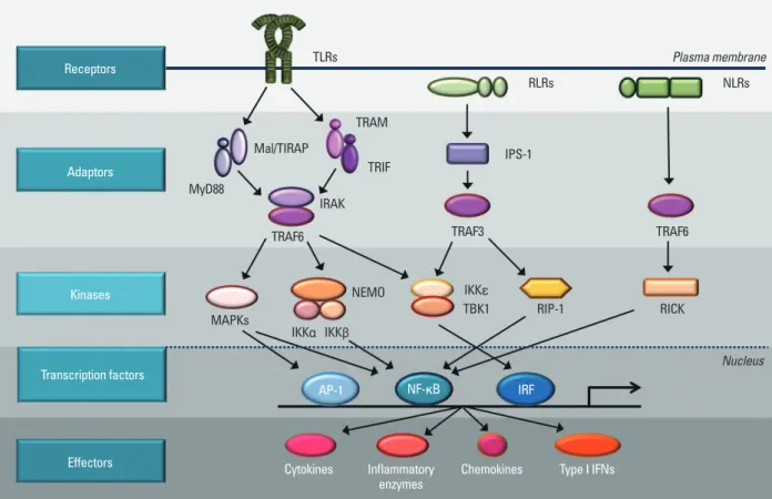
Fig. 1. Intracellular signaling pathways of pattern-recognition receptors. TLRs are mostly present on the membrane. RLRs and NLRs are located in the cyto- sol. TLRs send signal through Mal/TIRAP and MyD88 or TRAM/TRIF to IRAK/TRAF6 to activate downstream kinases. RLRs use IPS-1 and TRAF3 as adaptor molecules and NLRs transmit activation signals through TRAF6. The signals from TLRs, RLRs, and NLRs are delivered to kinases such as MAPKs, IKKs, TBK1, RIP-1, and RICK to activate transcription factor, AP-1, NF-κB, and IRF. Transcription factors bind to specific DNA sequences and produce effecter molecules such as cytokines, inflammatory enzymes, chemokines, and type I interferons (IFNs).
Receptors
Adaptors
Kinases
Transcription factors
Effectors
TLRs
RLRs
MyD88
Mal/TIRAP
TRAM
TRIF
IRAK
IPS-1
TRAF3
IKKε
TBK1 RIP-1
TRAF6
RICK
Nucleus TRAF6
MAPKs
AP-1 NF-κB
Cytokines Inflammatory Chemokines Type I IFNs enzymes
IRF IKKα IKKβ
NEMO
NLRs Plasma membrane
TLR. MyD88 has an N-terminal death domain (DD), an in- termediary domain (ID), and a C-terminal Toll-interleukin 1 receptor (TIR) domain. The TIR domain of MyD88 can bind to the TIR domain of TLR directly or indirectly.91 The N-terminal death domain of MyD88 binds to the death do- mains of other proteins, through homophilic DD-DD inter- action, leading to the activation of NF-κB and JNK.92 In a past study, where MyD88 was knocked-out, treatment with ligands of TLR2, TLR5, TLR7, and TLR9 did not exhibit the proper immune responses.93 However, unlike other TLRs, TLR4 signals still exist in MyD88-deficient mice.
This study led to the search for a MyD88-independent adap- tor molecule, as it was suggested that TLR4 has another adaptor molecule, which was later discovered to be TRIF.
TRIF (TIR-domain containing adaptor protein inducing interferon-β) is another adaptor molecule associated with TLR signaling TRIF was found by database screening dur- ing the search for a TIR domain containing protein. TRIF interacts with TLRs through TIR-TIR interaction. In con- trast with MyD88, which is broadly used as an adaptor molecule in TLR signaling, TRIF is only involved in the leading to the transcription of the proinflammatory genes.
INTRACELLULAR SIGNALING COMPONENTS OF PRR
Adaptor molecules of PRRs
TLRs, RLRs and NLRs act through adaptor molecules to activate various kinases and transcription factors. Adaptor molecules are very important messengers that deliver sig- nals from the receptors to protect the host from infection.
MyD88 (Myeloid differentiation primary response gene 88) is one of the representative adaptor molecules in TLR signaling. ‘MyD’ refers to myeloid differentiation and ‘88’
stands for the number of the gene. MyD88 is a protein that is induced by terminal differentiation of M1D+ myeloid precursors and responses to IL-6.90 MyD88 is located in the cytosol near the cytosolic part of TLRs and delivers an acti- vation signal that is initiated by receptor activation. MyD88 is used by all TLR family members, except TLR3, to acti- vate NF-κB. The structure of MyD88 is similar to that of
Fig. 2. Endogenous and exogenous regulators of TLR activation. Endogenous negative regulators of TLRs are revealed. The short form of MyD88 (sMyD88) sub- stitutes MyD88 but cannot send signals downstream. Tollip interacts with IRAK to decrease phosphorylation. A20 deubiquitylates TRAF6. SOCS1 regulates phos- phoryltation of IκBα, p38, and JNK. Exogenous TLR regulators have been identified. Small molecules such as surforaphane, cinnamaldehyde, and curcumin block oligomerization of the receptor, whereas resveratrol, EGCG, and certain flavonoids such as lutelolin, quercetin, chrysin, and eriodictyol have the ability to decrease kinase activity of TBK1. In addition, ligand binding to the receptor complex can be another target for regulation of the TLR signaling pathway.
Endogenous inhibitors Exogenous inhibitors
sMyD88
TLRs
Mal/TIRAP
TRAM
TRIF
IRAK
TRAF6
NEMO MAPKs
AP-1 NF-κB IRF
IKKα IKKβ TBK1 IKKε MyD88
Oligomerization
Surforaphane Cinnamaldehyde
Curcumin
Resveratrol LuteolinEGCG Quercetin
Chrysin Eriodictyol Tollip
A20
SOCS1
which is required for the adaptor molecule to be localized within plasma membrane. Mutation of the myristoylation motif in TRAM abolishes the activation signal. Further- more, protein kinase Cε (PKCε) phosphorylates TRAM. If phosphorylation is blocked, TRAM signals are impaired, which proves that PKCε is an essential component of the LPS-induced signaling pathway in macrophages.97
SARM (sterile α- and armadillo-motif-containing protein) consists of a sterile α motif (SAM) and a TIR domain. SARM has been shown to be a negative regulator of NF-κB and IRF in TLR signaling.98 Knockdown of SARM expression in primary human peripheral-blood mononuclear cells led to increased poly I:C- and LPS-induced chemokine and cy- tokine expression. Treatment of cells with LPS increased SARM protein levels, indicating negative feedback regula- tion of the TLR4/TRIF pathway. Since it is unclear how SARM inhibits TRIF function, it will be important to clari- fy this mechanism.
Among Toll-like receptors, endosomal TLRs require traf- ficking proteins, which transport TLRs from ER to endo- somes. UNC93B ER membrane protein carries TLR3, TLR7 and TLR9. In addition to UNC93B, Adaptor protein 3 (AP-3) was also involved in TLR9 trafficking. AP-3 en- hances the formation of the TLR9 complex with TRAF3 and IRF7. In the absence of AP-3, CpG-A DNA-induced type I IFN production through the stimulation of TLR9 is impaired.96
Interferon beta promoter stimulator-1 (IPS-1) contains an N-terminal CARD domain which is homologous with the domain in RIG-I. IPS-1 is localized in mitochondria, and initiates a signaling process that activates IRF3 and NF-κB, via TBK1/IKKε and IKKα/IKKβ, respectively. IPS-1 binds to RIG-I through CARD-CARD interaction. IPS-1-defi- cient mice fail to activate NF-κB and IRF3, with concomi- tant loss of type I IFN and inflammatory cytokine induc- tion, after infection.99
Main kinases in PRR signaling pathways
Phosphorylation is one of the typical mechanisms that acti- vate signaling cascades. Signals from adaptor molecules ac- tivate kinases, which can phosphorylate downstream mole- cules to regulate transcriptional factors.
A multiprotein complex, termed the IKK (IκB kinase) complex, consists of two catalytic components, IKKα and IKKβ, and a regulatory component, NF-κB essential modifi- er (NEMO, also known as IKKγ). IKKα and IKKβ are structurally similar, having a kinase domain, a leucine zipper signaling pathways of TLR3 and TLR4. TRIF is considered
to be closely related to anti-viral signaling, since signals mediated by TRIF are linked to IRF activation and produc- tion of IFN.94 While TLR3 only uses TRIF as its adaptor molecule, TLR4 uses TRIF under limited conditions in a MyD88-independent manner. It has been questioned as to whether there is any regulatory mechanism for the prefer- ential activation between MyD88- and TRIF-dependent signaling pathways in TLR4 signaling. A recent study sug- gests that LPS structure, and its relationship with CD14, could provide the answer. LPS structure can be differentiat- ed into ‘smooth LPS’ and ‘rough LPS’. Full-length O- chains render the ‘smooth LPS’ structure, whereas the re- duction of O-chains produces the ‘rough LPS’ structure.
‘Rough LPS’ can bind to the TLR4/MD2 complex, while CD14 is required for ‘smooth LPS’ to bind to TLR4/MD2.
When ‘rough LPS’ engages with a TLR4/MD2 complex in the absence of CD14, the complex initiates only MyD88- dependent responses. On the other hand, either ‘smooth’ or
‘rough LPS’, bound to TLR4, initiates both MyD88-depen- dent and MyD88-independent responses, in a CD14-depen- dent manner.95 TRIF recruits TRAF3 and TBK1 (TNF re- ceptor-associated factor (TRAF) family member associated NF-κB activator binding kinase 1) in order to phosphory- late IRF3. A knockout study, using TRIF-deficient mice, re- vealed that production of type I IFNs, through TLR3 or TLR4, requires the presence of TRIF.94
MAL/TIRAP (MyD88-adapter-like protein/TIR domain- containing adapter protein) is an adaptor molecule essential to the TLR2 and TLR4 signaling pathways. MAL/TIRAP acts as a bridge between MyD88 and TLR. MAL/TIRAP has an N-terminus binding domain that binds to phosphati- dylinositol-4,5-bisphosphate; this process mediates the re- cruitment of MAL/TIRAP to the plasma membrane and, in particular, to the microdomains that contain TLR4. MyD88 does not bind directly to TLR4, but instead interacts with MAL/TIRAP in association with TLR4.90 TLR2 and TLR4 signaling is impaired in cytokine production in MAL/TI- RAP-deficient mice; however, TLR2 response is affected to a greater extent than TLR4 response.
TRAM (TRIF-related adaptor molecule) also known as TICAM2 plays an essential role in the MyD88-independent signaling pathway of TLR4. TRAM has a TIR domain, and acts as a bridge connecting TLR and TRIF, which allows for the activation of the TRIF dependent pathway in response to LPS.96 The activation of TRAM affects IRF3 and NF-κB activation as well. TRAM is regulated by myristoylation,
RIP-1 is also involved in the TRIF pathway of TLR3 and TLR4.109 TRIF recruits RIP-1 upon TLR3 and TLR4 acti- vation. In the absence of RIP-1, TLR3-induced NF-κB sig- naling is abolished.
The NLR proteins NOD1 and NOD2 interact with the serine-threonine kinase RICK (receptor-interacting protein (RIP)-like interacting caspase-like apoptosis regulatory pro- tein kinase; also known as Ripk2 or RIP2), to induce NF- κB and MAPK signaling. Direct or indirect ligand recogni- tion by NOD1 and NOD2 induces recruitment of RICK through CARD-CARD interactions.110 This CARD-con- taining serine-threonine kinase directly binds and promotes K63-type polyubiquitylation of the regulator IKKγ and ac- tivation of the kinase TAK1,111 a prerequisite for activation of the IKK complex. These events result in the degradation of the NF-κB inhibitor IκBα and the subsequent transloca- tion of NF-κB to the nucleus, where transcription of the NF-κB-dependent target gene occurs.
Major transcription factors of PRRs
The stimulation of TLRs, RLRs or NLRs delivers signals through adaptor molecules and kinases. Ultimately, tran- scription factors, which trigger target gene transcription, are activated in the nucleus.
NF-κB is present in the cytoplasm, in an inactive form, captured by an inhibitor of NF-κB (IκB) proteins. Upon stimulation with various TLR ligands, IκBs are phosphory- lated at serine residues by IKK complexes, which consist of IKKα and IKKβ protein kinases and a regulatory molecule, IKKγ/NEMO. Phosphorylation targets IκBs for ubiquitina- tion and degradation, performed by the 26S proteasome, al- lowing NF-κB to be released into the nucleus and to bind to a response element, which starts transcription of the target genes.
AP-1 (Activator protein 1) has a dimeric basic region com- posed of members of the Jun, Fos, activating transcription factor (ATF), and Maf subfamilies. AP-1 may bind to TPA- response elements or cAMP-response elements. Among the AP-1 family proteins, c-Jun is thought to play a central role in inflammatory responses. AP-1 activation, in the TLR sig- naling pathway, is mostly mediated by MAP kinases, such as JNK, p38 and ERK, through phosphorylation. Many TLR li- gands activate MAP kinases with similar kinetics.112
TBK1 and IKKε have central roles in the induction of type I IFN through phosphorylation and activation of its transcription factors, IRF3 and IRF7. In a resting state, IRF3 is located in the cytoplasm in an inactive form; how- domain, helix-loop-helix structures and a NEMO-binding
domain (NBD). The IKK complex has a role in phosphory- lating IκB. Phosphorylated IκB is degradated by ubiquitina- tion. Then, NF-κB, which had been inhibited by IκB, is re- leased to translocate into nucleus. The IKK complex is a common factor for activating NF-κB, while the regulator of the IKK complex is different in each pathway.100
In addition to IKKs, MAPKs act as important kinases.
The expression of IL-6, IL-8, IL-12p40, and MCP-1 is reg- ulated by MAPK signaling.101 There are three groups of MAPKs in mammals: extracellular signal regulated kinase 1/2 (ERK1/2), p38 proteins (α, β, γ, and δ), and c-Jun N- terminal kinases (JNKs). The upstream MAPK kinases (MAPKKs or MEKs) are MEK1/2, MKK3/6, MKK4/7, and MEK5, respectively.102 ERK1/2, p38, and JNK are acti- vated by various TLR ligands. Through MyD88, TRAF6 activates a MAPK kinase kinase (MAPKKK) called trans- forming growth factor-activated kinase (TAK1). Activated TAK1 can phosphorylate MKK3 and MKK6, the kinases upstream of p38 MAPKs and JNK.103 TAK1 can also acti- vate the IKK complex. The activation of the IKK complex by TAK1 appears to be indirect, and the identity of the ki- nase that is responsible for direct phosphorylation of the IKK complex remains unidentified.
TBK1 (TRAF family member-associated NF-κB activa- tor (TANK) binding kinase-1) and IKKε (also known as IKKι) were initially implicated in IRF3 phosphorylation and activation, to produce type I IFN in the anti-viral re- sponse. Overexpression of IKKε and TBK1 markedly acti- vates NF-κB, as IKKε and TBK1 also regulate NF-κB, in addition to IRF3. IKKε was originally isolated as an LPS- inducible protein in mouse macrophages and was shown to exhibit a similar sequence to canonical IKKs.104 TBK1 was identified as a protein kinase that interacts with TANK (also known as I-TRAF).105 TBK1 deficiency in mice results in embryonic lethality, around day 14.5, because of liver weak- ness.106 Given that the lethality of TBK1-deficient mice is nullified when TNFR is absent, TBK1 might be involved in TNFR signaling to NF-κB, especially in the liver.107
IPS-1 interacts with receptor-interacting protein-1 (RIP-1), which was originally shown to be associated with the TNF receptor family of death receptors. RIP-1 is a death domain kinase, and is implicated in virus infection-induced type I IFN induction.108 IPS-1 interacts with RIP-1 via the non- CARD region to facilitate NF-κB activation, rather than IRF3 activation. RIP-1 action is also facilitated by IPS-1 to activate NF-κB through activation of the IKK complex.
been detected in the spleen and, less strongly, in the brain.
However, expression of MyD88s was upregulated in the human monocytic cell line (THP-1) following 16 hours of stimulation with LPS.117 In the presence of MyD88s, IRAK1 was still recruited, through a death-domain interaction with MyD88s, but was no longer phosphorylated.118 The pres- ence of MyD88s prevents IRAK4 from phosphorylating IRAK1, since D88s cannot associate with IRAK4. This in- dicates that MyD88s might be involved in a negative-feed- back regulatory mechanism that controls excessive TLR activation.119
Macrophages from SOCS1-deficient mice exhibited en- hanced phosphorylation of STAT1, IκBα, p38, and JNK, and produced high levels of nitric oxide and pro-inflamma- tory cytokines, in response to the presence of TLR4 and TLR9 ligands, LPS and CpG DNA, respectively.120 SOCS1- deficient mice die within three weeks of birth from multi- organ inflammation and high susceptibility to sepsis. Fur- thermore, LPS and CpG DNA induced SOCS1 expression in macrophages, which indicates that SOCS1 is a non-re- dundant negative regulator of TLR signaling, and may par- ticipate in the termination and resolution processes of in- flammation.
Tollip (Toll-interacting protein) was originally identified through a yeast-two-hybrid screening process. The process used the cytoplasmic tail of the IL-1R associated protein (residues 385-570) as bait to isolate a murine complemen- tary DNA, which encodes a protein of 274 amino acids, in order to find a new component or IL-1R pathway. Further study showed that Tollip was also able to interact with sev- eral members of the TIR superfamily, including TLR2 and TLR4.121 Overexpression of Tollip has been shown to result in inhibition of TLR2- and TLR4-mediated NF-κB activa- tion. Tollip interacts with IRAK1, and the level of IRAK1 autophosphorylation is reduced in the presence of Tollip.
IRAK1 causes phosphorylation of Tollip upon TLR stimu- lation. Although the physiological importance of this is un- clear, it is possible that phosphorylation of Tollip facilitates the ubiquitinylation of IRAK1 and its subsequent degrada- tion. In addition, Tollip expression is elevated in intestinal epithelial cells that are hypo-responsive to TLR2 ligands.
Therefore, phosphorylation and dephosphorylation of Toll- ip and IRAK, in the TLR signaling pathway, may be a switch for TLR4- and TLR2-mediated responses.
A20 was initially identified as a TNF-induced zinc-finger protein that suppresses TNF-mediated NF-κB activation.122 A20 expression is rapidly induced by both TLR4 ligands, ever, either TLR3 and TLR4 ligands or viral infection cause
TBK1- and IKKε-mediated phosphorylation of the C-ter- minal region of IRF3. This allows IRF3 to form a homodi- mer and translocate into the nucleus, where it can bind to the promoter regions of its target genes, such as the IFN- stimulated response element. Embryonic fibroblast cells from TBK1-deficient mice exhibit reduced IRF3 activation and IFN-β induction after stimulation with TLR3 and TLR4 ligands.113 While IKKε-deficient mice show no obvi- ous changes with respect to IRF3 activation and IFN-β in- duction, cells deficient in both TBK1 and IKKε exhibit a complete loss of IRF3 activation and IFN-β induction, indi- cating a possible role of IKKε in IRF3 activation.107 Akt also participates in activation of IRF3 in TLR3 and -4 sig- naling pathways as Akt knockdown by siRNA resulted in the diminishment of IRF3 phosphorylation and dimeriza- tion.114 As TBK1 is able to enhance phosphorylation of Akt in response to TLR3 or -4 agonist, the interaction between TBK1 and Akt promotes IRF3 activation and IFNβ expres- sion in TLR/TRIF-pathway. Notably, IRF3 activation by stimulation with TLR3 and TLR4 ligands is impaired in TRIF-deficient mice, but it is intact in MyD88-deficient mice, which suggests that IRF3 activation is controlled by the TRIF-dependent pathway. TBK1 and IKKε can also phosphorylate and activate IRF7, which is the member of the IRF family most closely related to IRF3.115 Whereas IRF3 is ubiquitously expressed and not inducible, IRF7 is expressed at low levels in most types of cells but is strongly induced in response to various stimuli.116 Therefore, IRF7 might be involved in positive feedback regulation of type I IFN induction.
INTRINSIC AND EXTRINSIC REGULATION OF PRR SIGNALING
Endogenous regulators
There exists a cellular device to prevent over- or unneces- sary activation of PRRs. Several intracellular negative regu- lators include MyD88s (the short form of MyD88), SOCS1, TOLLIP, A20, and CYLD.
The most universal adaptor molecule in TLR signaling is MyD88, which is employed by TLR2, TLR4, TLR5, TLR7, TLR8 and TLR9. MyD88s lacks the intermediary domain that mediates DD interaction between IRAK4 and MyD88, which is present in wild-type MyD88. Although MyD88 is ubiquitously expressed, expression of MyD88s has only
known anti-inflammatory agents. This leads us to search for new therapeutic targets for the treatment of inflammatory diseases and immune disorders.
Receptor oligomerization is an initial step of TLR signal- ing, which triggers the association of intracellular domains to provide a platform for the recruitment of downstream molecules. When dimerization is blocked, the signal cannot be delivered to the adaptor molecules and downstream sig- naling cascades. Recently, the suppression of TLR dimer- ization has been suggested as the inhibitory target for small molecules such as curcumin, cinnamaldehyde, and sulfora- phane, which have been reported to have anti-inflammatory effects.126,127 Thiol-modifying activity appears to be related to the action of these phytochemicals since a supplement of thiol-donors reversed the inhibitory effects of the phyto- chemicals on TLR4 activation. Indeed, the study using LC- MS/MS analysis has revealed that sulforaphane binds di- rectly to cysteine residues in the TLR4 extracellular domain and inhibits TLR4-TLR4 interaction. These results suggest that receptor clustering, especially the dimerization step, could be a novel target for TLR regulators, and that the mod- ification of cysteine residues could be a promising strategy for modulating TLR activation.127
The representative kinase found in TRIF-dependent TLR signaling is TBK1. TBK1 acts as a critical kinase for IRF3 activation and type I IFNs production by phosphorylating IRF3. Resveratol and its structural analog stilbene specifi- cally inhibit TRIF signaling in the TLR3 and TLR4 path- way by targeting TBK1. Resveratol directly blocks TBK1 kinase activity, as demonstrated by an in vitro kinase as- say.128 Certain flavonoids such as EGCG, luteolin, querce- tin, chrysin, and eriodictyol also inhibit TBK1 kinase activ- ity, resulting in a decrease in IRF3 activation and target gene expression, while naringenin and hesperetin had no such effect. This proves that kinases, especially TBK1, can be a regulatory target in TLR signaling, and provide a po- tential base for developing an inflammation inhibitor.129,130
In case of TLR4, MD2 is the essential partner in receptor cluster forming two TLR4-MD2 complexes upon engage- ment of LPS. Understanding the structure of MD2 and the interaction between MD2 and LPS can suggest a therapeu- tic strategy for regulating TLR4 activation. A free cysteine residue at the 133 position within a binding pocket of MD2 has been suggested as an important site for modulating the interaction between MD2 and LPS. Binding of MD2 Cys133 by thiol-reactive compounds decreases LPS signaling, such as NO production and NF-κB activation, possibly by pre- LPS and TNF, and is expressed in many cell types, which
suggests that it is involved in regulating TLR function. Mac- rophages from A20-deficient mice produced elevated levels of pro-inflammatory cytokines when stimulated with the TLR2 ligands (peptidoglycan and lipoteichoic acid), the TLR3 ligand (poly I:C), and the TLR9 ligand (CpG DNA).123 A20 is important in preventing the host from developing endotoxic shock; however, A20 deficiency does not play an important part in LPS tolerance. A20 is a cysteine protease de-ubiquitylating enzyme that blocks TLR mediated signal- ing by deubiquitylating TRAF6. A20 is a negative regulator that can control both MyD88-dependent and MyD88-inde- pendent TLR-signaling pathways.
The tumor suppressor CYLD (cylindromatosis) is a neg- ative regulator of the RIG-I-mediated innate antiviral re- sponse.124 Ectopic expression of CYLD inhibits both the IRF3 signaling pathway and IFN production triggered by RIG-I; conversely, CYLD knockdown enhances the RIG-I- induced IFN production. CYLD is closely related, in its function, to a deubiquitinating enzyme that removes Lys 63-linked polyubiquitin chains, which suggests a functional association between the two molecules. CYLD removes polyubiquitin chains from RIG-I as well as TBK1, which is the kinase that phosphorylates IRF3, inhibiting the IRF3 signaling pathway. Furthermore, CYLD protein level is re- duced by tumor necrosis factor or viral infection, concomi- tant with enhanced IFN production.
Poly(ADP-ribose) polymerases (PARPs), which regulate cell survival, cell death, and other biological functions, con- sist of at least 17 members. Among them, PARP-13 (ZAP) is known to be involved in IFN production against viral infec- tion. The shorter form of PARP-13 (ZAPS) is an especially strong stimulator of the RIG-I-signaling pathway, as it re- sponds to 5’-triphosphate-modified RNA (3pRNA). ZAPS promotes the activation of IRF3 and NF-κB through its asso- ciation with RIG-I. The production of not only IFN but also of other inflammatory cytokines such as IL-6, TNF-α and CXCL10 is regulated in a ZAPS-dependent manner.125 Regulation of PRR activation by exogenous substances Since the activation of PRRs is closely associated with the risk of chronic inflammatory diseases and immune disor- ders, the identification of therapeutic target points in PRR signaling could provide critical information for the preven- tion and treatment of these diseases. IKKβ and NF-κB have long been popular targets for anti-inflammation studies.
However, there remain unrevealed mechanisms for well-
downstream signal activation mechanisms, are essential in order to understand how to efficiently regulate the immune system and, eventually, to advance the quality of human life. As the effort to develop new therapeutic agents modu- lating PRRs is being extensively pursued,137 identification of novel regulatory targets provides an important informa- tion on constructing beneficial therapeutic strategies.
ACKNOWLEDGEMENTS
This work was supported by a grant from the Korea Food and Drug Administration for Studies on the Standardization of Herbal Medicine (2009, 2010).
REFERENCES
1. Janeway CA Jr. The immune system evolved to discriminate in- fectious nonself from noninfectious self. Immunol Today 1992;
13:11-6.
2. Medzhitov R, Janeway CA Jr. Innate immunity: impact on the adaptive immune response. Curr Opin Immunol 1997;9:4-9.
3. Medzhitov R, Janeway CA Jr. Decoding the patterns of self and nonself by the innate immune system. Science 2002;296:298- 4. Medzhitov R. Toll-like receptors and innate immunity. Nat Rev 300.
Immunol 2001;1:135-45.
5. Akira S, Takeda K, Kaisho T. Toll-like receptors: critical proteins linking innate and acquired immunity. Nat Immunol 2001;2:675- 80.
6. Hoffmann JA. The immune response of Drosophila. Nature 2003;426:33-8.
7. Meylan E, Tschopp J. Toll-like receptors and RNA helicases: two parallel ways to trigger antiviral responses. Mol Cell 2006;22:
561-9.
8. Yoneyama M, Fujita T. RNA recognition and signal transduction by RIG-I-like receptors. Immunol Rev 2009;227:54-65.
9. Lee JY, Zhao L, Hwang DH. Modulation of pattern recognition receptor-mediated inflammation and risk of chronic diseases by dietary fatty acids. Nutr Rev 2010;68:38-61.
10. O’Neill LA, Bryant CE, Doyle SL. Therapeutic targeting of Toll- like receptors for infectious and inflammatory diseases and can- cer. Pharmacol Rev 2009;61:177-97.
11. Lee JY, Hwang DH. The modulation of inflammatory gene ex- pression by lipids: mediation through Toll-like receptors. Mol Cells 2006;21:174-85.
12. Lemaitre B, Nicolas E, Michaut L, Reichhart JM, Hoffmann JA.
The dorsoventral regulatory gene cassette spätzle/Toll/cactus controls the potent antifungal response in Drosophila adults. Cell 1996;86:973-83.
13. Medzhitov R, Preston-Hurlburt P, Janeway CA Jr. A human ho- mologue of the Drosophila Toll protein signals activation of adaptive immunity. Nature 1997;388:394-7.
14. Akira S, Uematsu S, Takeuchi O. Pathogen recognition and in-
venting LPS access to the MD2 pocket.131 Other small mol- ecules that occupy this binding pocket in MD2 will prevent the ligand from engaging the receptor and the subsequent activation of intracellular signaling pathways.
CONCLUSION
The search for new innate immune receptors, and their sig- naling pathways, is still ongoing. The PYHIN proteins, AIM2 and IFI16, have been proposed as members of the AIM-like receptor (ALRs) family, which senses bacterial and viral DNA in the cytoplasm.132,133 In addition, LRRFIP1 has been identified as another cytosolic sensor for intracel- lular DNA.134 The three receptors have different intracellu- lar signaling pathways: AIM2 couples with ASC and cas- pase-1 to cleave pro-IL-1β producing mature IL-1β; IFI16 associates with STING and TBK1 to activate IRF3 and NF- κB; LRRFIP1 promotes phosphorylation of β-catenin, which activates IRF3 and produces IFNβ. Along with the discov- ery of new receptors, new regulators of immune response are also being revealed. TREX1 (Three prime repair exonu- lease 1) degrades IFN-stimulatory DNA derived from virus, which blocks the recognition of viral DNA by sensors and suppresses anti-viral immunity.135
Some PRRs are coupled with other receptors. The in- flammasome system, which activates caspase-1, requires a primary signal from other PRRs in order to initiate the tran- scription of pro-IL-1β, the caspase-1 substrate. It is well recognized that inflammasome plays a critical role in sens- ing danger signals. Nalp3 inflammasome is activated by en- dogenous and environmental toxicants such as uric acid, amyloid protein, alum, asbestos, and silica. Therefore, it is expected that inflammasomes regulate autoimmune diseas- es. Representative diseases that occur due to abnormalities in inflammasome function are systemic onset juvenile idio- pathic arthritis (SOJIA), urate crystal arthritis (Gout), and type 2 diabetes.136 The cause of these diseases is a mutation in the inflammasome-related genes. Malfunction of the in- flammasome results in inappropriate regulation of the im- mune system and the production of excess IL-1β, which leads to the development of chronic inflammatory diseases such as arthritis.
Dysregulation of innate immune receptors leads to un- controlled and improper immune responses against infec- tion and danger signals, and causes severe diseases. There- fore, discovery of new receptors and investigation into their
genase-2 mediated through Toll-like receptor 4. J Biol Chem 2001;276:16683-9.
33. Stewart CR, Stuart LM, Wilkinson K, van Gils JM, Deng J, Halle A, et al. CD36 ligands promote sterile inflammation through as- sembly of a Toll-like receptor 4 and 6 heterodimer. Nat Immunol 2010;11:155-61.
34. Schmidt M, Raghavan B, Müller V, Vogl T, Fejer G, Tchaptchet S, et al. Crucial role for human Toll-like receptor 4 in the devel- opment of contact allergy to nickel. Nat Immunol 2010;11:814-9.
35. Wieland CW, Knapp S, Florquin S, de Vos AF, Takeda K, Akira S, et al. Non-mannose-capped lipoarabinomannan induces lung inflammation via toll-like receptor 2. Am J Respir Crit Care Med 2004;170:1367-74.
36. Campos MA, Almeida IC, Takeuchi O, Akira S, Valente EP, Procópio DO, et al. Activation of Toll-like receptor-2 by glyco- sylphosphatidylinositol anchors from a protozoan parasite. J Im- munol 2001;167:416-23.
37. Ozinsky A, Underhill DM, Fontenot JD, Hajjar AM, Smith KD, Wilson CB, et al. The repertoire for pattern recognition of patho- gens by the innate immune system is defined by cooperation be- tween toll-like receptors. Proc Natl Acad Sci U S A 2000;97:
13766-71.
38. Bieback K, Lien E, Klagge IM, Avota E, Schneider-Schaulies J, Duprex WP, et al. Hemagglutinin protein of wild-type measles virus activates toll-like receptor 2 signaling. J Virol 2002;76:
8729-36.
39. Sato M, Sano H, Iwaki D, Kudo K, Konishi M, Takahashi H, et al. Direct binding of Toll-like receptor 2 to zymosan, and zymo- san-induced NF-kappa B activation and TNF-alpha secretion are down-regulated by lung collectin surfactant protein A. J Immunol 2003;171:417-25.
40. Frasnelli ME, Tarussio D, Chobaz-Péclat V, Busso N, So A.
TLR2 modulates inflammation in zymosan-induced arthritis in mice. Arthritis Res Ther 2005;7:R370-9.
41. Takeuchi O, Sato S, Horiuchi T, Hoshino K, Takeda K, Dong Z, et al. Cutting edge: role of Toll-like receptor 1 in mediating im- mune response to microbial lipoproteins. J Immunol 2002;169:
10-4.
42. Takeuchi O, Kawai T, Mühlradt PF, Morr M, Radolf JD, Zych- linsky A, et al. Discrimination of bacterial lipoproteins by Toll- like receptor 6. Int Immunol 2001;13:933-40.
43. Hoebe K, Georgel P, Rutschmann S, Du X, Mudd S, Crozat K, et al. CD36 is a sensor of diacylglycerides. Nature 2005;433:523-7.
44. Gantner BN, Simmons RM, Canavera SJ, Akira S, Underhill DM. Collaborative induction of inflammatory responses by dec- tin-1 and Toll-like receptor 2. J Exp Med 2003;197:1107-17.
45. Scheibner KA, Lutz MA, Boodoo S, Fenton MJ, Powell JD, Horton MR. Hyaluronan fragments act as an endogenous danger signal by engaging TLR2. J Immunol 2006;177:1272-81.
46. Lee JY, Zhao L, Youn HS, Weatherill AR, Tapping R, Feng L, et al. Saturated fatty acid activates but polyunsaturated fatty acid in- hibits Toll-like receptor 2 dimerized with Toll-like receptor 6 or 1.
J Biol Chem 2004;279:16971-9.
47. West XZ, Malinin NL, Merkulova AA, Tischenko M, Kerr BA, Borden EC, et al. Oxidative stress induces angiogenesis by acti- vating TLR2 with novel endogenous ligands. Nature 2010;467:
972-6.
48. Hayashi F, Smith KD, Ozinsky A, Hawn TR, Yi EC, Goodlett DR, et al. The innate immune response to bacterial flagellin is mediated by Toll-like receptor 5. Nature 2001;410:1099-103.
nate immunity. Cell 2006;124:783-801.
15. Kawai T, Akira S. The roles of TLRs, RLRs and NLRs in patho- gen recognition. Int Immunol 2009;21:317-37.
16. Poltorak A, He X, Smirnova I, Liu MY, Van Huffel C, Du X, et al. Defective LPS signaling in C3H/HeJ and C57BL/10ScCr mice: mutations in Tlr4 gene. Science 1998;282:2085-8.
17. Hoshino K, Takeuchi O, Kawai T, Sanjo H, Ogawa T, Takeda Y, et al. Cutting edge: Toll-like receptor 4 (TLR4)-deficient mice are hyporesponsive to lipopolysaccharide: evidence for TLR4 as the Lps gene product. J Immunol 1999;162:3749-52.
18. Miyake K. Roles for accessory molecules in microbial recogni- tion by Toll-like receptors. J Endotoxin Res 2006;12:195-204.
19. Park BS, Song DH, Kim HM, Choi BS, Lee H, Lee JO. The structural basis of lipopolysaccharide recognition by the TLR4- MD-2 complex. Nature 2009;458:1191-5.
20. Gazzinelli RT, Ropert C, Campos MA. Role of the Toll/interleu- kin-1 receptor signaling pathway in host resistance and pathogen- esis during infection with protozoan parasites. Immunol Rev 2004;201:9-25.
21. Burzyn D, Rassa JC, Kim D, Nepomnaschy I, Ross SR, Piazzon I. Toll-like receptor 4-dependent activation of dendritic cells by a retrovirus. J Virol 2004;78:576-84.
22. Kurt-Jones EA, Popova L, Kwinn L, Haynes LM, Jones LP, Tripp RA, et al. Pattern recognition receptors TLR4 and CD14 mediate response to respiratory syncytial virus. Nat Immunol 2000;1:398- 401.
23. Ohashi K, Burkart V, Flohé S, Kolb H. Cutting edge: heat shock protein 60 is a putative endogenous ligand of the toll-like recep- tor-4 complex. J Immunol 2000;164:558-61.
24. Asea A, Rehli M, Kabingu E, Boch JA, Bare O, Auron PE, et al.
Novel signal transduction pathway utilized by extracellular HSP70:
role of toll-like receptor (TLR) 2 and TLR4. J Biol Chem 2002;277:15028-34.
25. Vabulas RM, Braedel S, Hilf N, Singh-Jasuja H, Herter S, Ah- mad-Nejad P, et al. The endoplasmic reticulum-resident heat shock protein Gp96 activates dendritic cells via the Toll-like re- ceptor 2/4 pathway. J Biol Chem 2002;277:20847-53.
26. Smiley ST, King JA, Hancock WW. Fibrinogen stimulates mac- rophage chemokine secretion through toll-like receptor 4. J Im- munol 2001;167:2887-94.
27. Termeer C, Benedix F, Sleeman J, Fieber C, Voith U, Ahrens T, et al. Oligosaccharides of Hyaluronan activate dendritic cells via toll-like receptor 4. J Exp Med 2002;195:99-111.
28. Okamura Y, Watari M, Jerud ES, Young DW, Ishizaka ST, Rose J, et al. The extra domain A of fibronectin activates Toll-like recep- tor 4. J Biol Chem 2001;276:10229-33.
29. Johnson GB, Brunn GJ, Platt JL. Cutting edge: an endogenous pathway to systemic inflammatory response syndrome (SIRS)- like reactions through Toll-like receptor 4. J Immunol 2004;172:20-4.
30. Vogl T, Tenbrock K, Ludwig S, Leukert N, Ehrhardt C, van Zoel- en MA, et al. Mrp8 and Mrp14 are endogenous activators of Toll- like receptor 4, promoting lethal, endotoxin-induced shock. Nat Med 2007;13:1042-9.
31. Miller YI, Viriyakosol S, Worrall DS, Boullier A, Butler S, Witz- tum JL. Toll-like receptor 4-dependent and -independent cytokine secretion induced by minimally oxidized low-density lipoprotein in macrophages. Arterioscler Thromb Vasc Biol 2005;25:1213-9.
32. Lee JY, Sohn KH, Rhee SH, Hwang D. Saturated fatty acids, but not unsaturated fatty acids, induce the expression of cyclooxy-