Myocardial infarction is the leading cause of morbidity and mortality in industrialized countries. The critical prognosis factor of myocardial infarction is the size of the myocardial infarction, which is directly correlated with heart dilation and cardiac failure. Despite current pharmacotherapy, interventional procedures and surgical therapeutic methods preventing ventricular remodeling is limited due to their inability to repair damaged myocardium. Recent animal and clinical studies show that various types of stem cells can decrease cardiac infarction size and improve cardiac function .1-4
Previous studies have focused on the use of embryonic stem cells, cord blood derived stem cells, and skeletal muscle myoblasts. However, the clinical use of these cells has presented problems, including low cell number upon harvest and an ethical controversy.
Mesenchymal stem cells (MSCs) from adult tissues provide an attractive and alternative source of cells for tissue engineering. In addition, adult MSCs are relatively easily harvested from bone marrow, skin, muscle and adipose tissue.5-7 Currently, bone marrow is the primary source of adult mesenchymal stem cells.
Therapeutic Potential of Human Adipose Stem Cells in a Rat Myocardial Infarction Model
Seal Hwangbo,
1Jongok Kim,
2Sungho Her,
3Hyekyung Cho,
5and Jongho Lee
4Departments of 1Radiology, 2Pathology, 3Cardiology, and 4Thoracic and Cardiovascular Surgery, 5Clinical Research Institute, Daejeon St. Mary’s Hospital, College of Medicine, The Catholic University of Korea, Daejeon, Korea.
Purpose:Stem cell transplantation is expected to have good effects in the treatment of myocardial infarction (MI).
We tested the effect of the transplantation of human adipose-derived cells (ASCs) in Sprague-Dawley (SD) rats with myocardial infarctions. Materials and Methods:ASCs were isolated from the waste of elective abdominal surgery. The MI model was set up in SD rats by permanent ligation of the left anterior descending coronary artery.
One week after MI, either 1 x 10⁶ ASCs or an equal volume of phosphate-buffered saline (PBS) was injected into the infarct zone. Cardiac function was assessed by echocardiography, 1 day, 1 week, 2 weeks, and 4 weeks after treatment. Four weeks after transplantation, immunohistochemistry was performed. Results:Left ventricular function, including fractional shortening (FS), and ejection fraction (EF) showed a significant improvement in the ASCs transplantation group compared to the PBS group 4 weeks after treatment (p < 0.05). The anterior wall thickness of the left ventricle was significantly thicker in the ASCs transplantation group compared to the PBS group (p < 0.01). Multiple troponin T staining, and irregular, small amounts of connexin 43 expression also was observed in the ASCs transplantation group. Infarcted myocardium showed higher capillary density in the ASCs transplantation group than in the PBS injected group (p < 0.01). Conclusion:This study provides encouraging evidence that transplantation of ASCs can improve cardiac function of infarct myocardium in rat models with a limitation of cardiac remodeling, improved wall thickness, and increased neovascularization.
Key Words: Myocardial infarction, stem cells, transplantation
Received: July 9, 2009 Revised: August 18, 2009 Accepted: August 21, 2009
Corresponding author: Dr. Jongho Lee, Department of Thoracic and Cardiovascular Surgery, Daejeon St. Mary’s Hospital, College of Medicine, The Catholic University of Korea, 520-2 Daeheung-dong, Jung-gu,
Daejeon 301-723, Korea.
Tel: 82-42-220-9570, Fax: 82-42-222-7925 E-mail: [email protected]
∙The authors have no financial conflicts of interest.
© Copyright:
Yonsei University College of Medicine 2010
INTRODUCTION
However, the low number of cells necessitates in vitro culture expansion to obtain sufficient numbers of cells for clinical applications.8In addition, bone marrow stem cells are not readily available for the immediate treatment of acute myocardial infarction.
Mesenchymal stem cells derived from adipose tissue, adipose-derived stem cells (ASCs), were first identified by Zuk, et al.9as a source of adult mesenchymal stem cells.
After lineage-specific stimulation, ASCs show multiple- lineage differentiation potentials. They can differentiate into adipogenic, chondrogenic, myogenic, cardiomyogenic, osteogenic, endothelial, and neurogenic lineages.10,11
Adipose tissue is an abundant, expandable, and easily accessible source of mesenchymal stem cells. In our labo- ratory, we have established a method that readily isolates and expands stem cells from human adipose tissue. The purpose of this study is to investigate whether transplanta- tion of ASCs could improve cardiac function in a rat myo- cardial infarction model.
ASCs preparation and labeling
We used human waste adipose tissues which were obtain- ed after elective surgery and donated upon informed consent of the patients from our hospital.
The isolation of cells was performed as described prev- iously by Zuk, et al.12 In brief, adipose tissue was stored in sterile phosphate-buffered saline (PBS) at 4˚C. Then, the tissue was washed extensively with PBS and mechanically chopped before processing. To isolate the stromal vascular fraction (SVF), the tissue was enzymatically digested with PBS containing 0.1% collagenase A (Sigma Aldrich, St.
Louis, MO, USA) for 30 to 60 minutes at 37˚C with inter- mittent shaking.
The digested tissue was then washed with Dulbecco’s modified Eagles medium (DMEM) (Sigma Aldrich, St.
Louis, MO, USA) containing 10% fetal bovine serum (FBS), following centrifugation for 10 min at 200 g to remove mature adipocytes. The cell pellet was resuspensed in PBS and passed through a 100-µm mesh (BD, Falcon, Franklin Lakes, NJ, USA) to remove suspension.
Cells from the SVF were seeded and cultured for several passages in DMEM supplemented with 10% FBS, 100 U/mi penicillin, 100 µg/mL streptomycin in a 37˚C incubator with 5% CO2. Cells reaching 90% confluency were deta- ched with 0.5 mM ethylenediaminetetraacetic acid (EDTA) /0.05% trypsin for 10 min at 37˚C and then replated. Pas- sages p3 through p5 were used for all in vivo experiments.
Cell viability was assessed using the trypan blue exclusion assay with 0.2% trypan blue.
In culture, ASCs express cell-surface markers similar to those expressed by bone marrow MSCs, including CD117 (stem cell factor receptor), CD29 (β1-intergrin), CD 105 (mutilineage differentiation markers), CD-90, CD54 [inter- cellular adhesion molecule-I (ICAM-I)] and CD44. 5,7
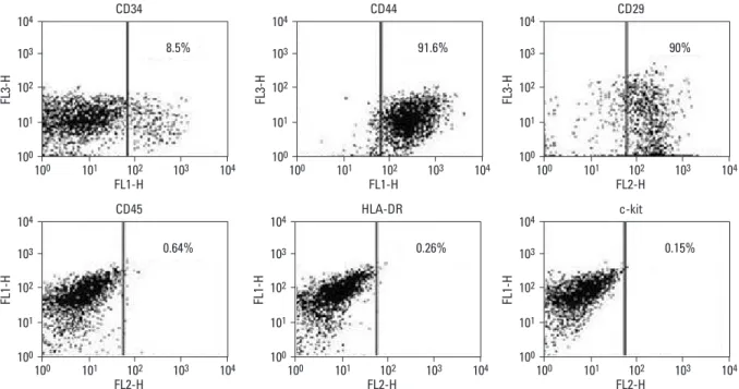
In addition, ASCs, like bone marrow MSCs, do not express the endothelial marker CD31, or the hematopoietic marker CD45. There is some controversy in the literature regarding the expression of CD34 that is widely used as a marker of hematopoietic stem cells but is also highly ex- pressed in vascular endothelial cell.13,14Using flow cytome- tric analysis, we analyzed several stem cell markers on ADCs including CD34, CD44, CD29, and CD45. HLA DR and c-kit (all, BD PharMingen, San Diego, CA, USA) expressions were also observed.
One day before cell transplantation, ASCs were labeled with 4,6-diamidino-2-phenylindole (DAPI) (Sigma Aldrich, St. Louis, MO, USA) for 12 hours in the ASC transplan- tation group.
Myocardial infarction model and stem cell transplantation
All animal studies were performed in accordance with the Animal Studies Committee guidelines, The Catholic Univer- sity of Korea, Korea.
Male Sprague-Dawley (SD) rats weighing 230-260 g at the beginning of the study were used. SD rats were anes- thetized with ketamine (80 mg/kg, intraperitoneally) and xylazine (8 mg/kg, intraperitoneally) and were placed in a supine position with all four paws taped. Tracheostomy and mechanical ventilation was maintained with room air by using a small animal ventilator (Harvard Apparatus, Holliston, MA, USA) and a 20-guage IV catheter as the tracheostomy tube throughout the procedure. A thoraco- tomy was performed through the fifth intercostal space and ribs were retracted. After opening the pericardium, the left anterior descending artery of heart (LAD) was permanently ligated with a 7-0 polypropylene suture 5 mm from the left atrial appendage. Infarction of the anterior wall of the left ventricle was confirmed by the presence of a pale myocar- dium after LAD ligation. The chest wall, muscle layers and skin were then closed with 4-0 silk sutures in three layers. The tracheostomy tube was removed and the suture of the tracheostomy site with 7-0 polypropylene was done.
Three or four days later, surviving rats with fractional shortening less than 40% were included in this study. One week after myocardial infarction, 30 rats were divided randomly into two groups: the ASCs transplantation group (n = 17, group 1) and the PBS injection control group (n = 13, group 2). The rats were anesthetized and ventilated as described above. Their chest was opened and ASCs (1×
106cells) in 40 µL medium or same volume of PBS were
MATERIALS AND METHODS
injected in the margin and in the center of infarction with four separate injections using a 30-guage needle.
The sham operation group (n = 10, group 3) underwent thoracotomy and cardiac exposure with neither coronary artery ligation nor cell transplantation.
Echocardiographic analysis
Echocardiography was performed 1 day, 1 week, 2 weeks and 4 weeks after cell transplantation in all 3 groups.
Rats were anesthetized with ketamine (50 mg/kg, intraperi- toneally) and xylazine (5 mg/kg, intraperitoneally). Echocar- diography was performed with a commercially available echocardiography system (IU22, Philips, Bothell, MA, USA) with 17-5 MHz small linear array transducer (hockey stick).
All measurements were taken over 2 consecutive cardiac cycles and averaged. All measurements were performed by an experienced cardiologist who was blind to the study group. Left ventricular end systolic diameter (LVESD), left ventricular end diastolic diameter (LVEDD), and left ventricular anterior wall thickening at the end-diastole data were obtained by a two dimensional targeted M-mode view. The percentage of fractional shortening (FS) was computed as representative of systolic function: FS(%) = (LVEDD-LVESD) / LVEDD ×100. Ejection fraction (EF) was calculated as: EF(%) = (Voldia- Volsys) / Voldia. End- diastolic volumes (Voldia) and end-systolic volumes (Volsys) were calculated by manually drawing endocardial contours at end-diastolic and end-systolic phases in the apical two chamber view using the modified Simpson’s rule.15
Histology and immunohistochemistry
Four weeks after cell transplantation, all rats underwent the final echocardiography and were then sacrificed with an overdose of ketamine and xylazine. The hearts were remov- ed and fixed with 4% phosphate-buffered formalin solution.
The following day, the hearts were sectioned into two parts from mid-ventricular level to apex and then embedded in paraffin. Paraffin sections (4 µm thickness) were mounted on coated slides were dried, deparaffinized, rehydrated and stained with hematoxylin-and-eosin staining for an evalua- tion of morphology. Sections were then used to identify the transplanted ASCs labeled with DAPI by fluorescent microscopy. One of the adjacent the ASCs positive sections preceded immunohistochemical stainings. Representative paraffin-embedded sections were immunostained with anti-α-smooth muscle actin (1 : 400, Chemicon, Temecula, CA, USA), anti-troponin T (Sigma Chemical, St. Louis, MO, USA), anti-connexin-43 (Invitrogen) and anti-collagen I (Santa Cruz Biotechnology, Santa Cruz, CA, USA).
Blood vessel density was measured at 400× magnifica- tion in three microscopic fields after immunostaining with anti-α-smooth muscle actin (1 : 400, Chemicon, Temecula, CA, USA).
Statistical analysis
Results are reported as mean
±
standard deviation. Statisti- cal analysis was performed with the SPSS program package (SPSS version 12.0; SPSS, Chicago, IL, USA). Differences between the two groups were compared with the Student’s t-test. Groups were compared with one-way analysis ofFig. 1. Flow-cytometric analysis of adult adipose cells expanded to three passages. Most of the ASCs expressed CD44 and CD29, and did not express for CD34, CD45, HLADR and c-kit. ASCs, adipose-derived cells.
variance (ANOVA) test. A value of p < 0.05 was consi- dered statistically significant.
Characterization of ASCs
CD44 and CD29 expressions were observed in 92% and 90%, respectively. CD45, HLA DR and c-kit expressions were observed in less than 1% (Fig. 1). CD34 was expressed in 8.3 % in our study.
Assessment of echocardiographic evaluation
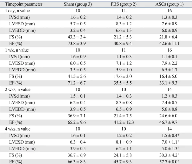
Table 1 shows the results of echocardiographic function in three groups. Left ventricular function, including fractional shortening (FS) and ejection fraction (EF) at 1 day, 1 week, and 2 weeks after treatment, did not differ between group 1 and group 2. However, results after 4 weeks showed a significant improvement of cardiac function in group 1
compared to group 2 (Fig. 2). The FS was 30.3 5 ± 4.2%
in group 1 and 24.15 ± 5.8% in group 2 and the EF was 57.75 ± 8.0% in group 1 and 45.75 ± 9.3% in group 2 four weeks after treatment (p < 0.05). However, EF and FS in group 1 were lower than in group 3, which suggests that group 1 did not recover normal function fully.
The anterior wall thickness was significantly thicker in group 1 than in group 2 four weeks of treatment (1.55 ± 0.4 mm and 1.25 ± 0.2 mm, respectively) (p < 0.01).
Myocyte differentiation of human ASCs
To identify human ASCs in vivo, DAPI labeled ASCs were observed at the transplant area. DAPI labeled ASCs were expressed in the rat heart 4 weeks after cell treatment.
DAPI labeled ASCs were observed near the infracted myocardium, but development into the multinucleated cell was not observed in our study. However, troponin T staining was observed near the cell transplantation area but not at the PBS injection area (Fig. 3). Troponin T is the early
Table 1. Echocardiographic Data
Timepoint parameter Sham (group 3) PBS (group 2) ASCs (group 1)
1 day, n value 10 11 16
IVSd (mm) 1.6 ± 0.2 1.4 ± 0.2 1.3 ± 0.3
LVESD (mm) 5.7 ± 0.5 8.3 ± 1.2 7.6 ± 0.9
LVEDD (mm) 3.2 ± 0.4 6.6 ± 1.3 6.0 ± 0.9
FS (%) 43.3 ± 3.4 21.2 ± 5.3 21.8 ± 6.4
EF (%) 73.8 ± 3.9 40.8 ± 9.4 42.6 ± 11.1
1 wk, n value 10 11 16
IVSd (mm) 1.6 ± 0.9 1.1 ± 0.3 1.1 ± 0.1
LVESD (mm) 6.0 ± 0.5 7.1 ± 1.2 7.9 ± 2.2
LVEDD (mm) 3.5 ± 0.5 5.9 ± 1.0 6.5 ± 1.7
FS (%) 41.5 ± 5.6 17.6 ± 3.0 16.4 ± 5.0
EF (%) 71.2 ± 6.7 35.5 ± 5.5 33.1 ± 9.3
2 wks, n value 10 10 14
IVSd (mm) 1.5 ± 0.1 1.4 ± 0.3 1.2 ± 0.3
LVESD (mm) 6.2 ± 0.4 8.3 ± 0.8 7.4 ± 0.7
LVEDD (mm) 3.9 ± 0.5 6.5 ± 0.9 5.6 ± 0.8
FS (%) 36.9 ± 7.1 21.4 ± 7.5 24.6 ± 6.0
EF (%) 65.2 ± 9.6 41.2 ± 12.3 46.7 ± 9.7
4 wks, n value 10 10 14
IVSd (mm) 1.6 ± 0.1 1.2 ± 0.2 1.5 ± 0.4*
LVESD (mm) 6.3 ± 0.4 8.1 ± 0.9 7.0 ± 1.1�
LVEDD (mm) 3.9 ± 0.5 6.2 ± 1.1 5.0 ± 1.3�
FS (%) 36.7 ± 6.9 24.1 ± 5.8 30.3 ± 4.2�
EF (%) 66.3 ± 8.3 45.7 ± 9.3 57.7 ± 8.0�
IVSd, interventricular septal thickness at end-diastolic; LVESD, left ventricular end-systolic dimension; LVEDD, left ventricular end-diastolic dimension; FS, fractional shortening; EF, ejection fraction; PBS, phosphate-buffered saline.
*p < 0.01 vs. PBS (group 2).
�p < 0.05 vs. PBS (group 2).
RESULTS
marker of myocyst.
Group 3 showed parallel alignment of cardiomyocysts with regular expression of connexin 43. However, group 1 showed irregular expression and small amounts of connexin 43 and group 2 showed little expression of connexin 43 (Fig. 3).
Assessment of neovascularization
Capillary density increased more in group 1 and group 2, than in group 3, as determined by α-smooth muscle actin immunostaining (Fig. 3).
The vascular densities were in 2.2 ± 0.3 vessels/mm2, 3.5 ± 0.4 vessels/mm2, and 6.8 ± 1.3 vessels/mm2in group 3, 2, and 1, respectively.
Assessment of collagen expression
In group 1 and group 2, collagen volume increased marke- dly, compared to group 3. In group 1, less increased colla- gen volume was observed than in group 2. Group 3 showed scanty and uniformly distributed collagen, while group 1 showed clumpy distribution.
Various cell types are currently under investigation for their therapeutic potential in cardiac infarction. Several clinical trials have reported encouraging results with stem cell transplantation to myocardial infarction patients.16,17
DISCUSSION
Fig. 2. Echocardiographic images after treatment. M-mode echocardiographic images demonstrating an increase in anterior wall thickening in the left ventricle and an improvement of ventricular function in the ASC transplantation group at the top images. Similar images representing post-infarction remodeling with left ventricular dilatation of the PBS group in middle. Echocardiographic images in the control sham operation group at the bottom. ASCs, adipose-derived cells;
PBS, phosphate-buffered saline.
2 weeks 4 weeks
ASCPBSSham
Fig. 3. H & E staining (A,×400) and immunohistochemical staining of connexin 43 (B,×100), troponin T (C,×100) and α-smooth muscle actin (D,×100) of the three groups.
ASCs, adipose-derived cells; PBS, phosphate-buffered saline.
(A) (B) (C) (D)
ASCPBSSham
Having an optimal source of cells to be transplanted is important and human adult stem cells are an attractive source. Bone marrow stem cells have been used in hema- tological disorders and were studied in cardiac infarction models. However, findings of these studies are controversial and no definite conclusion can be drawn from them.18-20In addition, obtaining the therapeutic quantity of these cells requires general anesthesia and hospitalization. A study using autologous skeletal myocytes showed clear benefit, but also showed concerns of arrhythmia.21-23
Brown adipose tissue also expresses stem cell markers of cardiomyocytes and differentiates into cardiomyocytes.24 Brown adipose tissue is uncommon in adults and using brown adipose tissue in cell transplantation has limitations.
Adult human adipose stem cell (ASC) is likely a feasible source of stem cells because it is abundant, it lacks donor limitation and obtaining it has a low risk of side effects.
After lineage-specific stimulation, ASCs show multiple- lineage differentiation including adipogenic, chondrogenic, myogenic, cardiomyogenic, osteogenic, endothelial and neurogenic lineages, similar to bone marrow stem cells.10,11 Transplantation of these cells can decrease the size of a myocardial infarction by promoting angiogenesis and myogenesis.25-28
Our echocardiography results revealed better heart func- tion in the ASCs transplantation group than in the PBS injection group. By the fourth week after PBS injection, definite left ventricular functional improvement was not observed and dilatation of the left ventricle without wall thickening in the infracted area continued. In contrast, the ASC transplantation group showed improvement of left ventricular function 4 weeks after transplantation with abundant neovascularization. Neovascularization in the infracted areas is an important component in the cardiac remodeling process.14,29Miranville, et al.30showed that ASCs with CD34+/CD31- are capable of in vivo and in vitro endo- thelial differentiation. Mirencille, et al.30also showed that delivery of ACSs accelerates perfusion in the limb ischemic model. Rehman, et al.31suggested that improvement of perfusion in the ischemic region was due to the vascular endothelial growth factor (VEGF) and other angiogenic factors expressed by ASCs. In the present study, DAPI labeled ASCs were observed near and in the arterioles walls of the infarct myocardial area. This strongly suggests that ADCs have the ability to differentiate into endothelial cells, and thus play a potential role in neovascularization. Al- though the precise mechanism by which ASCs transplan- tation limits the myocardial remodeling is unknown, ASCs can improve tissue ischemia in part through paracrine mechanisms.
Planat-Bérnaed, et al.32 observed spontaneous differen- tiation of rat ASCs into cardic myocytes after three weeks of
culture in methycellulose-based culture media.30 In this study, the ASC transplantation group expressed large quantities of troponin I, the cardiac transcription factor and expressed small quantities of connexin 43, a gap junction protein, at contact points with native cardiac myocytes and with ASCs in cell transplantation areas. These results are consistent with an in vivo study of differentiation of ASCs into cardiomyocysts.33However, we were not able to ob- serve direct transformation of the DAPI labeled ASCs into multinucleated cells or myocysts.
The most striking observation in this study is the increase in the anterior wall thickness of the left ventricle in the ASC transplantation group. The maintenance of the thickness of infarcted myocardium may be due to the alteration of colla- genase activity or to other enzymatic pathways responsible for cardiac remodeling and fibrosis. Collagen composes most of the extracellular matrix in infarcted myocardium.
Because the loss of cardiomyocytes and the replacement of collagen are responsible for ventricular remodeling after myocardial infarction, preventing cell apoptosis and fibrosis is a major mechanism of cell therapy in the cardiac infarc- tion model. Our results demonstrated a lack of collagen replacement in the cell transplantation group compared to the PBS injection group.
In this study, we observed three cases which represent multifocal intramyocardial calcifications in the ASC trans- plantation site. These results demonstrate that direct trans- plantation of unselected stem cells into infarcted myocar- dium may induce intramyocardial calcification.34We spec- ulated that unselected ASCs contained other progenitor cells that might differentiate into bone or cartilage creating an island of calcification. ASCs cultured in a specific culture medium would differentiate into myocytes more efficiently in cell transplantation.
In this study, we delivered cells 1 week after myocardial infarction to avoid cell loss due to inflammation and wash- out at the infarct area.35-37Existing techniques for tracking stem cells include fluorescent transfection, transfection of fluorescence in situ hybridization, using specific surface markers of transplanted cells and labeling cells with iron- oxide nanoparticles or radiotracers.38-40In this study, we used commercially available fluorescent dyes to label cell nuclei (DAPI). This dye was easy to handle and resulted in a 100%
labeling of cells. However this agent intercalates into DNA and may interfere with normal cell function, one of the limi- tations of our study. Several other limitations in our study were also revealed. The number of animals in each group was relatively small. Echocardiography to evaluate heart function is controversial in small animal assessment due to operator dependant procedures.
In conclusion, the present study provides encouraging evidence that transplantation of ASCs can improve cardiac
function in infarct myocardium in a rat model. Limitation of cardiac remodeling, improved wall thickness and increased neovascularization results in improved cardiac function.
Many issues must be explored before the safe application of these cells in the clinical setting. However, with appropriate validation of cell types and optimal performance, they should achieve a demonstrable benefit in cell therapy.
This work was supported by the Clinical Research Institute, Catholic University, Daejeon St. Mary’s Hospital, Korea.
1. Dimmeler S, Zeiher AM. Wanted! The best cell for cardiac regeneration. J Am Coll Cardiol 2004;44:464-6.
2. Dimmeler S, Zeiher AM, Schneider MD. Unchain my heart:
scientific foundations of cardiac repair. J Clin Invest 2005;
115:572-83.
3. Laflamme MA, Murry CE. Regenerating the heart. Nat Biotechnol 2005;23:845-56.
4. Lee RH, Kim B, Choi I, Kim H, Choi HS, Suh K, et al. Charac- terization and expression analysis of mesenchymal stem cells from human bone marrow and adipose tissue. Cell Physiol Biochem 2004;14:311-24.
5. Pittenger MF, Mackay AM, Beck SC, Jaiswal RK, Douglas R, Mosca JD, et al. Multilineage potential of adult human mesen- chymal stem cells. Science 1999;284:143-7.
6. Vats A, Bielby RC, Tolley NS, Nerem R, Polak JM. Stem cells.
Lancet 2005;366:592-602.
7. Fraser JK, Wulur I, Alfonso Z, Hedrick MH. Fat tissue: an underappreciated source of stem cells for biotechnology. Trends Biotechnol 2006;24:150-4.
8. Caplan AI. Review: mesenchymal stem cells: cell-based recon- struction therapy in orthopedics. Tissue Eng 2005;11:1198-211.
9. Zuk PA, Zhu M, Ashjian P, De Ugarte DA, Huang JI, Mizuno H, et al. Human adipose tissue in a source of multipotent stem cells.
Mol Biol Cell 2002;13:4279-95.
10. Oedayrajsingh-Varma MJ, van Ham SM, Knippenberg M, Helder MN, Klein-Nulend J, Schouten TE, et al. Adipose tissue- derived mesenchymal stem cell yield and growth characteristics are affected by the tissue-harvesting procedure. Cytotheraphy 2006;8:166-77.
11. Prunet-Marcassus B, Cousin B, Caton D, André M, Pénicaud L, Casteilla L. From heterogeneity to plasticity in adipose tissues:
site-specific differences. Exp Cell Res 2006;312:727-36.
12. Zuk PA, Zhu M, Mizuno H, Huang J, Futrell JW, Katz AJ, at al.
Multilineage cells from human adipose tissue: implications of cell-based therapies. Tissue Eng 2001;7:211-28.
13. Trischmann TM, Schepers KG, Civin CI. Measurement of CD34+ cells in bone marrow by flow cytometry. J Hematother 1993;2:305-13.
14. Davani S, Marandin A, Mersin N, Royer B, Kantelip B, Hervé P, et al. Mesenchymal progenitor cells differentiate into an endo- thelial phenotype, enhance vascular density, and improve heart
function in a rat cellular cardiomyoplasty model. Circulation 2003;108 Suppl 1:II253-8.
15. Jensen-Urstad K, Bouvier F, Höjer J, Ruiz H, Hulting J, Samad B, et al. Comparison of different ehocardiographic methods with radionuclide imaging for measuring left ventricular ejection fraction during acute myocardial infarction treated by throm- bolytic therapy. Am J Cardiol 1998;81:538-44.
16. Assmus B, Schächinger V, Teupe C, Britten M, Lehmann R, Döbert N, et al. Transplantation of progenitor cells and regenera- tion enhancement in acute myocardial infarction (TOPCARE- AMI). Circulation 2002;106:3009-17.
17. Britten MB, Abolmaali ND, Addmus B, Lehmann R, Honold J, Schmitt J, et al. Infarct remodeling after intracoronary progenitor cell treatment in patients with acute myocardial infarction (TOPCARE-AMI): mechanistic insights from serial contrast- enhanced magnetic resonance imaging. Circulation 2003;
108:2212-8.
18. Kawada H, Fujita J, Kinjo K, Matsuzaki Y, Tsuma M, Miyatake H, et al. Nonhematopoietic mesenchymal stem cells can be mobilized and differentiate into cardiomyocytes after myocardial infarction. Blood 2004;104:3581-7.
19. Murry CE, Soonpaa MH, Reinecke H, Nakajima H, Nakajima HO, Rubart M, et al. Haematopoietic stem cells do not trans- differentiate into cardiac myocysts in myocardial infarcts. Nature 2004;428:664-8.
20. Balsam LB, Wagers AJ, Christensen JL, Kofidis T, Weissman IL, Robbins RC. Haematopoietic stem cells adopt mature haematopoietic fates in ischaemic myocardium. Nature 2004;
428:668-73.
21. Menasché P, Hagége AA, Vilquin JT, Desnos M, Abergel E, Pouzet B, et al. Autologous skeletal myoblast transplantation for severe postinfarction left ventricular dysfunction. J Am Coll Cardiol 2003;41:1078-83.
22. Hagége AA, Carrion C, Menasché P, Vilquin JT, Duboc D, Marolleau JP, et al. Viability and differentiation of autologous skeletal myoblast grafts in ischaemic cardiomyopathy. Lancet 2003;361:491-2.
23. Fernandes S, Amirault JC, Lande G, Nguyen JM, Forest V, Bignolais O, et al. Autologous myoblast transplantation after myocardial infarction increases the inducibility of ventricular arrhythmias. Cardiovasc Res 2006;69:348-58.
24. Yamada Y, Yokoyama S, Wang XD, Fukuda N, Takakura N.Cardiac stem cells in brown adipose tissue express CD133 and induce bone marrow nonhematopoietic cells to differentiate into cardiomyocytes. Stem Cells 2007;25:1326-33.
25. Asahara T, Masuda H, Takahashi T, Kalka C, Pastore C, Silver M, et al. Bone marrow origin of endothelial progenitor cells responsible for postnatal vasculogenesis in physiological and pathological neovascularization. Circ Res 1999;85:221-8.
26. Itescu S, Kocher AA, Schuster MD. Myocardial neovasculari- zation by adult bone marrow-derived angioblast: strategies for improvement of cardiomyocyte function. Heart fail revi 2003;
8:253-8.
27. Orlic D, Kajstura J, Chimenti S, Jakoniuk I, Anderson SM, Li B, et al. Bone marrow cells regenerate infracted myocardium. Nature 2001;410:701-5.
28. Kajstura J, Rota M, Whang B, Cascapera S, Hosoda T, Bearzi C, et al. Bone marrow cells differentiate in cardiac cell lineages after infarction independently of cell fusion. Circ Res 2005;96:127-37.
29. Jackson KA, Majka SM, Wang H, Pocius J, Hartley CJ, Majesky
REFERENCES
ACKNOWLEDGEMENTS
MW, et al. Regeneration of ischemic cardiac muscle and vascular endothelium by adult stem cells. J Clin Invest 2001;107:1395-402.
30. Miranville A, Heeschen C, Sengenès C, Curat CA, Busse R, Bouloumié A. Improvement of postnatal neovascularization by human adipose tissue-derived stem cells. Circulation 2004;
110:349-55.
31. Rehman J, Traktuev D, Li J, Merfeld-Clauss S, Temm-Grove CJ, Bovenkerk JE, et al. Secretion of angiogenic and antiapoptotic factors by human adipose stromal cells. Circulation 2004;109:
1292-8.
32. Planat-Bénard V, Menard C, André M, Puceat M, Perez A, Garcia-Verdugo JM, et al. Spontaneous cardiomyocyst differen- tiation from adipose tissue stroma cells. Circ Res 2004;94:223-9.
33. Strem BM, Zhu M, Alfonso Z, Daniels EJ, Schreiber R, Beygui R, et al. Expression of cardiomyocytic markers on adipose tissue- derived cells in a murine model of acute myocardial injury.
Cytotherapy 2005;7:282-91.
34. Yoon YP, Park JS, Tkebuchava T, Luedeman C, Losordo DW.
Unexpected severe calcification after transplantation after tran- splantation of bone marrow cells in acute myocardial infarction.
Circulation 2004;109:3154-7.
35. Dow J, Simkhovich BZ, Kedes L, Kloner RA. Washout of transplanted cells from the heart: a potential new hurdle for cell transplantation therapy. Cardiovasc Res 2005;67:301-7.
36. Reinecke H, Zhang M, Bartosek T, Murry CE. Survival, integra- tion, and differentiation of cardiomyocyte grafts: a study in normal and injured rat hearts. Circulation 1999;100:193-202.
37. Li RK, Mickle DA, Weisel RD, Rao V, Jia ZO. Optimal time for cardiomyocyte transplantation to maximize myocardial function after left ventricular injury. Ann Thorac Surg 2001;72:1957-63.
38. Potapova I, Plotnikov A, Lu Z, Danilo P Jr, Valiunas V, Qu J, et al. Human mesenchymal stem cells as a gene delivery system to create cardiac pacemakers. Circ Res 2004;94:952-9.
39. Kraitchman DL, Heldman AW, Atalar E, Amado LC, Martin BJ, Pittenger MF, et al. In vivo magnetic resonance imaging of mesen- chymal stem cells in myocardial infarction. Circulation 2003;
107:2290-3.
40. Barbash IM, Chouraqui P, Baron J, Feinberg MS, Etzion S, Tessone A, et al. Systemic delivery of bone marrow-derived mesenchymal stem cells to the infarcted myocardium: feasibility, cell migration, and body distribution. Circulation 2003;108:863-8.