Korean J Radiol 12(2), Mar/Apr 2011 www.kjronline.org 216
INTRODUCTION
Nutritional support is an essential part in the overall management of patients with head and neck cancer (1).
Gastrostomy is often required to provide enteral nutrition in this group of dysphagic patients during combined
Modifi ed Radiology-Guided Percutaneous Gastrostomy (MRPG) for Patients with Complete Obstruction of the Upper Digestive Tract and Who are without Endoscopic or Nasogastric Access
Siu-Cheung Chan, MD
1, 5, Winnie Chiu-Wing Chu, MD
2, Kar-Wai Liu, MD
1, 5, Chun-Ta Liao, MD
3, 5, Tsung-Shih Lee, MD
4, Shu-Hang Ng, MD
1, 51Department of Diagnostic Radiology, Chang Gung Memorial Hospital, Linkou Medical Center and Keelung Hospital, Taoyuan, Taiwan; 2Department of Diagnostic Radiology and Organ Imaging, The Chinese University of Hong Kong, Hong Kong SAR, China; 3Department of Otorhinolaryngology, Chang Gung Memorial Hospital, Linkou Medical Center, Taoyuan, Taiwan; 4Department of Gastroenterology, Chang Gung Memorial Hospital, Linkou Medical Center and Keelung Hospital, Taoyuan, Taiwan; 5College of Medicine, Chang Gung University, Taoyuan, Taiwan
Objective: We wanted to report on our experience with modified radiology-guided percutaneous gastrostomy (MRPG) without endoscopic or nasogastric access for treating patients with complete obstruction of the upper digestive tract.
Materials and Methods: Fourteen oncology patients (13 had hypopharyngeal cancer and 1 had upper esophageal cancer) with complete obstruction of the upper digestive tract were recruited. Conventional percutaneous endoscopic gastrostomy (PEG) and radiologic (fl uoroscopy-guided) percutaneous gastrostomy (RPG) were not feasible in all the patients. An MRPG technique (with a combination of ultrasound, an air enema and fl uoroscopic guidance) was performed in these patients.
Results: We achieved successfully percutaneous gastrostomy using the modifi ed technique in all patients without any major or minor complications after the procedure.
Conclusion: A modifi ed radiology-guided percutaneous gastrostomy technique can be safely performed in patients who failed to receive conventional PEG or RPG due to the absence of nasogastric access in the completely obstructed upper digestive tract.
Index terms: Percutaneous gastrostomy; Complete obstruction; Upper digestive tract
Received September 13, 2010; accepted after revision December 23, 2010.
Corresponding author: Winnie Chiu-Wing Chu, MD, Department of Diagnostic Radiology and Organ Imaging, Prince of Wales Hospital, Shatin, Hong Kong SAR, China.
• Tel: (852) 2632-2290 • Fax: (852) 2636-0012
• E-mail: [email protected]
This is an Open Access article distributed under the terms of the Creative Commons Attribution Non-Commercial License (http://creativecommons.org/licenses/by-nc/3.0) which permits unrestricted non-commercial use, distribution, and reproduction in any medium, provided the original work is properly cited.
Original Article
DOI: 10.3348/kjr.2011.12.2.216 pISSN 1229-6929 · eISSN 2005-8330 Korean J Radiol 2011;12(2):216-219
treatment of chemotherapy and radiotherapy due to an obstructed upper digestive tract (2, 3). Percutaneous endoscopic gastrostomy (PEG) and radiologic percutaneous gastrostomy (RPG) are two currently established methods to provide enteral feeding to this group of patients (4-9).
However, PEG is not feasible in patients who have high- grade narrowing or obstruction of the upper digestive tract and this precludes the passage of an endoscope.
For conventional fl uoroscopy-guided RPG, the stomach is fi rst distended with air introduced via the nasogastric tube. Percutaneous access to the stomach is achieved by the use of fl uoroscopy to puncture the gasfi lled stomach.
In some diffi cult cases where the esophageal narrowing is tight, a fi ne bore catheter is introduced via a coaxial guidewire system as a substitute for a nasogastric tube for the purpose of air insuffl ation. However, in the cases with complete esophageal obstruction that prevents
Korean J Radiol 12(2), Mar/Apr 2011
www.kjronline.org 217
Modifi ed Radiology-Guided Percutaneous Gastrostomy in Complete Obstruction of Upper Digestive Tract
MATERIALS AND METHODS
From December 2005 to June 2010, fourteen patients with complete obstruction of the pharynx or esophagus and who were unable to obtain PEG or conventional fl uoroscopy-guided percutaneous gastrostomy underwent modifi ed radiology-guided percutaneous gastrostomy (MRPG). This cohort included two female patients and placement of either a nasogastric tube or a guidewire, the
percutaneous access to the stomach has to be monitored with a combined approach using ultrasound, an air enema and fl uoroscopic guidance.
In this report, we describe a modifi ed combined radiology-guided approach to perform percutaneous gastrostomy in patients with complete obstruction of the upper gastrointestinal tract.
C D
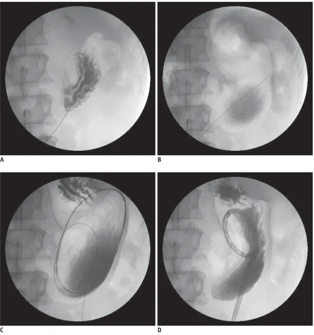
Fig. 1. Modifi ed radiology-guided percutaneous gastrostomy technique.
A. 21G fi ne needle punctured localized collection of air, which was visible in collapsed stomach under fl uoroscopy-guided gastrostomy. Needle tip is then gradually withdrawn while injecting small amounts of water-soluble contrast medium. Location of stomach is confi rmed by visualization of opacifi ed gastric rugae. B. Stomach was infl ated with approximately 600-800 mL of room air through 21G fi ne needle. C. 100-cm stainless steel guide wire is inserted through needle, and gastro-percutaneous tract is gradually dilated. D. Insertion of 14-Fr pigtail gastrostomy catheter and injection of small amount of water-soluble contrast medium via pigtail catheter confi rmed that gastrostomy catheter is correctly placed within stomach.
A B
Korean J Radiol 12(2), Mar/Apr 2011 www.kjronline.org 218
Siu-Cheung Chan et al.
twelve male patients. The age range was 41-68 years (mean age 49.8 years). Thirteen patients had hypopharyngeal cancers and one had upper thoracic esophageal cancer. The Institutional Review Board of our hospital approved this retrospective study and informed consent was waived due to the retrospective and anonymous nature of the analysis.
In this new approach, ultrasound was fi rst performed to outline the margin of the left lobe of the liver. The liver margin was marked on the skin. Then the patients received intravenous administration of 20 mg of hyoscine-N-
butylbromide (Buscopan, Boehringer, Ingelheim, Germany).
Simultaneously, a fl uoroscopically-controlled air enema using 300 mL of room air via the rectum was performed, which outlined the anatomic position of the transverse colon. Next, the position of the collapsed stomach was estimated by the presence of any localized collection of air at the left subdiaphragmatic region. The presumed collapse stomach was then punctured using a 21G fi ne needle under fl uoroscopic guidance with special care to avoid puncturing the liver or transverse colon. The needle was then gradually withdrawn under fl uoroscopic control while a small amount of water-soluble contrast medium (Iothalamate meglumine 60%; Conray 60, Mallinckrodt Canada Incorporated, QB, Canada) was continuously injected until the needle tip was confi rmed to be within the stomach. Entry of the needle tip within the stomach was recognized when the gastric rugae were opacifi ed by the pooling of contrast medium.
The stomach was then insuffl ated under fl uoroscopic control with approximately 600-800 mL of room air through the 21G fi ne needle (Fig. 1A). After this point, the procedure was similar to what has been described for RPG. In addition to local anesthesia, intravenous sedation with 5 mg midazolam (Dormicum, Roche, Basel, Switzerland) and 50 mg pethidine (Demerol, Roche, Basel, Switzerland) was given to the patients. Gastropexy was performed using two T-fasteners (Cope gastrointestinal suture anchor set;
Cook Incorporated, Bloomington, IN). An 18-gauge, 8 cm catheter needle was punctured directly through the gastric wall via a small incised area at the center of the skin between the two gastropexy fasteners. A 100 cm stainless steel guide wire was inserted through the needle and gradual dilation of the tract was carried out by insertion a 14-Fr locking gastrostomy catheter (Wills-Oglesby percutaneous gastrostomy set, Mallinckrodt Institute Modifi cation, Cook Incorporated, Bloomington, IN).
Technical success was checked at the end of the procedure with 10 mL of water-soluble contrast medium injected via
the pigtail catheter to ensure the gastrostomy catheter was correctly placed within the stomach (Fig. 1B-D). The T-fasteners were cut 14 days after the catheter insertion.
RESULTS
Successful insertion of gastrostomy was achieved in all the patients without any procedural complications. An oral diet was successfully started after 24 hours for all the patients. No complications were attributed to the procedure on the 14-, 30- and 60-day follow up.
DISCUSSION
Radiologic percutaneous gastrostomy has been performed since the early 1980s (10-12). Although PEG is a currently acceptable method to construct an enteral access, RPG offers both the highest technical success rate and the lowest cost (13). However, both techniques are not feasible in cancer patients who have high grade narrowing of the oropharynx and/or upper esophagus and for whom endoscopic access or placement of catheter was not possible. In the conventional fl uoroscopy-guided RPG, the stomach is directly distended with air by a nasogastric catheter or using the snare method as described by Rosenzweig et al. (14). In our series, gastric insuffl ation was achieved via a percutaneously placed catheter. To ensure the safety of the puncture, important adjacent structures were outlined using ultrasound (for the outline of liver) and an air enema (for the outline of the transverse colon).
This was our initial experience, and this technique was only applied to patients who had complete upper digestive tract obstruction, loss of nasogastric access and no previous gastric surgery. Anatomic change and the small volume of the gastric remnants will make the procedure more diffi cult. However, this method can also be applied to those patients with incomplete high grade upper digestive tract obstruction. Although insertion of a nasogastric tube is potentially possible in this group of patients for the purpose of conventional RPG, this new technique can avoid both tumor bleeding and lengthy manipulations to pass the nasogastric tube through the obstructed segment of the digestive tract and provide an alternative approach for the insertion of a feeding gastrostomy tube.
In conclusion, MRPG using combined ultrasound, an air enema and fl uoroscopy-guided procedures can be safely,
Korean J Radiol 12(2), Mar/Apr 2011
www.kjronline.org 219
Modifi ed Radiology-Guided Percutaneous Gastrostomy in Complete Obstruction of Upper Digestive Tract
effectively and successfully performed in patients who have complete obstruction of the upper digestive tract without endoscopic or nasogastric access.
REFERENCES
1. Copeland EM 3rd, Daly JM, Dudrick SJ. Nutritional concepts in the treatment of head and neck malignancies. Head Neck Surg 1979;1:350-365
2. Pezner RD, Archambeau JO, Lipsett JA, Kokal WA, Thayer W, Hill LR. Tube feeding enteral nutritional support in patients receiving radiation therapy for advanced head and neck cancer. Int J Radiat Oncol Biol Phys 1987;13:935-939 3. Lee JH, Machtay M, Unger LD, Weinstein GS, Weber RS,
Chalian AA, et al. Prophylactic gastrostomy tubes in patients undergoing intensive irradiation for cancer of the head and neck. Arch Otolaryngol Head Neck Surg 1998;124:871-875 4. Wollman B, D’Agostino HB. Percutaneous radiologic and
endoscopic gastrostomy: a 3-year institutional analysis of procedure performance. AJR Am J Roentgenol 1997;169:1551- 1553
5. Laasch HU, Wilbraham L, Bullen K, Marriott A, Lawrance JA, Johnson RJ, et al. Gastrostomy insertion: comparing the options--PEG, RIG or PIG? Clin Radiol 2003;58:398-405 6. Chan SC, Ko SF, Ng SH, Cheung YC, Chang JT, Liao CT, et
al. Fluoroscopically guided percutaneous gastrostomy with
modifi ed gastropexy and a large-bore balloon-retained catheter in patients with head and neck tumors. Acta Radiol 2004;45:130-135
7. Seo TS, Song HY, Lee JH, Ko GY, Sung KB, Lim JO, et al.
Newly designed sheaths for gastroduodenal intervention: an experimental study in a phantom and dogs. Korean J Radiol 2004;5:114-120
8. Given MF, Hanson JJ, Lee MJ. Interventional radiology techniques for provision of enteral feeding. Cardiovasc Intervent Radiol 2005;28:692-703
9. Laasch HU, Martin DF. Radiologic gastrostomy. Endoscopy 2007;39:247-255
10. Wills JS, Oglesby JT. Percutaneous gastrostomy. Radiology 1983;149:449-453
11. Ho CS. Percutaneous gastrostomy for jejunal feeding.
Radiology 1983;149:595-596
12. Tao HH, Gillies RR. Percutaneous feeding gastrostomy. AJR Am J Roentgenol 1983;141:793-794
13. Barkmeier JM, Trerotola SO, Wiebke EA, Sherman S, Harris VJ, Snidow JJ, et al. Percutaneous radiologic, surgical endoscopic, and percutaneous endoscopic gastrostomy/gastrojejunostomy:
comparative study and cost analysis. Cardiovasc Intervent Radiol 1998;21:324-328
14. Rosenzweig TB, Palestrant AM, Esplin CA, Gilsdorf RB.
A method for radiologic-assisted gastrostomy when
percutaneous endoscopic gastrostomy is contraindicated. Am J Surg 1994;168:587-590