25(4) : 348-353 (2019)
https://doi.org/10.20307/nps.2019.25.4.348
348
Identification of Soluble Epoxide Hydrolase Inhibitors from the Seeds of Passiflora edulis Cultivated in Vietnam
To Dao Cuong
1,2,3,*, Hoang Thi Ngoc Anh
1, Tran Thu Huong
1, Pham Ngoc Khanh
1, Vu Thi Ha
1, Tran Manh Hung
5, Young Ho Kim
6, and Nguyen Manh Cuong
1,4,*
1
Institute of Natural Products Chemistry, Vietnam Academy of Science and Technology (VAST), 18 Hoang Quoc Viet, Cau Giay, Hanoi, Vietnam
2
Faculty of Pharmacy, Phenikaa University, Yen Nghia, Ha Dong, Hanoi 12116, Vietnam
3
Phenikaa Research and Technology Institute (PRATI), A&A Green Phoenix Group JSC, No.167 Hoang Ngan, Trung Hoa, Cau Giay, Hanoi 11313, Vietnam
4
Graduate University of Science and Technology, Vietnam Academy of Science and Technology, Hanoi, 100803, Vietnam
5
Department of Biomedical Sciences, Institute for Research and Executive Education (VNUK), The University of Danang, 158A Le Loi Street, Hai Chau district, Da Nang 551000, Vietnam
6
College of Pharmacy, Chungnam National University, Daejeon 34134, Republic of Korea
Abstract − Soluble epoxide hydrolases (sEH) are enzymes present in all living organisms, metabolize epoxy fatty acids to 1,2-diols. sEH in the metabolism of polyunsaturated fatty acids plays a key role in inflammation. In addition, the endogenous lipid mediators in cardiovascular disease are also broken down to diols by the action of sEH that enhanced cardiovascular protection. In this study, sEH inhibitory guided fractionation led to the isolation of five phenolic compounds trans-resveratrol (1), trans-piceatannol (2), sulfuretin (3), (+)-balanophonin (4), and cassigarol E (5) from the ethanol extract of the seeds of Passiflora edulis Sims cultivated in Vietnam.
The chemical structures of isolated compounds were determined by the interpretation of NMR spectral data, mass spectra, and comparison with data from the literature. The soluble epoxide hydrolase (sEH) inhibitory activity of isolated compounds was evaluated. Among them, trans-piceatannol (2) showed the most potent inhibitory activity on sEH with an IC
50value of 3.4 µM. This study marks the first time that sulfuretin (3) was isolated from Passiflora edulis as well as (+)-balanophonin (4), and cassigarol E (5) were isolated from Passiflora genus.
Keywords − Passiflora edulis, Passifloraceae, Phenolic, Stilbene, sEH
Introduction
Soluble epoxide hydrolase (sEH, EC 3.3.2.10) is the major enzyme responsible for the hydrolysis of epoxy fatty acids (EpFAs) to their corresponding vicinal diols in humans and other mammals.
1These EpFAs include the epoxides of linoleic, arachidonic, eicosapentaenoic, and docosahexaenoic acid that are produced primarily by cytochrome P450s. These natural molecules are pleiotropic endogenous mediators with key functions in inflamma-
tion,
1pain,
2and blood pressure regulation.
3Increasing the levels of endogenous EpFAs by inhibiting sEH has been shown to block and resolve inflammation,
4reduce pain,
2lower blood pressure, and prevent cardiovascular diseases.
5To overcome these problems, finding new inhibitors from natural plants has been investigated. A few sEH inhibitors from natural products have been identified. The results showed that natural compounds found to inhibit sEH were diverse including biflavonoids,
6selaginellin,
7stilbenes,
8anthraquinone derivatives,
9carbazole-type alkaloids (iso- mahanine, bisisomahanine),
10alkylphloroglucinol deri- vatives, and triterpenoids.
11These results encourage us to continue our studies in discovery of sEH inhibitors from natural sources.
From our screening results, we found that the ethanol extract of Passiflora edulis Sims had appreciable inhibi- tory activity. Passiflora edulis Sims (Passifloraceae), a
*Author for correspondence
To Dao Cuong, Faculty of Pharmacy, Phenikaa University, Yen Nghia, Ha Dong, Hanoi 12116, Vietnam.
Tel: +84-971-886-989; E-mail: [email protected] Nguyen Manh Cuong, Institute of Natural Products Chemistry, Viet- nam Academy of Science and Technology (VAST), 18 Hoang Quoc Viet, Cau Giay, Hanoi, Vietnam.
Tel: +84-24-3791-1812; E-mail: [email protected]
popular tropical fruit throughout the world, is usually used for juice production,
12and widely cultivated in South America, Africa, and Asia. In Vietnam, P. edulis is popularly cultivated in Tay Nguyen, Nghe An and Son La with areas of over ten thousand hectares. P. edulis was found to possess biological activities including anti- inflammatory,
13antihypertensive,
14anti-oxidant,
15anti- tumor,
16anti-anxiety,
17antifungal,
18and found to inhibit melanogenesis and promote collagen synthesis.
19Previous studies on chemical constituents of P. edulis showed the presence of triterpenoids,
20flavonoids,
21alkaloids,
22caro- tenoids,
23stilbenoids,
19,24oil, and tocopherols.
25In spite of the number of studies that have been performed,
19,24,25there has been no investigation of chemical constituents and sEH inhibitory activity of P. edulis seeds cultivated in Vietnam. Therefore, this paper described the isolation and structural elucidation of these compounds as well as the evaluation of their inhibitory activity on sEH.
Experimental
General experimental procedures –
1H-NMR (500 MHz) and
13C-NMR (125 MHz) were measured on a Bruker Avance 500 MHz spectrometer. ESI-MS was obtained from a Varian FT-MS spectrometer and MicroQ- TOF III (Bruker Daltonics, Ettlingen Germany). Optical rotations were measured on P-2000 polarimeter (JASCO, Tokyo, Japan). Column chromatography was carried out on silica gel (Si 60 F
254, 40-63 mesh, Merck, St. Louis, MO, USA). All solvents were redistilled before use. Pre- coated TLC plates (Si 60 F
254) were used for analytical purposes. Compounds were visualized under UV radiation (254, 365 nm) and by spraying plates with 10% H
2SO
4followed by heating with a heat gun.
Plant materials – The seeds of Passiflora edulis Sims were provided by Nafoods Group JSC (Nghe An Province, Vietnam) in 2016 and identified by botanist Dr.
Nguyen Quoc Binh, Vietnam National Museum of Nature, VAST, Hanoi, Vietnam. A voucher specimen (C- 573) was deposited in the Herbarium of the Institute of Natural Products Chemistry, VAST, Vietnam.
Extraction and isolation – The dried powdered seeds (1.0 kg) of P. edulis were extracted three times with n- hexane (3 × 4.0 L) at room temperature for 3 days, filtered, and then concentrated under decreased pressure to give n-hexane extract (200 g) and residue. The dried residue (700 g) was then extracted three times with ethanol (3 × 3.0 L) by sonication for 6 hours. The ethanol extract (60 g) was suspended in hot-water (0.3 L) and partitioned with dichloromethane (CH
2Cl
2, 3 × 3.0 L) and
ethyl acetate (EtOAc, 3 × 3.0 L) successively. The resulting fraction was concentrated under decreased pressure to give CH
2Cl
2(5.2 g) and EtOAc (20 g) fractions, respec- tively. By the guided-fractionation activity, the EtOAc soluble fraction was chromatographed on a silica gel column chromatography (CC) eluting with a gradient of CHCl
3–MeOH (20:1 to 0:1) to afford eight fractions (Fr.
E1 to Fr. E8). Fraction E3 (820 mg) was subjected on a silica gel CC eluting with a gradient of n-hexane–acetone (4:1 to 0:1) to afford compounds 1 (6.2 mg) and 2 (200 mg). Fraction E4 (3.5 g) was also subjected to silica gel CC eluting with a gradient of CHCl
3-acetone (4:1 to 0:1) to afford six sub-fractions (E4.1 to E4.6). Compounds 3 (15.8 mg) and 4 (16.2 mg) were obtained from sub-fraction E4.3 (250 mg) by using C18-RP silica gel CC and eluting with a gradient of MeOH–H
2O (1:2 to 2:1). Fraction E5 (6.5 g) was also subjected to silica gel CC eluting with a gradient of CHCl
3–MeOH (5:1 to 0:1) to afford eight sub- fractions (E5.1 to E5.8). The sub-fraction E5.6 (320 mg) was further subjected to C18-RP silica gel CC, eluted with a gradient of MeOH–H
2O (1:3 to 1:1) to afford compound 5 (10.2 mg).
Trans-resveratrol (1) − Ivory amorphous powder;
1H- NMR (500 MHz, Methanol-d
4) δ
H(ppm): 6.47 (2H, d, J = 2.0 Hz, H-2/H-6), 6.18 (1H, t, J = 2.0 Hz, H-4), 7.36 (2H, d, J = 8.5 Hz, H-2'/H-6'), 6.78 (2H, d, J = 8.5 Hz, H- 3'/H-5'), 6.97 (1H, d, J = 16.0 Hz, H-8), 6.81 (1H, d, J = 16.0 Hz, H-7);
13C-NMR (125 MHz, Methanol-d
4) δ
C(ppm): 159.6 (C-3/C-5), 158.3 (C-4), 141.3 (C-1), 130.4 (C-1'), 129.3 (C-8), 128.7 (C-2'/C-6'), 127.0 (C-7), 116.4 (C-3'/C-5'), 105.7 (C-2/C-6), 102.6 (C-4); ESI-MS m/z 229.09 [M+H]
+(Calcd. for C
14H
12O
3).
Trans-piceatannol (2) − Ivory amorphous powder;
1H- NMR (500 MHz, Acetone-d
6) δ
H(ppm): 8.36 (2H, s, 3/5- OH), 8.16 (1H, br s, 4'-OH), 8.06 (1H, br s, 3'-OH), 7.07 (1H, d, J = 2.0 Hz, H-2'), 6.93 (1H, d, J = 16.5 Hz, H-8), 6.89 (1H, dd, J = 8.5, 2.0 Hz, H-6'), 6.82 (1H, d, J = 8.5 Hz, H-5'), 6.81 (1H, d, J = 16.5 Hz, H-7), 6.53 (2H, d, J = 2.0 Hz, H-2/6), 6.25 (1H, s, H-4);
13C-NMR (125 MHz, Acetone-d
6) δ
C(ppm): 159.5 (C-3/C-5), 146.0 (C- 3'/C-4'), 140.7 (C-1), 130.5 (C-1'), 129.2 (C-8), 126.8 (C- 7), 119.8 (C-6'), 116.1 (C-5'), 113.7 (C-2'), 105.5 (C-2/C- 6), 102.6 (C-4); ESI-MS m/z 245.08 [M+H]
+(Calcd. for C
14H
12O
4).
Sulfuretin (3) − Yellow amorphous solid;
1H-NMR
(500 MHz, Methanol-d
4) δ
H(ppm): 7.63 (1H, d, J = 8.5
Hz, H-5), 7.54 (1H, d, J = 2.0 Hz, H-8), 7.26 (1H, dd,
J = 8.5, 2.0 Hz, H-6), 6.86 (1H, d, J = 8.0 Hz, H-5'), 6.73
(1H, dd, J = 8.0, 2.0 Hz, H-6'), 6.72 (1H, d, J = 2.0 Hz, H-
2'), 6.71 (1H, s, H-2);
13C-NMR (125 MHz, Methanol-d
4)
δ
C(ppm): 184.5 (C-4), 169.8 (C-7), 168.4 (C-9), 149.3 (C-4'), 147.7 (C-3), 146.7 (C-3'), 126.8 (C-5), 126.3 (C- 6'), 125.5 (C-1'), 118.9 (C-2'), 116.7 (C-5'), 114.8 (C-10), 114.6 (C-6), 114.1 (C-2), 99.3 (C-8); ESI-MS m/z: 271.06 [M+H]
+(Calcd. for C
15H
10O
5).
(+)-Balanophonin (4) − Yellow amorphous powder;
+16.3° (c 0.05, MeOH);
1H-NMR (500 MHz, Methanol- d
4) δ
H: 9.60 (1H, d, J = 8.0 Hz, H-9'), 7.61 (1H, d, J = 15.5 Hz, H-7'), 7.31 (1H, s, H-6'), 7.25 (1H, s, H-2'), 6.97 (1H, s, H-2), 6.85 (1H, d, J = 8.0 Hz, H-5), 6.80 (1H, d, J = 8.0, H-6), 6.70 (1H, dd, J = 15.5, 8.0 Hz, H-8'), 5.63 (1H, d, J = 6.5 Hz, H-7), 3.92 (3H, s, 3'-OCH
3), 3.91-3.87 (2H, m, H-9), 3.84 (3H, s, 3-OCH
3), 3.59 (1H, q, J = 6.5 Hz, H-8);
13C-NMR (125 MHz, Methanol-d
4) δ
C(ppm): 196.1 (C-9'), 156.8 (C-4'), 156.1 (C-3'), 152.9 (C-4), 149.2 (C-3), 146.0 (C-7'), 133.9 (C-1), 131.3 (C-5), 129.6 (C-1'), 127.1 (C-8'), 120.0 (C-6), 119.8 (C-6'), 116.2 (C-5), 114.3 (C-2'), 110.6 (C-2), 90.1 (C-7), 64.5 (C-9), 56.8 (3-OCH
3), 56.4 (3'-OCH
3), 54.6 (C-8); ESI-MS m/z:
357.14 [M+H]
+(Calcd. for C
20H
20O
6).
Cassigarol E (5) − Brown amorphous powder;
-56.4° (c 0.12, MeOH);
1H-NMR (500 MHz, Methanol- d
4) δ
H(ppm): 7.15 (1H, d, J = 2.0 Hz, H-2), 7.09 (1H, dd, J = 8.5, 2.0 Hz, H-6), 6.99 (1H, d, J = 16.0 Hz, H-7), 6.96 (1H, d, J = 8.5 Hz, H-5), 6.88 (1H, d, J = 16.0 Hz, H-8), 6.68 (1H, d, J = 2.0 Hz, H-2'), 6.67 (1H, d, J = 8.5 Hz, H- 5'), 6.50 (1H, dd, J = 8.5, 2.0 Hz, H-6'), 6.48 (2H, overlap, H-12/H-12'), 6.19 (1H, t, J = 2.0 Hz, H-14'), 6.17 (1H, t, J = 2.0 Hz, H-10'), 6.12 (2H, d, J = 2.0 Hz, H-10/H-14), 4.75 (2H, d, J = 2.5 Hz, H-7'/H-8');
13C-NMR (125 MHz, Methanol-d
4) δ
C(ppm): 159.6 (C-11/C-11'), 159.2 (C-13/
C-13'), 146.6 (C-4'), 146.1 (C-3'), 145.4 (C-4), 145.0 (C- 3), 141.0 (C-9), 140.1 (C-9'), 132.6 (C-1), 129.4 (C-7), 129.0 (C-1'), 128.4 (C-8), 121.0 (C-6'), 120.7 (C-6), 118.1 (C-5), 115.9 (C-2), 115.8 (C-5'), 115.6 (C-2'), 107.4 (C- 10'/C-14'), 105.9 (C-10/C-14), 103.6 (C-12'), 102.9 (C- 12), 82.2 (C-8'), 81.8 (C-7'); ESI-MS m/z: 487.13 [M+
H]
+(Calcd. for C
28H
22O
8).
sEH Inhibitory Activity Assay − The sEH assay was performed as described previously.
8,11Briefly, 130 µL of sEH in 25.0 mM Bis-Tris-HCl buffer (pH 7.0) and 20.0 µL of the compounds (1 – 0.06 mM concentration) diluted in methanol, were added in 96-well plate, to which 50.0 µL of 20.0 µM PHOME was added in the mixture. After initiating the enzyme reaction at 37
oC, the products by hydrolysis of the substrate were monitored at excitation and emission of 330 and 465 nm for one hour.
Inhibitory activity (%) = 100 − [(C
40− C
0) − (S
40− S
0) / (C
40− C
0)] × 100
where C
40and S
40were the fluorescence of the control
and inhibitor, respectively, after 40 min, S
0and C
0is the fluorescence of inhibitor and control, respectively, at 0 min. 12-(3-adamantan-1-yl-ureido)dodecanoic acid (AUDA) was used as a positive control.
Statistical Analysis − sEH inhibitory activity assay was performed in triplicate. The results are presented as the means ± standard error of the mean.
Result and Discussion
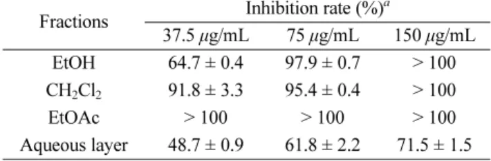
After removing the oil (vegetable oil) from the seeds of P. edulis by n-hexane, the residue was extracted with ethanol to obtain ethanol extract. In the search for sEH inhibitors from natural sources, we found that the ethanol extract of the seeds of P. edulis inhibited 64.7% of sEH activity at a concentration of 37.5 µg/mL. This extract was then partitioned with dichloromethane (CH
2Cl
2)
,ethyl acetate (EtOAc) fractions and aqueous residue. In the preliminary experiment, we tested the inhibitory activity of these fractions at 37.5, 75.0 and 150.0 µg/mL.
Based on the results in Table 1, CH
2Cl
2- soluble fraction showed 91.8% inhibition at the concentration of 37.5 μg/
mL, which was approximately 2.0-fold more potent than aqueous layer (48.7%). Interestingly, the EtOAc-soluble fraction exhibited very potently with > 100% sEH activity at the same concentration. Considering that EtOAc-soluble fraction showed the strongest action, our subsequent studies focused on the isolation of active components.
This sub-fraction was subjected to column chromato- graphy on a silica gel and C18-RP silica gel column to obtain five compounds (1 – 5) (Fig. 1).
Compound 1 was obtained as an ivory amorphous powder. The
1H-NMR of 1 showed signals the presence of 1,3,5-trisubstituted benzene ring characterized with AB
2system [ δ
H6.47 (2H, d, J = 2.0 Hz, H-2/H-6) and 6.18 (1H, t, J = 2.0 Hz, H-4)], and a 1,4-disubstituted benzene ring characterized with an A
2B
2system [ δ
H7.36 (2H, d, J = 8.5 Hz, H-2'/H-6') and 6.78 (2H, d, J = 8.5 Hz, H-3'/H-5') together with trans-olefinic protons [ δ
H6.97 (1H, d, J = 16.0 Hz, H-8) and 6.81 (1H, d, J = 16.0 Hz, H-7)] (Fig. 1). The
13C-NMR spectrum of 1 exhibited 14 carbon signals including 12 aromatic carbons [δ
C102.6 – 159.6], belonging to two benzene rings, and two olefinic carbons [δ
C129.3 (C-7), and 128.7 (C-8)] (Fig. 1). The ESI-MS data of 1 indicated the pseudo molecular ion at m/z 229.09 for the [M+H]
+, indicating a molecular weight of 228. Its molecular formula was determined to be C
14H
12O
3, according to ESI-MS,
1H- and
13C-NMR spec- troscopic data. Based on the above evidence and com- parison with reported data,
26compound 1 was identified α
[ ]
D25α
[ ]
D25as trans-resveratrol. Compound 2, a derivative of 1, which the
1H- and
13C-NMR spectra were similar to those of 1 except for the presence of 1,3,4-trisubstituted benzene ring characterized with ABX system [ δ
H7.07 (1H, d, J = 2.0 Hz, H-2'), 6.89 (1H, dd, J = 8.5, 2.0 Hz, H-6'), and 6.82 (1H, d, J = 8.5 Hz, H-5')] in 2 (Fig. 1). The ESI-MS spectrum of compound 2 showed the pseudo molecular ion at m/z 245.08 [M+H]
+, indicating the molecular formula C
14H
12O
4. Thus, compound 2 was identified as trans-piceatannol in comparison with literature data.
27These compounds (1 – 2) possess the basics of stilbene skeleton and are known to inhibit anti-oxidant, anti- inflammatory, anti-diabetes and anticancer.
28Compound 3 was obtained as yellow amorphous solid.
The
1H-NMR of 3 showed signals of 1,2,4-trisubstituted benzene rings characterized with two ABX systems [δ
H7.63 (1H, d, J = 8.5 Hz, H-5), 7.54 (1H, d, J = 2.0 Hz, H- 8), and 7.26 (1H, dd, J = 8.5, 2.0 Hz, H-6); δ
H6.86 (1H, d, J = 8.5, Hz, H-5'), 6.73 (1H, d, J = 2.0 Hz, H-2'), and 6.72 (1H, dd, J = 8.5, 2.0 Hz, H-6')], together with an olefinic proton at δ
H6.71 (1H, s, H-2) (Fig. 1). The
13C- NMR and distortionless enhancement by polarization transfer (DEPT) spectra of 3 showed signals for six aromatic quaternary carbon [δ
C169.8 (C-7), 168.4 (C-9), 149.3 (C-4'), 146.7 (C-3'), 125.5 (C-1'), 114.8 (C-10)] and six aromatic carbons [δ
C126.8 (C-5), 126.3 (C-6'), 118.9 (C-2'), 116.7 (C-5'), 114.6 (C-6) and 99.3 (C-8)] (Fig. 1).
Furthermore, a carbonyl carbon at δ
C184.5 (C-4), together with two olefinic carbons [δ
C147.7 (C-3), and 114.1 (C- 2)] were also observed in the
13C NMR spectrum indicated that 3 was aurone skeleton (Fig. 1).
29The ESI-MS spectrum of compound 3 showed the pseudo molecular ion at m/z 271.06 [M+H]
+, indicating the molecular formula C
15H
10O
5. Based on the above evidence and comparison with reported data,
30compound 3 was identified as sulfuretin, which was isolated from P. edulis for the first time. This compound possessed anti-inflammatory,
31anticancer,
32and neuroprotective activities.
33Compound 4 was isolated as yellow amorphous powder. The
1H-NMR spectrum of 4 showed an aldehyde proton at δ
H9.60 (1H, d, J = 8.0 Hz, H-9'), trans-olefinic
protons at δ
H6.70 (1H, dd, J = 15.5, 8.0 Hz, H-8') and 7.61 (1H, d, J = 15.5 Hz, H-7'), which were assigned to a trans-cinnamaldehyde moiety. In addition, the
1H-NMR of 4 showed two sets of aromatic protons signal [δ
H6.97 (1H, s, H-2), 6.85 (1H, d, J = 8.0 Hz, H-5), 6.80 (1H, d, J = 8.0, H-6); δ
H7.31 (1H, s, H-6'), 7.25 (1H, s, H-2') arising from 1,3,4-trisubstituted and 1,3,4,5-tetrasubstituted benzene rings, respectively (Fig. 1). A stereochemistry of the dihydrobenzofuran ring [δ
H5.63 (1H, d, J = 6.5 Hz, H-7), δ
H3.59 (1H, q, J = 6.5 Hz, H-8) and δ
H3.91-3.87 (2H, m, H-9)], and two singlets of methoxy groups [δ
H3.92 (3H, s, 3'-OCH
3), and 3.84 (3H, s, 3-OCH
3)] were also observed in the
1H-NMR spectrum. The large proton coupling constant between H-7 and H-8 (J
7,8= 6.5 Hz) suggested the dihydrofuran ring has a trans-configuration.
The
13C-NMR and DEPT spectra of 4 revealed the presence of twelve aromatic carbons, two olefinic carbons, an aldehyde carbon [δ
C196.1 (C-9')], a hydroxymethyl carbon [δ
C64.5 (C-9)] and two methoxy carbons [δ
C56.8 (3-OCH
3), and 56.4 (3'-OCH
3)] (Fig. 1). The HMBC correlations of proton H-2 at δ
H6.97 (1H, s) and methoxy protons at δ
H3.84 (3H, s) to carbon signal at δ
C149.2 (C- 3), as well as proton H-2' at δ
H7.25 (1H, s) and methoxy protons at δ
H3.92 (3H, s) to carbon signal at δ
C156.1 (C- 3'), suggested that two methoxy groups were located at C- 3 and C-3' (Fig. 1). Further analysis of these signals by the COSY, HMQC and HMBC spectra led to the partial structures of 4. The ESI-MS data of 4 indicated the pseudo molecular ion at m/z 357.14 for the [M+H]
+, indicating a molecular weight of 356. Its molecular formula was determined to be C
20H
20O
6, according to ESI-MS,
1
H- and
13C-NMR spectroscopic data. Therefore, com- pound 4 was identified as (+)-balanophonin,
34which was isolated from Passiflora genus for the first time. This compound was known to possess anti-oxidant,
35,36anti- cholinesterase,
36anti-inflammatory, anticancer, and anti- neurodegenerative activities.
37Compound 5 was isolated as brown amorphous powder.
The
1H-NMR spectrum of 5 showed the presence of two
ABX system signals for the A ring [δ
H6.68 (1H, d,
J = 2.0 Hz, H-2'), 6.67 (1H, d, J = 8.5 Hz, H-5'), 6.50 (1H,
Fig. 1. The structures of isolated compounds (1 - 5) from the seeds of P. edulis.
dd, J = 8.5, 2.0 Hz, H-6')] and the D ring [δ
H7.15 (1H, d, J = 2.0 Hz, H-2), 7.09 (1H, dd, J = 8.5, 2.0 Hz, H-6), 6.96 (1H, d, J = 8.5 Hz, H-5)], two sets of AB
2system signals for the B and C rings [6.48 (2H, overlap, H-12/H-12'), 6.19 (1H, t, J = 2.0 Hz, H-14'), 6.17 (1H, t, J = 2.0 Hz, H- 10'), 6.12 (2H, d, J = 2.0 Hz, H-10/H-14)], two doublets for the trans-olefinic protons [δ
H6.99 (1H, d, J = 16.0 Hz, H-7), 6.88 (1H, d, J = 16.0 Hz, H-8)], and two equivalent oxybenzyl methine protons [δ
H4.75 (2H, d, J = 2.5 Hz, H-7'/H-8')] (Fig. 1). The
13C-NMR and DEPT spectra of 5 exhibited 28 carbon signals including 24 aromatic carbons [δ
C102.9 – 159.6], belonging to four benzene rings, and two olefinic carbons [δ
C129.4 (C-7), and 128.4 (C-8)]. In addition, the carbon signals [δ
C82.2 (C-8') and 81.8 (C- 7')] indicate the presence of two equivalent oxybenzyl methine carbons. Analysis of these signals by the COSY, HMQC and HMBC spectra led to the partial structures of compound 5 (Fig. 1). The ESI-MS data of 5 indicated the pseudo molecular ion at m/z 487.13 for the [M+H]
+, indicating a molecular weight of 486. Its molecular formula was determined to be C
28H
22O
8, according to ESI-MS,
1H- and
13C-NMR spectroscopic data. The relative configuration between the C-7' and the C-8' positions was concluded to be cis from the coupling constant of the two oxybenzylmethine protons (J = 2.5 Hz). Based on the above evidence and comparison with reported data,
38compound 5 was identified as cassigarol E, which was isolated from Passiflora genus for the first time. Cassigarol E possessed anti-HIV-1,
39antidiabetic,
40and anticancer activities.
41The effect of isolated compounds (1 – 5) from P. edulis on sEH inhibitory activities was evaluated. The sEH inhibitory activities were determined using recombinant human sEH incubated with PHOME, an artificial substrate for fluorescence detection with AUDA (IC
50value 4.4 nM), a sEH inhibitor as the positive control. The result showed that compound 4 had no inhibitory (N.I) effect on the activity of enzyme sEH at a concentration of 100 μM, while compounds 1 and 2 at a concentration of 100 μM exhibited the highest sEH inhibitory activity (>100%) (Table 1). Sulfuretin (3), and cassigarol E (5) also showed a strong inhibitory effect on sEH with their inhibition values in the range of 74.6 and 77.7%, respectively (Table 1). Compounds 1 - 3, and 5 showed inhibitory rates over 50% and were further evaluated at concentrations ranging from 6.2 to 100 μM, to elucidate the IC
50values. These inhibitors (1 - 3, and 5) showed dose-dependent inhibition, with IC
50values of 14.2 ± 0.6, 3.4 ± 4.8, 15.8 ± 1.0, and 14.4 ± 0.8 μM, respectively (Table 2).
Previously, natural plant components with stilbene
skeletons from other plants have also shown inhibitory effects on sEH activity. Rhapontigenin, isorhapontin and astringin from Rheum undulatum (Polygonaceae),
8and 2- isopropyl-5-[(E)-2- phenylvinyl]benzene-1,3-diol have displayed potent sEH inhibitory activity.
42Several stilbene from Polygonum multiflorum (Polygonaceae), such as (E)-2,3,5,4'-tetrahydroxystilbene-2-O- β-D-glucoside, (E)- 2,3,5,4'-tetrahydroxystilbene-2-O- β-D-xyloside, and (E)- 2,3,5,4'-tetrahydroxystilbene-2-O- β-D-(6''-O-acetyl)-glu- coside completely inhibited sEH in a dose-dependent manner, with low IC
50values.
43As in this study, some isolated stilbenes from P. edulis can manifest sEH inhi- bitory activity. Such compounds have been purified from natural medicinal plants for many years all over the world. In addition to other medicinal properties, such as anti-inflammatory, anticancer, and antioxidant activities, the sEH inhibitory activity is worthy of notice, because low-molecular-weight materials can easily reach the site of action following oral administration since they cross the blood-brain barrier.
44Acknowledgments
We would like to thank Vietnam Academy of Science and Technology (VAST), Vietnam for financial support (under grand No. VAST.CTG.03/17-18) and Nafoods Group JSC for providing materials.
Table 1. Effects of extracts of P. edulis on sEH inhibitory activity Fractions Inhibition rate (%)
a37.5 µg/mL 75 µg/mL 150 µg/mL EtOH 64.7 ± 0.4 97.9 ± 0.7 > 100 CH
2Cl
291.8 ± 3.3 95.4 ± 0.4 > 100
EtOAc > 100 > 100 > 100
Aqueous layer 48.7 ± 0.9 61.8 ± 2.2 71.5 ± 1.5
a
Extracts and fractions were tested three times.
Table 2. The sEH inhibitory activities of isolated compounds from P. edulis
Compound 100 µM (%) IC
50value (µM)
a1 >100 14.2 ± 0.6
2 >100 3.4 ± 4.8
3 74.6 ± 0.4 15.8 ± 1.0
4 N.I N.T
5 77.7 ± 1.3 14.4 ± 0.8
AUDA
b68.9 ± 0.3 4.4 ± 0.1 (nM)
N.T: Not tested.
N.I: Not inhibition.
a
Compounds were tested three times.
b