리포좀을 이용한 분유 내 크로노박터 신속검출기술
Rapid Detection of Cronobacter Species in Infant Formula Powder using Liposomes
* Corresponding author: Myunghee Kim
Department of Food Science and Technology, Yeungnam University
280 Daehak-ro, Gyeongsan-si, Gyeongsangbuk-do 712-749, Republic of Korea Tel: +82-53-810-2950
Fax: +82-53-810-4662 E-mail: [email protected]
이기백
1, 슈리티 슈클라, 송신지에, 박선현
2, 오영숙, 김명희*
Gibaek Lee
1, Shruti Shukla, Xinjie Song, Sunhyun Park
2, Youngsook Oh, Myunghee Kim*
1서울우유 생산1팀, 2한국식품연구원 식품표준연구센터, 영남대학교 식품공학과
1 Production 1 Team, Seoul Dairy Cooperative
2Food Standard Research Center, Korea Food Research Institute Department of Food Science and Technology, Yeungnam University
I. 서론
식품은 인간의 기본 욕구를 해결하기 위한 중 요한 자원이면서 동시에 인간의 삶을 영위하는 데 필요한 기본 자원이다. 국민 소득이 증가하고 삶의 질이 향상되면서 건강 및 보건에 대한 국민 의 관심이 점점 증가하고 있다. 최근에는 분석기 술이 발달하여서 극미량의 식품 유해물질 검출 도 가능하게 되었고 동시에 새로운 식중독 원인 물질이 출현하면서 식품안전에 대한 정부, 산업 체, 국민의 관심이 더욱 커지고 있다.
Cronobacter 속 세균들은 자연에 널리 분포하고
있는 세균의 일종으로서 다양한 식품에 존재하 며 특히, 유아용 조제분유를 통해 영유아에게 감 염되어 괴사성 장염, 뇌수막염, 패혈증 등 심각한 증상을 야기 할 수 있다는 사실이 알려지면서 국
내외적으로 큰 관심을 받고 있다( Ivernsen et al., 2003; Muytjens et al., 1983: Jung and Lee, 2010;
Kim et al., 2008). Cronobacter sakazakii (C. sakaza-
kii)는 Enterobacteriaceae에 속하는 그람음성 간균이며, 포자를 형성하지 않는 통성혐기성 균으로 1980년 Enterobacter sakazakii 라고 명명되었으나, 2008년에 Cronobacter species로 재분류되면서 C.
sakazakii로 학명이 변경되었다(Friedemann, 2007;
Iversen et al., 2008). C. sakazakii는 1958년 수막염 에 걸린 신생아 환자에서 최초로 보고되었으며, 모든 연령대에서 질환을 일으킬수 있으나, 특히 영유아에게 수막염, 패혈증, 신생아 괴사성 장염 과 같은 증상을 일으키는 매우 위험한 기회감염균 이다(FAO/WHO, 2008; Muytjens and Kollee, 1990).
신생아는 분유 이외의 식품에 대한 선택의 폭이
극히 제한되어 있으며, 실제로도 Cronobacter 속
세균이 분유에서 검출된 사례가 국외에서 많이 보고되어(Choi et al., 2008), 한국을 비롯한 각국 에서는 유아용 식품에 대한 엄격한 기준을 정하 고 있다(Commission Regulation, 2007). 신생아 및 유아의
C. sakazakii 감염은 치사율이 40-80%에도달할 만큼 매우 치명적이며, 지난 10여 년 동안
C. sakazakii에 의한 감염사례가 세계적으로 증가하는 추세이다(Derzelle et al., 2007).
C. sakazakii의 검출은 세균 배양, 생화학적 검
사, 혈청학적 분석, 유전자 분석을 통하여 수행되 는데 이러한 방법들은 시간이 많이 소요되고 정 확하지 않다는 문제점이 있다(FDA, 2002). Poly- merase chain reaction (PCR), real-time PCR 방법은 세균 배양법에 비하여 민감도와 특이도가 좋은 반면 DNA를 증폭 확인하기 위한 분자생물학적 실험과 전문적인 기술을 필요로 하기 때문에 보 편적으로 사용하기 어려운 단점이 있다( Orlandi and Lampel, 2000). 이를 보완하기 위해서는 기존 분석 방법의 단점들을 보완할 수 있는 식중독균 검출기술의 개발이 시급하다.
항원과 항체의 특이 반응을 이용한 진단방법은 과거 수십년 동안 세균의 동정 및 subtyping에 이 용되어 왔다( Kim et al., 2003). 형광물질과 결합 한 항체를 이용하여 세균을 분석하는데 이용할 수 있다는 보고 이후, 면역형광기술은 특히 식품 에서의 식중독균 검출에 이용되어 왔다(Coons et
al., 1942; Haglund et al., 1964; Swamunathan et al.,1978; Thomason 1981). 면역형광기술은 PCR과 같 은 유전자 증폭기술과는 달리 고가의 장비가 필 요하지 않으며 숙련된 인력이 요구되지 않고, 다 량의 시료를 처리할 수 있는 민감성 등 다양한 장 점을 가지고 있다(Leem, 2012).
리포좀은 수용액상에서 자발적으로 형성되는 인지질 이중층의 소포로서(Bangham et al., 1965) 내부 공간에 형광물질, 효소, 약물을 포집할 수 있으므로 Escherichia coli(E. coli) O157:H7, Salmo-
nella spp. 와 같은 식중독 세균의 진단시약으로서개발되고 있다(Park and Kim, 2008; Shukla et al., 2011). 최근에는 시료에서 세균의 분리 효율을 높
이기 위한 방법으로서 면역자성분리법 등이 분 석에 활용되고 있다(Shukla et al., 2016).
따라서 본 연구실에서 수행하고 있는 고감도, 신속성, 재현성을 갖춘 Cronobacter 검출 기술의 예 로써 나노자성입자와 항체, 리포좀을 이용하여 개 발한 신속, 간편 검출법을
Cronobacter muytjensii(
C. muytjensii)와 C. sakazakii 중심으로 정리하고자한다. 한편, 서론에서 언급한 내용과 이하 방법 및 결과, 결론은 Park 등(2012), Song 등(2015), Shukla 등 ( 2016)의 연구논문에서 발췌한 것임을 밝혀둔다.
II. 재료 및 방법
1) 면역원의 준비
C. muytjensii와 C. sakazakii를 배양 후 cell을 얻
고 여기에 formalin을 처리한 후(formalin-killed cell: FKCs), 토끼의 면역에 이용하였다(Park et
al., 2012).2) 면역
New Zealand산 흰색 암컷토끼를 이용하여 Cana- dian Council on Animal Care의 표준과 규격에 따라 면역을 실시하였다( Canadian Council on Animal Care, 2002). FKCs에 Freund’s incomplete adjuvant를 1:1의 비율로 혼합한 후 토끼에 접종하였다(Park et
al., 2012).3) 면역글로블린 G(IgG)의 정제
면역기간 동안 채취한 혈액 시료는 면역혈청을
분리하기 위하여 4℃에서 30분 동안 원심분리
( 10,000 × g)하였다(Song and Kim, 2013). 원심분
리 후 얻어진 상등액에 caprylic acid와 ammonium
sulfate 침전을 이용하여 토끼 혈액에서 분리한 혈
청에서 IgG 항체를 정제하였다(McKinney and Par-
kinson, 1987). 정제된 항체의 역가를 측정하기 위
하여 indirect noncompetitive enzyme-linked immu-
nosorbent assay(INC-ELISA: 간접비경합효소면역 분석법)를 실시하였다(Song and Kim, 2013).
4) 리포좀의 제조
리포좀은 Shukla 등(2011)의 방법에 따라 1, 2 -dipalmitoyl-sn-glycero-3-phosphoethanolamine (DPPE, 7.2 μmol), 1, 2-dipalmitoyl-sn-glycero-3-phosphocho- line (DPPC, 14.3 μmol), 1, 2-dipalmitoyl-sn–glycero- 3-[phospho-rac-(1-glycerol)] (DPPG, 40.3 μmol), cho- lesterol (40.9 μmol), 그리고 100 mM sulforhodamine B(SRB)를 이용하여 제조하였다.
5) 면역리포좀의 제조
SH-tagged 리포좀과 maleimide-derivatized IgG를 섞고 하루 동안 반응시킨 용액에 100 mM의 eth- ylmaleimide를 포함하는 0.02 M TBS buffer (0.15 M NaCl, 0.01% NaN
3포함, pH 7.0)를 DPPE-ATA 1 mol 당 ethylmaleimide 10 mol의 비율로 가한 후 실온의 암실에서 70 rpm으로 30분 반응시켰다.
Sepharose CL-4B column에 통과시켜 면역리포좀 분획을 얻은 후 4℃ 암실에서 하루 동안 투석하 였다.
6) 리포좀의 특성
리포좀의 크기는 Malvern Nano-Zs particle size analyzer (Malvern, Worcestershire, UK)을 이용하여 측정하였다.
7)
C. muytjensii분석
C. muytjensii 배양액을 96-well microtiter plate에
분주 하여 37℃에서 2시간 코팅시켰다. 코팅이 끝난 후 0.01 M PBS buffer를 이용하여 세척 후, 5% skim milk를 포함하는 0.01 M PBS buffer를 첨 가하여 37℃에서 1시간 동안 blocking 시키고 다 시 0.01 M PBS buffer로 세척하였다. 그 후 anti-C.
muytjensii IgG-tagged 리포좀을 96-well microtiter
plate에 분주 후 37℃에서 1시간 동안 반응시켰다.
다시 한번 5% skim milk를 포함하는 0.01 M PBS buffer를 이용하여 각각의 well을 2회씩 세척하여 결 합되지 않은 면역리포좀을 제거하였다. 다음, 계면 활성제의 일종인 30 mM octyl-β-D-glucopyranoside ( OG) 280 μL를 넣고 리포좀을 파괴시켰다. 방출된 SRB는 흡수파장 550 nm, 여기파장 585 nm로 형광도 를 측정하였다,
8) 나노자성입자를 이용한
C. sakazakii분석
Borosilicate tube에 C. sakazakii 배양 희석액을 농도별로 1 μL씩 가한 후 각각의 tube에 20 μL의 면역나노자성입자를 주입하여 1시간 동안 반응 시켰다. 반응 후 0.01 M PBS buffer로 세척하고, 4% skim milk를 포함하는 0.02 M TBS buffer로 다 시 세척하여 결합되지 않은
C. sakazakii를 제거하였다. 형성된 면역나노자성입자-C. sakazakii 복 합체에 20배 희석한 면역리포좀 용액을 70 μL 씩 tube에 주입 후 1시간 동안 반응시켜 면역리포 좀-면역나노자성입자-C. sakazakii 복합체를 형 성시켰다. 이 복합체를 0.02 M TBS buffer로 세척 한 다음 계면활성제의 일종인 30 mM OG 280 μL 를 넣고 리포좀에 포집된 SRB들을 방출시켰다.
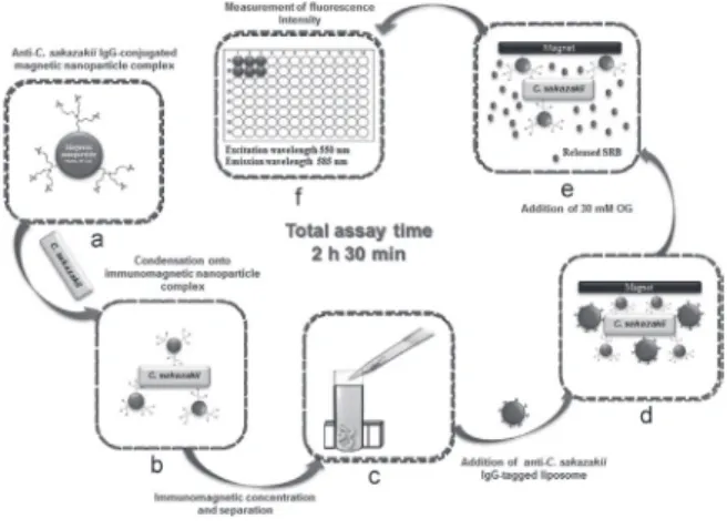
Fig. 1. Concept of immunoliposome based immunomagnetic
concentration and separation assay.
방출된 SRB를 흡수파장 550 nm, 여기파장 585 nm에서 형광도를 측정하였다(Fig. 1).
9) INC-ELISA와의 비교
나노자성입자를 이용한 C. sakazakii 분석 결과를 INC-ELISA assay와 비교하였다(Song and Kim, 2013).
10) 교차반응 시험
교차반응 시험은 Cronobacter spp. 6종(C. saka-
zakii, C. dublinensis, C. malonaticus, C. universalis, C. condiment, C. muytjensii)과 주요 병원균인 Sal- monella enterica subsp. enterica serovar Enteritidis, E. coli O157:H7, Bacillus cereus, Enterobacter aero- genes, 그리고 C. freundii와의 교차반응성을 측정하였다.
11) 분유적용 시험
유아용 조제분유를 식품시료 적용시험을 위한 sample matrix로 이용하였다(Cho and Irudayaraj, 2013).
III. 결과
1. 항체의 역가
C. muytjensii, C. sakazakii에 대한 항원을 면역한
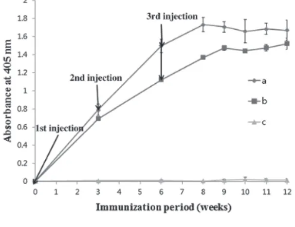
후 12주까지 항혈청의 역가를 INC-ELISA로 측정 한 결과, 면역기간에 따른 항혈청 역가는 두 균종 모두 면역 8-9주까지 증가하는 것으로 나타났다 ( Fig. 2).
Fig. 2에 나타난 C. muytjensii 항원에 대한 항혈 청 역가 추이를 보면 anti-C. muytjensii IgG는 1차 면역 후 증가하는 양상을 보였다. 2차, 3차 면역 은 1차 접종 후 3주 간격으로 수행되었다. 일반적 으로 2차, 3차 면역은 항체 역가의 정체 또는 감 소를 방지한다고 보고되므로(Leenaars and Hen- driksen, 2005) 1차 면역 이후 3주 간격의 추가 면 역으로 고역가 항체 생산을 유도하였다.
2. SRB 포집 리포좀의 특성
실험에 사용한 리포좀은 Table 1에서 보는 바와 같이 206 nm의 직경을, 리포좀을 이루는 이중 지 질막 안쪽의 부피는 4.06 × 10
-12μL, 리포좀 입자 1개당 4.06 × 10
-13μmol의 SRB를 포집하는 것으 로 나타났다.
또한 생산한 리포좀 용액 1 mL 당 5.74 × 10
11개 의 리포좀 입자가 분포하는 것으로 계산되었다.
유사 연구로써 Ho 등(2008)이 개발한 리포좀은 3.65 × 10
12개의 입자와 236 nm의 크기를 가지며 methyl blue를 포집하었고 Salmonella 신속 검출을 위한 시약으로 사용되었다. Shukla 등(2014)은 223 nm 크기의 SRB 포집 리포좀을 Salmonella 검출을
Fig. 2. Titer of developed rabbit anti-C. muytjensii IgG during immunization period. Each arrow indicates immunogen injection time. a and b: antibody titer from two indepen- dent immunization experiments; c: antibody titer from non-immunized rabbit as a control.
Table 1. Characterization of SRB-encapsulated liposomes Mean diameter (nm)
Inner volume (entrapped) of liposomes (μL) SRB concentration (mM)
Amount of SRB/liposome (μmol) Concentration of liposomes (particles/mL)
206
4.06×10
-12100
4.06×10
-135.74×10
11위한 면역크로마토그래피 분석에 응용하였다.
3. 리포좀 integrity 측정
리포좀을 이용한 식중독 세균의 검출의 원리는 리포좀에 포집되어 있는 형광물질의 세기를 측 정하여 목표세균을 정량적으로 분석하는 것이 다. 따라서 리포좀에 형광물질이 온전하게 포집 되었는지 확인하는 것이 필요한데, 이를 위하여 리포좀 희석액에 계면 활성제의 일종인 OG를 넣 기 전과 후의 형광도를 측정하였다( Fig. 3). 측정 결과, Fig. 3에서 보는 바와 같이 1:10
3으로 희석 한 리포좀 용액에서 OG를 넣기 전과 후에 10배 정도의 형광도 차이가 있는 것으로 나타났다. 리 포좀 내부에 높은 농도로 포집되어 있는 SRB가 quenching effect로 인해 낮은 형광도를 보이다가 OG 첨가 후 리포좀이 깨지면서 포집되었던 SRB 가 수용액 상에 분산되어 높은 형광도를 보이는 것을 확인할 수 있었다.
4.
C. muytjensii분석
Anti-C. muytjensii IgG-tagged 리포좀을 이용하 여 순수배양에서 C. muytjensii를 검출한 결과, 검 출감도가 6.3 × 10
4CFU/mL로 나타났다(Fig. 4).
C. muytjensii 검출에 필요한 시간은 13시간으로
sandwich ELISA와 비교하여 더 빠르고 간단하게 검출되는 것을 알 수 있었다. 하룻밤 동안의 코 팅, 6시간의 분석시간으로 0.4-14.4 × 10
5CFU/
mL의 검출한계를 나타낸 Hochel과 Skvor(2009) 의 간접경합효소면역분석법과 비교해 볼 때, 여 기에서 개발한 분석법은 민감도와 시간에서 더 향상된 결과를 보였다. 이 결과는
Cronobacterspp.를 검출하기 위해 리포좀을 이용한 최초의 연 구로써 C. muytjensii를 신속, 간편, 민감하게 검출 하는데 있어서 리포좀의 이용 가능성을 보여주 고 있다.
Fig. 3. Size distribution and integrity measurement of SRB- tagged liposomes. Values are the mean of three mea- surements. Error bars represent±S.D. The coefficient of variation(% CV) for fluorescence intensity(n=6) is below 15%.
Fig. 4. Standard curve for detection of C. muytjensii. All experi- ments were conducted three times. Data represent mean
± SD.
Fig. 5. Specificity of developed fluorescence-based liposome
immunoassay. All experiments were conducted three
times. Data represent mean ± SD.
개발된 anti-C. muytjensii IgG는 C. muytjensii ATCC 51329와 C. muytjensii CDC 3523-75에 높은 반응성을 나타내었다( Fig. 5). 즉, C. muytjensii 이 외의 균들에 대한 교차반응 없이 C. muytjensii 만 을 검출하는 높은 효율성을 나타내었다.
5. 나노자성입자와 항체의 conjugation 확인
Fig. 6(a)는 anti-C. sakazakii IgG가 나노자성입자 에 conjugation 되는 과정을 나타낸 모식도이다.
자성입자와 항체 간의 결합유무를 파악하기 위 하여 Bradford assay를 이용하여 각각의 과정마다 항체 농도를 측정 하였다. 이 실험에서 반응에 사 용한 항체 농도는 8.35±0.01 mg/mL 이었다. 1시 간 반응 후 자성입자와 항체가 결합하여 만들어 진 면역자성 입자를 확인한 결과 반응에 참여하 지 않은 항체는 1.15 mg/mL 임을 알 수 있었다.
초기 항체농도에서 결합하지 않고 남은 항체의 농도를 빼준 결과, 나노자성입자와 결합한 항체 의 농도는 7.20±0.01 mg/mL 이었으며, 결합률은 86.23±0.59% 임을 알 수 있었다(Table 2).
6.
C. sakazakii분석
개발된 검출법을 이용하여 C. sakazakii 배양액 을 0, 10
1- 10
8CFU/mL의 농도로 조절하여 실험을 수행하였다. 농도별로 희석된 C. sakazakii 1 mL 에 20 μL의 면역자성나노입자를 분주한 후 1시간 동안 반응시켜 면역 자성 나노입자에 C. sakazakii 를 선택적으로 결합하게 하였다. 여기에 면역리 포좀을 가하여 C. sakazakii 양과 상관관계를 보이 는 형광도를 측정하였다. 그 결과, 10
3CFU/mL 부터 시그널을 확인할 수 있었고, 검출한계는 3.3
× 10
3CFU/mL(Fig. 7)인 것을 확인할 수 있었다.
나노자성입자를 이용하여 개발된 분석법과 기 존에 사용되고 있는 검출법의 성능을 비교하기 위해 신속검출법으로 알려진 INC-ELISA와의 검 출감도를 비교한 결과, 순수한 배지에서의 검출 시 INC-ELISA는 6.2 × 10
5CFU/mL에서부터 signal 이 나타났고( Fig. 7), 새롭게 개발된 면역 리포좀 기반의 면역자성분리법은 3.3 × 10
3CFU/mL의 검출한계를 보여 100배 정도 민감한 것을 확인하 였다. 이때 검출한계는 음성대조군의 형광도 평 균값으로 부터 3배의 표준편차에 해당하는 세균 수로 결정하였다(Hochel and Skvor, 2009). Kelley 와 DeSilva(2007)는 분석의 재현성과 정확성을 입 증하기 위한 %CV는 15-20%보다 낮아야 한다고 보고하였는데, 이 연구의 결과 % CV 15% 미만으 로 만족스런 결과를 보였다. 개발된 측정법의 검 출한계는 3.3 × 10
3CFU/mL 이고, C. sakazakii를 2 시간 30분 만에 검출할 수 있었으며 적정 검출범 위는 10
3– 10
7CFU/mL로 나타났다. 반면 INC-
Fig. 6. Detailed schematics of the procedure for (a)conjugation of magnetic nanoparticles with anti-C. sakazakii IgG; (b) reaction of immunomagnetic nanoparticles with C. saka- zakii cells; (c)formation of immunoliposome-C. sakaza- kii- immunomagnetic nanoparticle complex.
Table 2. Binding efficiency of rabbit anti-C. sakazakii IgG onto magnetic nanoparticle
Initial antibody(mg/mL) Free antibody(mg/mL) Bound antibody(mg/mL) Bounding efficiency(%)
8.35±0.01
1.15±0.01
7.20±0.01
86.23±0.01
Bounding efficiency(%)=Bound(initial-free) antibody/Ini-
tial antibody×100
ELISA의 검출한계는 6.2 × 10
5CFU/mL, 분석시 간을 제외하고도 증균배양 시간으로 17시간을 요구한다. 이러한 결과는 기존의 검출법과 비교 하여 면역자성나노입자의 적용으로 검출시간과 검출감도를 향상시킬 수 있다는 것을 시사한다 ( Shukla et al., 2016).
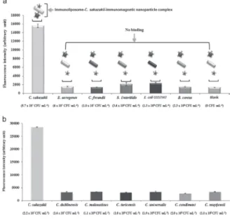
개발된 검출법의 교차반응성을 평가하기 위하 여
C. sakazakii와 다른 병원성 균들 간의 교차반응을 진행하였다. 결과에서도 C. sakazakii 제외한 6종 모두 10
7– 10
8CFU/mL의 높은 농도에서도 blank의 시그널과 거의 유사한 시그널을 나타냈 고, C. sakazakii에서만 높은 시그널이 나타났다 ( Fig. 7). 즉, C. sakazakii에만 특이적으로 반응하 는 특이성을 확인할 수 있었다.
7. 분유 적용시험
나노자성입자를 이용한 검출법을 식품 시료에 적용하기 위하여 10% 조제분유를 준비하여 C.
sakazakii 검사를 실시한 결과, 분유 시료 안에서 C. sakazakii 검출한계는 3.5 × 10³
CFU/mL 인 것 을 확인할 수 있었다. 기존의 다른 검출법의 검출 한계가 1 × 10
5CFU/mL 인 점을 감안한다면 새롭 게 개발된 면역리포좀 기반의 면역자성 분리법
은 분유 시료 내의 경쟁물질에 영향을 받지 않고
C. sakazakii를 특이적으로 검출할 수 있다는 것을증명할 수 있었다(Fig. 8).
IV. 결론
1. 리포좀을 이용한
C. muytjensii분석
이 연구는 리포좀, anti-C. muytjensii IgG을 이용 하여 C. muytjensii를 검출하는데 있어서 간편성, 신속성, 고감도 검출 방법에 초점을 두었다. 개발 된 방법은 순수배양액에서 6.3 × 10
4CFU/mL의 검출한계를 가지며 C. muytjensii에 대하여 높은 특이성을 나타내었다. 다른 배지배양법( 2-5일 필요)과 비교하였을 때 상대적으로 짧은 시간(13 시간) 안에 C. muytjensii를 검출할 수 있다. 즉, VRBG agar, Enterobacter sakazakii isolation agar와 Druggan–Forsythe–Iversen agar를 이용한 배지배양
Fig. 7. Detection sensitivities of developed immunoliposome-based immunomagnetic concentration and separation assay and INC-ELISA in the detection of C. sakazakii. All experiments were conducted three times, and the data represent the mean±S.D. The coefficient of variation(%CV) for fluores- cence intensity(n=6) is below 15%.
Fig. 8. (a) Cross-reactivity of the developed immunoliposome-
based immunomagnetic concentration and separation assay
with other genera of foodborne pathogens. (b) Cross-reac-
tivity with Cronobacter strains. All experiments were conduct-
ed three times, and the data represent the mean +S.D. The
coefficient of variation (%CV) for fluorescence intensity
( n=6) is below 15%.
방법은 24시간의 증균 배양 후 선택배양을 통한 균의 분리, 의심균의 생화학적 test, 혈청학적 test, 또는 real-time PCR 같은 분자생물학적 방법을 이 용한 확인이 필요하다. 그러나, 현재의 개발된 방 법은 간단하고 C. muytjensii를 항원-항체 반응의 원리를 이용하여 검출함으로써 간편성과 신속성 을 향상시킨 것이다.
2. 리포좀과 나노자성비드를 이용한
C. sakazakii분석
이 분석법은 면역자성비드로 C. sakazakii를 선 택적 분리 후 면역리포좀을 이용하여 시그널을 얻는 방법으로, 조제분유에서 C. sakazakii의 정확 한 검출을 위한 고감도, 재현성, 신속, 특이성을 보유한다는 것을 확인하였다. 이 방법은 순수배 지와 조제분유에서 약 10
3CFU/mL 의 검출한계 를 나타내었다. 개발된 방법은 다른 검출법과 비 교하였을 때 고가의 장비 없이 실험이 가능하며, 실험이 빠르고 간단하다는 장점이 있다. 따라서 이 기술을 산업화에 응용한다면 식품의 모니터 링 단계에서 오염이 의심되는 시료를 빠르게 제 거함으로써 신속한 위기 대처가 가능해질 것이 며, 모니터링 이후의 분석단계를 거치는 시료의 수를 현저하게 감소시킬 수 있으므로 분석에 소 요되는 경비와 노동력을 절감할 수 있을 것으로 기대된다.
사사
이 논문은 2014년도 교육부의 재원으로 한구연 구재단의 지원을 받아 수행된 기초연구사업임 ( NRF-2014R1A2A1A11053211).
Ⅴ. 참고문헌