대한외과학회지:제 65 권 제 4 호
□ 증 례 □ Vol. 65, No. 4, October, 2003
366 서 론
지방육종은 성인에서 발생하는 가장 흔한 연부조직 육종 으로 전체 육종의 9.8∼16%를 차지하며,(1) 호발부위로는 사지와 후복막강이 있고, 정삭(spermatic cord), 고환, 흉곽 및 유방, 종격동, 대망, 장간막 등에서 드물게 발생하는 것 으로 알려져 있다. 조직학적 분화도에 따라 점액성, 고분화 성, 원형세포성, 다형성, 역분화성으로 나누어지며,(2,3) 기
원에 따라 원발성과 전이성으로 분류된다. 예후는 지방육 종의 위치 및 조직학적 아형에 따라 영향을 받는데, 후복막 강이나 복부에 발생한 경우 그리고 역분화성으로 진단된 경우 국소재발 및 원격전이의 빈도가 높다고 알려져 있다.
본 증례는 장간막 거대 지방육종에 대한 것으로 국소 재발 및 원격 전이에 대한 주기적인 관찰이 의의가 있을 것으로 생각되며, 지방육종의 위치, 조직학적 분류 그리고 크기를 고려할 때 우리나라에서는 보고된 예가 없어 문헌고찰과 함께 보고하는 바이다.
증 례
57세 남자 환자가 수년 전부터 복부팽만감이 있었는데, 내원 20여일 전부터 그로 인한 불편감이 심화되어 입원하 였다. 과거력상 17년 전 복강 내 종괴로 인해 종괴적출술 및 우측 신장절제술을 받았으나, 정확한 정보는 알 수 없었 다. 복부신체검사상 전반적인 복부팽만 소견을 보였으며, 우상복부에 약간의 압통 이외에 만져지는 복부 종괴는 없 었다. 가족력상 특이소견은 없었다.
검사실소견상 말초혈액검사, 혈청화학검사, 혈청전해질 검사, 소변검사 및 종양표지자검사에서 모두 정상 소견을 보였다. 복부 전산화단층촬영상 장간막 혈관의 지배를 받 고 있어 장간막 기원으로 생각되는 지방성 종괴가 전복강 을 점유하고 있었다. 종괴의 내부는 조영증강이 잘 이루어 지는 섬유성 격막으로 구분되었고, 종괴 간의 밀도를 비교 할 때 비균일성이 관찰되었다(Fig. 1).
수술소견상 장간막에 위치하는 부드러우며 밝은 노란색 의 다발성의 종괴가 복강 전체를 덮고 있었으며, 국소적으 로 단단한 부분이 촉지되었다. 종괴 크기는 2×3 cm에서 20
×15 cm에 이르기까지 다양하였으며, 각각의 소엽은 피막 형성이 잘 이루어져 있었다. 주위 장기의 전위 이외의 특이 소견은 관찰되지 않았다. 종괴적출술을 시행하였고 상행결 장부위와는 유착이 심하여 우측 결장절제술을 함께 시행하 였다.
절제 후 육안소견상 종괴의 무게는 7 kg에 달하였고, 종 괴는 피막화 및 내부의 격막화가 잘 이루어진 상태였으며, 부분적으로 단단하게 만져지는 부분이 있었다(Fig. 2). 광학 현미경소견상 집적된 지방세포 사이에 비전형적 지방세포 (atypical lipocyte)가 관찰되고 소혈관들의 수지상 배열이 진
장간막에 발생한 거대 지방육종
조선대학교 의과대학 외과학교실
송근영․장정환․김권천․민영돈․조현진․김성환․김경종
Giant Liposarcoma Arising in the Mesentery
Keun-Yeong Song, M.D., Jeong-Hwan Chang, M.D., Kweon-Cheon Kim, M.D., Young-Don Min, M.D., Hyun-Jin Cho, M.D., Sung-Hwan Kim, M.D. and Kyung-Jong Kim, M.D.
A liposarcoma is the most common soft tissue sarcoma in adults with an incidence of betwen 9.8 and 16% of all soft tissue sarcomas. Among the various histological types, a dedifferentiated liposarcoma has rarely been reported. We experienced a case of a giant 7 kg dedifferentiated liposarcoma arising in the mesentery of a 57-year old male patient. Abdominal computed tomography showed a huge lipoid mass occupying the whole abdomen. A surgical excision was carried out. Histologically, the tumor was com- posed of a well-differentiated liposarcomatous area and a leiomyosarcoma-like dedifferentiated area. The authors re- viewed the prognostic factors and treatments for a lipo- sarcoma. (J Korean Surg Soc 2003;65:366-368)
Key Words: Dedifferentiated liposarcoma, Mesentery 중심 단어: 역분화성 지방육종, 장간막
ꠏꠏꠏꠏꠏꠏꠏꠏꠏꠏꠏꠏꠏꠏꠏꠏꠏꠏꠏꠏꠏꠏꠏꠏꠏꠏꠏꠏꠏꠏꠏꠏꠏꠏꠏꠏꠏꠏꠏꠏꠏꠏꠏꠏꠏꠏꠏꠏꠏꠏꠏꠏꠏ
Department of Surgery, Chosun University College of Me- dicine
책임저자:김경종, 광주광역시 동구 서석동 588번지 ꂕ 501-717, 조선대학교 의과대학 외과학교실 Tel: 062-220-3068, Fax: 062-228-3073 E-mail: kjkim@mail.chosun.ac.kr
접수일:2003년 4월 12일, 게재승인일:2003년 6월 13일
송근영 외:장간막에 발생한 거대 지방육종 367
ꠏꠏꠏꠏꠏꠏꠏꠏꠏꠏꠏꠏꠏꠏꠏꠏꠏꠏꠏꠏꠏꠏꠏꠏꠏꠏꠏꠏꠏꠏꠏꠏꠏꠏꠏꠏꠏꠏꠏꠏꠏꠏꠏꠏꠏꠏꠏꠏꠏꠏꠏꠏꠏꠏꠏꠏꠏꠏꠏꠏꠏꠏꠏꠏꠏꠏꠏꠏꠏꠏꠏꠏꠏꠏꠏꠏꠏꠏꠏꠏꠏꠏꠏꠏꠏꠏꠏꠏꠏꠏꠏꠏꠏꠏꠏꠏꠏꠏꠏꠏꠏꠏꠏꠏꠏꠏꠏꠏꠏꠏꠏꠏꠏꠏꠏ
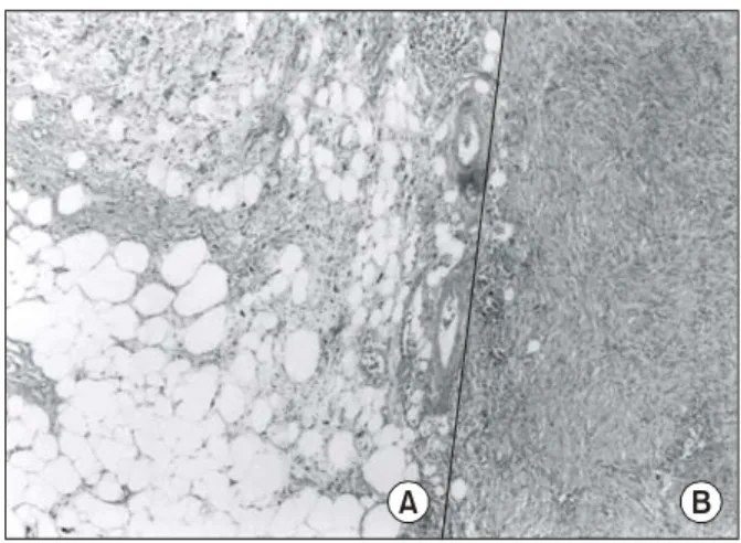
행되어 지방육종의 모습을 보이고 있었으며(Fig. 3), 각 종괴 들은 분화도에서 심한 차이를 보였다. 지방을 형성하지 않는 부분(nonlipogenic component)이 지방육종 부분과 인접하고 있었으며(Fig. 4), 면역조직화학염색상 actin과 S-100 protein 에 양성반응을 보여 평활근육종(leiomyosarcoma)으로 역분화 (dedifferentiation)가 이루어진 상태임을 알 수 있었다.
환자는 종괴적출술 및 우측 결장절제술(Mass excision &
Rt. hemicolectomy)을 받았으며, 정상적으로 회복되어 퇴원 하였고 보조적 항암요법을 시행하며 경과 관찰 중이다.
고 찰
역분화성 지방육종은 전체 지방육종의 10% 이하에서 발 생하고, 60대 장년층에서 호발하며, 남녀간 발생비율은 1.5:
1로 알려져 있다.(6-8) 복부에서 발생하는 경우 후복막강에 생기는 경우가 가장 흔하나, 지방성분이나 중배엽성 세포 가 존재하는 곳은 어디든지 생길 수 있다.(9)
발생기전은 이전에 존재하던 분화가 좋은 지방육종이 8 년 정도의 기간을 거쳐 줄기세포(stem cell)로 거슬러 역분 화하는 과정을 거치는 것으로 알려져 있으며, 그로 인해 지 방을 만들지 않는 부분(nonlipogenic area)이 함께 존재하게 된다.(10) 본 증례의 경우 17년 전 복강내 종괴에 대한 수술 후 추적검사가 전혀 이루어지지 않았고 의무기록도 소실된 상태로, 환자에게 시행한 문진상 지방육종절제술을 시행받 은 것으로 보이며, 17년이 경과하면서 역분화가 이루어진 것으로 추정된다. 역분화의 유형으로는 악성 섬유성조직구 종(malignant fibrous histiocytoma), 섬유육종(fibrosarcoma)이 흔하다고 보고되나,(7) 본 증례에서는 평활근육종(leiomy- osarcoma)으로 판명되었다.
Fig. 1. Abdominal computed tomography, enhanced. Bulky inho- mogeneously enhanced intraperitoneal mass displacing ad- jacent organs.
Fig. 2. Gross finding. Bright yellow colored soft masses with fibrous capsules.
Fig. 3. The groups of scattered lipoblasts with ramifying capillary networks and atypical lipocytes. H&E stain, ×200.
Fig. 4. Dedifferentiated nonlipogenic components (A) coexist with liposarcomatous components (B). H&E stain, ×100.
A B
368 대한외과학회지:제 65 권 제 4 호 2003
ꠏꠏꠏꠏꠏꠏꠏꠏꠏꠏꠏꠏꠏꠏꠏꠏꠏꠏꠏꠏꠏꠏꠏꠏꠏꠏꠏꠏꠏꠏꠏꠏꠏꠏꠏꠏꠏꠏꠏꠏꠏꠏꠏꠏꠏꠏꠏꠏꠏꠏꠏꠏꠏꠏꠏꠏꠏꠏꠏꠏꠏꠏꠏꠏꠏꠏꠏꠏꠏꠏꠏꠏꠏꠏꠏꠏꠏꠏꠏꠏꠏꠏꠏꠏꠏꠏꠏꠏꠏꠏꠏꠏꠏꠏꠏꠏꠏꠏꠏꠏꠏꠏꠏꠏꠏꠏꠏꠏꠏꠏꠏꠏꠏꠏꠏ 임상증상은 발생위치 및 크기에 따라 다양하나, 복강내
에 생기는 경우 무통성의 종괴로 발견되는 경우가 대부분 으로,(11) 이는 복강이 공간적 유순도가 높고 종괴발생이 심부연조직으로부터 서서히 이루어지기 때문인 것으로 생 각된다.
국내에 보고된 원발성 역분화성 지방육종의 예로는 정삭 (spermatic cord)과 우둔근에서 10×10 cm 이하로 발생한 예 와 후복막에서 32×22 cm의 크기에 4.25 kg의 무게로 발생 한 예에 불과하나,(12) 본 증례는 2×3 cm에서 20×15 cm에 이르는 다발성 종괴가 7 kg의 무게를 보여 국내에 발표된 원발성 역분화성 지방육종으로는 가장 거대하다고 할 수 있겠다.
지방육종의 치료로는 광범위 절제술이 원칙이며, 반복해 서 동결절편의 절제생검을 시행 후 안전한 절제범위를 결 정하는 방법을 사용한다. 본 증례의 경우 지방육종이 상장 간막혈관의 기시부까지 확대되고 소장 및 대장의 장간막 경계부에까지 유착되어 있어 소장 및 대장의 광범위 절제 가 필요하였으나, 주요혈관 인접부의 박리가 어려웠고, 단 장증후군 발생이 우려되어 종괴적출술과 우측 결장절제술 만 시행하였다.
보조적요법으로는 방사선치료와 항암화학요법이 있는 데, 술전 방사선 조사를 이용하여 지방육종의 파종 가능성 을 줄일 수 있다는 보고가 있으며, 주변정상장기의 손상을 최대한 막기 위한 술중 방사선조사법도 보고되고 있다. 항 암화학요법은 그 효용성은 떨어지나 MAID(Mesna, Adria- mycin, Ifosfamide, Dacarbazine)요법이 현재 주로 사용되고 있으나, 완전관해율은 10%에 불과하다.(11)
역분화성 지방육종의 국소재발률은 41∼52%, 원격전이 율은 17%, 질환으로 인한 사망률은 28%로 보고되고 있으 며,(13) 예후는 종양의 발생부위, 역분화의 정도 그리고 종 괴의 크기에 영향을 받는다고 알려져 있다.(14) 조직학적 분류(grading)법을 활용하여 재발률을 평가하고 있으며, 병 기결정법(staging)이 연구 중에 있으나, 사지에 발생한 경우 에만 활용될 뿐 복강 내에 발생한 지방육종의 경우엔 적용 이 불가능하다.(11) 최근 후복막강에 발생한 경우 의미있게 낮은 생존율을 보인다고 하여 발생부위가 예후에 미치는 비중이 높아져 가고 있다.
결 론
본 증례는 병력, 이학적 소견, 방사선 및 병리조직학적 검 사상 전형적인 역분화성 지방육종에 대한 것으로, 발생위 치와 종괴의 크기를 고려할 때 향 후 국소재발 및 원격전이 의 가능성이 높을 것으로 생각되어 앞으로의 예후에 대한 지속적인 관찰의 의의가 있을 것으로 생각되어 문헌고찰과 함께 보고하는 바이다.
REFERENCES
1) Russell WO, Cohen J, Enzinger FM, Hazolu SI, Heise H, Martin RG, et al. A clinical and pathological staging system for soft tissue sarcomas. Cancer 1977;40:1562.
2) Enzinger FM, weiss SW. Liposarcoma. In: Stama this G. ed.
Soft tissue tumors. 2nd ed. St. Louis: CV Mosby; 1988.
p.346-82.
3) Angelo P. Dei Tos MD. Liposarcoma: New entities and evolving concepts. Annals of Diagnostic Pathology. Phila- delphia: W.B. Saunder; 2000. p252-66.
4) Ehara S, Rosenberg AE, Kattapuram SV. Atypical lipomas, liposarcomas and other fat-containing sarcomas CT analysis of fat element. Clinical Imaging 1995;1:50-3.
5) Battaglia M, Tognetti A, Malaguti MC, Bacchini P, Monti C.
Anatomopathology, computerized tomography and magnetic resonance correlations in soft tissue liposarcoma. Radiology 1996;92:687-92.
6) Weiss SW, Goldblum JR. Soft tissue tumors St. Louis, Mosby, 4thed; 2001. p.641-70.
7) Hasegawa T, Seki K, Hasegawa F, Matsuno Y, Shimodo T, Hirose T, et al. Dedifferentiated liposarcoma of retroperi- toneum and mesentery: varied growth patterns and histological grade-a clinicopathologic study of 32 cases. Hum Pathol 2000;31:717-27.
8) Weiss SW, Rao VK. Well-differentiated liposarcoma (atypical lipoma) of deep soft tissue of the extremities, retroperitoneum and miscellaneous sites: a follow-up study of 92 cases with analysis of incidence of “dedifferentiation”. Am J Surg Pathol 1992;16:1051-8.
9) L. Lopez-Negrete, L. Luyando, J. Sala, C. Lopez, R. Menendez de Llano, J.L. Gomez. Liposarcoma of the stomach. Abdom- inal imaging. New York: Springer international 1997;22:373-5.
10) Bolen JW, Thorning D. Liposarcomas. A histogenetic approach to the classification of adipose tissue neoplasms. Am J Surg Pathol 1984;8:3-17.
11) Devita VT, Hellman S, Rosenberg SA. Cancer: principle &
practice of oncology. Philadelphia, Lippincott William &
Wilkins, 6th ed; 2001. p1841-91.
12) Cho YJ, Chun HJ, Park DK, Kim YB, Koh DW, Choung RS, et al. A case of dedifferentiated liposarcoma in retroperi- toneum. Korean Journal of Medicine 2002;62:552-6.
13) Henricks WH, Chu YC, Goldblum JR, Weiss SW. Dedif- ferentiated liposarcoma: a clinicopathological analysis of 155 cases with a proposal for an expanded definition of dedif- ferentiation. Am J Surg Pathol 1997;21:271-81.
14) Fang Z, Li J, Yan H. Pathological type of liposarcoma and its effects of clinical treatment. Zhonghua Wai Ke Za Zhi [Chinese Journal of Surgery] 1997;35:204-6.