486
서 론
B형 간염 바이러스에 의한 간염에 의한 간 질환은 감소 추세에 있지만 아직도 만성 간 질환의 상당한 부분을 차지 하는 심각한 질환이다. 담즙정체(cholestasis)는 간의 급성 및 만성질환에서 흔히 볼 수 있는 현상으로 임상의사가 흔 히 접할 수 있다. 임상에서 B형 간염바이러스에 의한 간염 에 의해 만성 간 질환이 있을 때 담도계질환에 의해서 황달 이 오는 경우도 흔히 접할 수 있는 경우이다. 이런 경우에 B형 간염 바이러스의 증식 및 소멸(clearance)이 황달에 의 해서 영향을 받을 것이라 생각된다. 실험적인 담즙정체는 총수담관을 결찰함으로서 만들 수 있는데 수많은 연구가 여러 가지 동물모델에서 1800년도부터 진행되어 왔다.
(1-4) Cameron(1)은 1866년부터 1931년까지 여러 동물에서 총수담관결찰모델을 정리하여 보고하였고 1957년에는
담즙정체가 B형 간염 바이러스의 증식에 미치는 영향에 대한 연구
한양대학교 의과대학 외과학교실
백 홍 규․최 동 호․이 광 수
The Effect of Cholestasis on Hepatitis B Virus Replications
Hong Kyu Baik, M.D., Dongho Choi, M.D. and Kwang Soo Lee, M.D.
Purpose: HBV infection acquired later in life elicits an in- flammatory response. The quality and intensity of the re- sponse determines whether virus clears or persists. Clear- ance is mediated through antigen-specific cytotoxic T-cells.
The immune response often precipitates cholestasis while releasing a number of inflammatory cytokines, such as tumor necrosis factor-α and interferon-γ, which are known to in- hibit HBV replication in vivo. Transgenic mice which replicate HBV provide a useful model for studying viral pathogenesis.
We try to determine whether cholestasis, apart from inflam- mation, modulates HBV replication.
Method: HBV-expressing female 9-wk old BALD/B6 mice matched for HBeAg titer were utilized. Cholestasis was achi- eved by mid-length ligation and transection of the common bile duct in anesthesized animals. Sham and ligated animals were sacrificed over at 4 h, 12 h, 24 h, 48 h, and 72 h after operation (4 animals/interval). Sera were assayed for ALT, GGT, and direct bilirubin. Histopathology was obtained.
Cytokine profiles for interferons, interleukins and tumor ne- crosis factor were monitored by RNase protection assay.
HBV replication was quantitated by measurement of HBV DNA and RNA using Southern and Northern blotting.
Results: Sham-operated animals remained without bioche- mical, pathological or serological changes. Operated animals demonstrated markedly elevated total bilirubin, ALT and GGT levels. Histologic examination showed marked periductular fibrosis and ductular proliferation and area of focal hepato- cellular necrosis. Ribonuclease protection assays demon-
책임저자:백홍규, 경기도 구리시 교문동 249-1 ꂕ 471-020, 한양대학교 구리병원 외과 Tel: 031-560-2294, Fax: 031-566-4409 E-mail: [email protected]
접수일:2002년 9월 2일, 게재승인일:2002년 9월 18일
본 연구는 Korea Science and Engineering Foundation (KOSEF)에서 연구비를 지원받았음.
본 논문의 주요내용은 2002년 춘계외과학회에서 구연되었음.
strated minimal infiltration of CD3 cells, and minimal to no migration of CD4 and CD8 cells. Interferon-γ mRNA was not detected. TNF-α peaked between 1 and 3 days post surgery, but to a much lesser extent than that found in naive virus-challenged animals. Both major HBV RNA species remained unchanged during the experiment. HBV DNA pro- duction demonstrated no changes in the quantity of the relaxed circular or single-stranded intermediates for the first 2 days.
However, by days 5 and 7, reduction in the quantity of viral intermediates were seen. This diminution did not appear to be due to the presence of inflammatory cytokines or CTLs (cyto- toxic T lymphocytes) previously implicated in viral clearance.
Conclusion: Whereas inflammatory cytokines and cellular immunity are essential for viral attenuation and clearance, Acute cholestasis does not appear to contribute indepen- dently to biological modulation of HBV replication. (J Korean Surg Soc 2002;63:486-492)
Key Words: Cholestasis, Hepatitis B virus 중심 단어: 담즙정체, B형 간염 바이러스
ꠏꠏꠏꠏꠏꠏꠏꠏꠏꠏꠏꠏꠏꠏꠏꠏꠏꠏꠏꠏꠏꠏꠏꠏꠏꠏꠏꠏꠏꠏꠏꠏꠏꠏꠏꠏꠏꠏꠏꠏꠏꠏꠏꠏꠏꠏꠏꠏꠏꠏ Department of Surgery, College of Medicine, Hanyang Uni- versity, Seoul, Korea
담관 결찰 후에 시간에 따라 변하는 간의 조직학적 특징 및 혈장의 생화학적 인자와 간 조직에서의 바이러스 DNA 와 RNA의 양의 변화의 특징에 대해서 알아보고자 하였다.
방 법
1) 동물
9주된 B형 간염 바이러스 transgene이 있는 암컷 BALB/
B6 F1 마우스(19∼23 gm)를 사용하였다. 마우스는 41% 습 도에서 70Fo로 항온항습조건에서 우리(cage)당 4 마리씩 12 시간 밤낮주기로 마우스사료를 이용하여서 키웠으며 수술 전 24시간 전부터 금식을 시켰다. 실험방법은 UCSD 의과 대학 및 Scripps research institute의 동물실험위원회에서 승 인한 방법을 사용하였고, Scripps research institute의 격리동 물실에서 사육되었다.
2) 수술방법
총수담관결찰은 수술현미경하에서 무균적으로 methoxy- flurane을 이용한 전신흡입마취하에서 시행되었다. 수술방 법은 정중절개로 복벽을 개복하고 십이지장을 아랫방향으 로 당겨서 간외담도를 노출시키는 동안 간의 가장자리는 부드러운 면봉으로 들었고 췌장의 위쪽 경계면과 간문부 사이의 간외담도를 확인하였다. 간외담도는 췌장위의 중간 부위를 prolene 9.0을 이용하여 중복결찰을 한 후에 분리하 였다. Sham 수술은 복부절개와 간외담도 주위의 박리를 한 후 결찰 및 분리를 시행하지 않았다. 실험동물은 생존여부 를 확인하기 위해서 매일 관찰하였다.
3) 실험방법
(1) 담즙울혈의 동물모델(중간부결찰, n=32): 총수담관의 중간부결찰을 이용한 담즙울혈모델을 사용하여 각 군당 4 마리씩 7개군으로 나누어 수술 후 4시간, 12시간, 1일, 2일, 3일, 5일, 7일에 희생하였다. 각 군당 Sham 수술을 시행한 마우스를 1마리씩 희생하여 간 조직을 얻어서 대조 군으로 사용하였다.
(2) 혈액채취와 분석: 혈액은 희생 시에 대정맥을 통해서 얻었고 total bilirubin, alanine aminotransferase (ALT), gamma-
특이한 32P-radiolabeled DNA probe를 이용하여 nylon filter에 hybridization시켰다.
(5) RNA 분석
① Northern blot analysis; 얼린 간 조직을 acid-guanidium phenol-chloroform method를 이용하여 mechanically puverize 시키고 extract하였다. Total RNA (20μg)이 Guidotti 등(7)의 방법에 의해서 B형 간염 바이러스와 GAPDH의 발현을 확 인하기 위해서 사용되었다.
② RNase protection assay; mRNA의 정량을 위한 RNase protection assay는 Hobbs 등(8)의 방법을 사용하였고 pGEM-4 transcription vector에 들어있는 mouse mIL-1, mIL-1, mIL-2, mIL-3, mIL-4, mIL-5, mIL-6, mIFN-γ, mTNF-α, mTNF-β, m L32 subclone을 사용하였다. mCD4, mCD3γ은 poly- merase chain reaction assisted cloning으로 얻었고 p- GEM-4에 들어있는 mCD8α는 Dr. Hyman (The Salk In- stitute, La Jolla, California)에게서 제공받았다. Subclone들은 EcoRI에 의해서 linearized 되었고 T7 polymerase에 의한 32P- labeled antisense RNA probes를 만들기 위한 template로 이 용되었다. Hybridization reaction, RNase treatment, RNA du- plexes의 분리, denaturating PAGE gel에서의 probe의 reso- lution 등은 Hobbs 등(8)의 방법을 이용하였다. 말린 gel은 film (XAR, Kodak) 위에 놓고 signal을 높이기 위해 inten- sifying screen을 이용하여 -70oC에 적당한 시간 동안 놔두 었다.
결 과
1) 간조직의 조직병리학적 관찰
담관결찰 후의 조직학적 변화는 Sham 수술을 시행한 마 우스에서는 발견되지 않았던 간세포의 괴사, 담도의 증식, 간의 섬유화 등이 관찰되었다. 주로 초기에는 간세포의 괴 사가 주로 생기다가 수술 후 3일 후부터는 담도의 증식이 심해졌다(Fig. 1).
2) 간기능검사
혈장이 생화학검사를 위해서 실험군과 대조군에서 얻어 졌다. Sham 수술을 시행한 대조군에서는 생화학적인 변화
Fig. 1. Histology of the liver in transgenic HBV mice following common bile duct ligation. H&E stain (×400). (A) Sham operated, (B) 4 hours. (C) 12 hours. (D) 1 day. (E) two days. (F) 3 days. (G) 5 days. (H) 7 days.
ꠏꠏꠏꠏꠏꠏꠏꠏꠏꠏꠏꠏꠏꠏꠏꠏꠏꠏꠏꠏꠏꠏꠏꠏꠏꠏꠏꠏꠏꠏꠏꠏꠏꠏꠏꠏꠏꠏꠏꠏꠏꠏꠏꠏꠏꠏꠏꠏꠏꠏꠏꠏꠏꠏꠏꠏꠏꠏꠏꠏꠏꠏꠏꠏꠏꠏꠏꠏꠏꠏꠏꠏꠏꠏꠏꠏꠏꠏꠏꠏꠏꠏꠏꠏꠏꠏꠏꠏꠏꠏꠏꠏꠏꠏꠏꠏꠏꠏꠏꠏꠏꠏꠏꠏꠏꠏꠏꠏꠏꠏꠏꠏꠏꠏꠏ
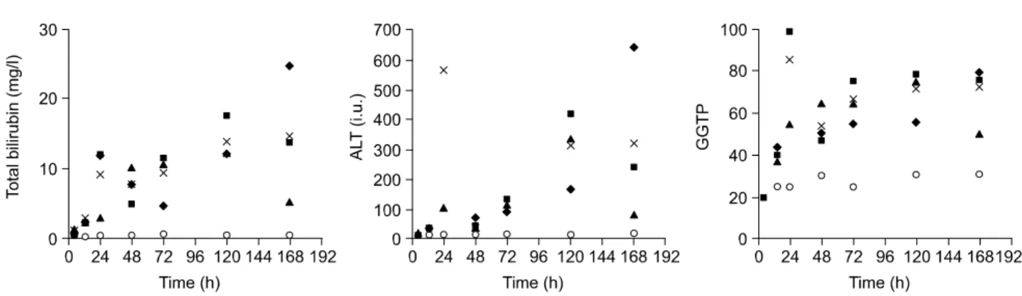
가 나타나지 않았지만 total bilirubin 수치는 수술 후 24시간 부터 점차적으로 증가해서 수술 후 7일째는 대조군보다 18 배 증가한 값을 보였다. ALT와 GGT는 편차가 많았으나 전 반적으로 증가하는 양상을 보였고 ALT는 수술 후 7일째 대 조군 보다 25배 증가한 값을 보였고 GGT는 3배 증가한 양 상을 보였다(Fig. 2).
3) DNA, RNA 분석
RNase protection assay는 CD3 cell의 침윤은 실험의 전 시 간대에서 나타나지 않았고 CD4와 CD8 cell은 아주 적은 양 만 확인할 수 있었으며(Fig. 3), cytokine 중에서는 IL-1α와 IL-1β가 수술 후에 12시간부터 증가된 양상을 보였고 In- terferon-γ의 mRNA는 확인되지 않았다. TNF-α는 수술 후 Fig. 2. Serum chemistries in bile-duct ligated and sham-operated mice at various times post-ligation. Sera were drawn at the time of sacrifice.
Top panel, total bilirubin. Middle panel, ALT. Botton panel, GGT. Open circles-sham animals. Average value of 3 bile-duct ligated animals at each time designated by X. Individual bile duct ligated animals at each time sacrificed designated by triangle, squares or diamond.
Fig. 3. RNase protection assay demonstrating presence of specific immune-cell markers. present in mouse liver tissue (containing HBV transgene) at designated times following common bile duct ligation. S = sham.
Fig. 4. RNase protection assay demonstrating quantity of specific cytokine mRNA species in transgenic mouse liver at designated times following common bile duct ligation. S = sham. Serum ALT measurements averaged for 3 animals are shown at the bottom.
ꠏꠏꠏꠏꠏꠏꠏꠏꠏꠏꠏꠏꠏꠏꠏꠏꠏꠏꠏꠏꠏꠏꠏꠏꠏꠏꠏꠏꠏꠏꠏꠏꠏꠏꠏꠏꠏꠏꠏꠏꠏꠏꠏꠏꠏꠏꠏꠏꠏꠏꠏꠏꠏꠏꠏꠏꠏꠏꠏꠏꠏꠏꠏꠏꠏꠏꠏꠏꠏꠏꠏꠏꠏꠏꠏꠏꠏꠏꠏꠏꠏꠏꠏꠏꠏꠏꠏꠏꠏꠏꠏꠏꠏꠏꠏꠏꠏꠏꠏꠏꠏꠏꠏꠏꠏꠏꠏꠏꠏꠏꠏꠏꠏꠏꠏ
1일에서 3일 사이에 가장 발현을 많이 하였다(Fig. 4). B형 간염바이러스 DNA와 RNA의 정량을 위해서 시행한 Southern 과 Nothern blot에서 B형 간염 바이러스 DNA의 생산은 B형 간염 바이러스 genomic transgene에 의해서 정량한 결과는 relaxed circular 혹은 single-stranded intermediates가 수술 후 처음 2 일간에는 변화가 없다가 수술 후 5일, 7일째 감소하 는 양상을 보였고 B형 간염 바이러스 RNA는 GAPDH에 의 해서 정량한 결과 수술 후에도 큰 차이가 없었다(Fig. 5).
고 찰
B형 간염 바이러스에 의한 만성간염 환자는 한국인에 있 어서 유병률이 상당히 높은 질환으로 임상의가 흔히 접하 고 있다. 이러한 만성간염 환자에서는 간염 자체나 혹은 다 른 양성 혹은 악성질환에 의해서 황달이 생기는 경우가 많 이 있는데 황달 자체에 의해서 B형 간염 바이러스 증식 및 소멸이 어떤 관계가 있는지는 정확히 알려진 바가 없다.
B형 간염 바이러스는 세포는 죽이지 않고 급성 혹은 만 성으로 간의 염증을 일으키고 간암을 일으키는 circular double-stranded DNA를 가지는 바이러스이다.(9) 어른이 B 형 간염바이러스에 감염되었을 경우에는 대부분 만성간염 이 되지 않고 치유가 되지만 흔치 않는 경우에 급성 전격성 간염으로 생명을 잃을 수가 있고 5% 정도에서는 만성간염 을 발전하여서 계속적으로 문제를 일으킨다. 특히 신생아 시기에 감염되었을 경우에는 90% 이상이 만성화되는 것으 로 알려져 있다. 더 문제가 되는 것은 간암과의 연관성인데 만성간염을 가지고 있는 사람은 정상인에 비해서 간암이 걸릴 확률이 100배가 더 높고 신생아기에 감염된 남자의 경 우 간암이 생길 확률이 40%에 육박하는 것으로 알려져 있 다.(10) 이러한 간염을 연구하기 위해서 많은 연구자들이 형질전환마우스를 만들었고 현재 활발한 연구가 진행 중이 다.(11-27) 본 연구에서도 Chisari 등이 확립한 B형 간염 형
질전환마우스를 이용하였다.
황달을 일으킬 수 있는 동물모델은 설치류에서의 총수담 관결찰이 간외담도협착의 좋은 동물모델로 여겨져왔다. 간 외담도결찰 후에 생기는 생화학적인 변화와 조직병리학적 변화에 대해서 구체적으로 확인하게 되었는데 세심하고 섬 세한 수술술기가 실험기간동안 생존하고 재현할 수 있는 동물모델을 만드는 데 아주 중요하기 때문에 백(28)은 세 군데의 결찰부위를 정하고 각 군마다 적당한 수의 마우스 를 사용하여 실험을 하였다. 원위부 및 근위부 총수담관을 결찰하는 경우는 담즙에 의한 복막염 및 췌장염 등으로 인 한 사망률의 증가로 선택되지 않았고 췌장의 바로 위와 담 낭관의 바로 아래의 총수담관의 중간부위를 결찰하는 모델 이 가장 낮은 사망률과 합병증의 비율이 관찰되어 선택되 었다. 시기적인 변화는 초기와 후기로 나뉘고 초기의 변화 는 세포질이나 세담도주위의 중심정맥주위 담즙정체, 문맥 부 부종, 백혈구의 적은 침윤, 문맥부의 원형화(rounding), 간의 소엽구조의 변화없는 간세포의 깃털 같은 변성(feathery degeneration), 호산성의 물질(acidophilic bodies)을 가지는 단 세포괴사(single cell necrosis), 간문맥부위의 가장자리에 있 는 초기 담도증식 등이었고 후기변화는 초기변화에서 진행 이 된 양상을 보이고 pseudoglandular formation을 동반한 liver plates의 파괴, bile infarct, 간세포의 confluent degeneration 및 lysis, macrophage collection, mixed portal inflammatory infiltrates, 문맥주위섬유화의 증가 등의 소견을 보였다. 본 연구에서는 만성간염모델에서 인위적으로 담즙정체을 만 들었을 때에도 초기에 간세포의 괴사와 후기의 담관주위의 섬유화의 증가 등의 소견을 보여서 간염이 없는 마우스의 실험결과(28)와 동일한 결과를 보였다.
B형 간염바이러스의 제거에 있어서 중요한 요소는 MHC Class-I-restricted CD8 cytotoxic T 임파구에 의한 간염된 간 세포의 제거와 T 임파구에서 분비되는 항바이러스성 사이 토카인인 IFN-γ와 TNF-α에 의해서 간염된 간세포에서 B Fig. 5. Quantitation of HBV RNA and DNA in liver tissue obtained from HBV-transgenic mice after common bile duct ligation. A Nothern
blot (NB) and Southern blot (SB) hybridized with radiolabelled HBV probes are shown. The transgene serves as an internal control for the Southern blot. RC-relaxed circle species. SS-single-stranded forms. The Northern blot can be normalized relative to GAPDH (bottom).
것을 확인하여서 이러한 간염 바이러스의 발현감소가 주입 된 cytotoxic T 임파구에 의한 사이토카인의 발현에 의한 것 이 중요한 기전이라는 것을 알았다. 본 실험에서는 급성답 즙정체후에 여러 가지 면역세포와 사이토카인을 확인한 결 과 CD8, CD4, CD3 등의 면역세포는 그 발현이 초기에 아주 미미하게 나타났고 사이토카인의 발현은 기존에 B형 간염 바이러스의 제거에 중요하다고 생각되는 INF-γ의 발현은 나타나지 않았고 TNF α와 β의 발현은 수술 후에 1일 과 3일 사이에 발현을 많이 하였으나 대조군에 비해서 적었으 며 IL1-α와 IL-β는 수술 후에 계속 증가된 양상을 보였다.
B형 간염 바이러스의 RNA는 수술 후에 계속적으로 발현의 차이를 발견할 수 없었고 B형 간염 바이러스의 DNA 는 relaxed circular 혹은 single-stranded form 모두 초기 2∼3일 까지는 발현의 차이가 없다가 5일에서 7일 사이에 발현이 감소하여서 사이토카인과 면역세포와는 관계가 없음을 보 여주어 담도결찰이 B형 간염 바이러스의 소멸이나 증식에 별 영향을 미치지 않았는 것을 알 수 있었다.
역으로 임상에서 흔히 볼 수 있는 담도계의 기계적인 폐 쇄 환자에서 활동성 간염을 동반한 경우에 기계적 폐쇄에 의한 황달이 활동성 간염을 악화시키지 않을 것으로 생각 된다.
결 론
담도결찰에 의한 담즙정체는 cytotoxic T 임파구에 의한 바이러스 소멸(viral clearance) 시에 B형 간염 바이러스 증식 을 억제하는 TNF와 IFN-γ 등의 여러 가지 사이토카인을 induction하지 않고 담도결찰 후에 초기 2일에서 3일에 생기 는 급성 담즙정체는 B형 간염 바이러스 RNA나 DNA의 감소의 효과를 나타내지 못하고 5일에서 7일 사이에 B형 간염 바이러스 RNA의 변화 없이 viral DNA의 소량의 감 소를 보이나 이 효과도 immune-mediated mechanism은 아 니다. 결론적으로 담즙정체는 B형 간염 바이러스 증식의 중요한 관련인자가 아닌 것을 본 실험에서 확인할 수 있 었다.
Bacteriol 1958;75:333-49.
5) Trams EG, Symeondis A. Morphologic and functional changes in the livers of rats after ligation or excision of the common bile duct. Am J Pathol 1957;33:13-25.
6) Chisari FV, Klopchin K, Moriyama T, Pasquinelli C, Dunsford HA, Sell S, et al. Molecular pathogenesis of hepatocellular carcinoma in hepatitis B virus transgenic mice. Cell 1989;59:
1145-56.
7) Guidotti LG, Ishikawa T, Hobbs MV, Matzke B, Schreiber R, Chisari FV. Intracellular inactivation of the hepatitis B virus by cytotoxic T lymphocytes. Immunity 1996;4:25-36.
8) Hobb MV, Weigle WO, Noonan DJ, Torbett BE, McEvilly RJ, Koch RJ, et al. Patterns of cytokine gene expression by CD4 T cells from young and old mice. J Immunol 1993;150:3602-14.
9) Chisari FV, Ferrari C. Immunobiology and pathogenesis of viral hepatitis. In: Nathenson N, Ahmed R, Gonzales-Scarano F, Griffin D, Holmes K, Murphy FA, et al. editors. Viral path- ogenesis. New York, NY, Raven Press, 1995.
10) Beasley RP, Lin CC, Hwang LY, Chen CS. Hepatocellular car- cinoma and hepatitis B virus: a prospective study of 22,707 men in Taiwan. Lancet 1981;2:1129-33
11) Chisari FV, Pinkert CA, Milich DR, Filippi P, McLachlan A, Palmiter RD, et al. A transgenic mouse model of the chronic hepatitis B surface antigen carrier state. Science 1985;230:
1157-60.
12) Burk RD, DeLoia JA, El Awady MK, Gearhart JD. Tissue preferential expression of the hepatitis B virus (HBV) surface antigen gene in two lines of HBV transgenic mice. J Virol 1988;62:649-54.
13) Araki K, Miyazaki JI, Hino O, Tomita N, Chisaka O, Mat- subara K, et al. Yamamura KI, Expression and replication of hepatitis B virus genome in transgenic mice. Proc Natl Acad Sci USA 1989;86:207-11.
14) Gilles PN, Fey G, Chisari FV. Tumor necrosis factor-alpha negatively regulates hepatitis B virus gene expression in trans- genic mice. J Virol 1992;66:3955-60.
15) Guidotti LG, Matzke B, Schaller, Chisari FV, High-level hepa- titis B virus replication in transgenic mice. J Virol 1995;69:
6158-69.
16) DeLoia JA, Burk RD, Gearhart JD. Developmental regulation of hepatitis B surface antigen expression in two lines of hep-
ꠏꠏꠏꠏꠏꠏꠏꠏꠏꠏꠏꠏꠏꠏꠏꠏꠏꠏꠏꠏꠏꠏꠏꠏꠏꠏꠏꠏꠏꠏꠏꠏꠏꠏꠏꠏꠏꠏꠏꠏꠏꠏꠏꠏꠏꠏꠏꠏꠏꠏꠏꠏꠏꠏꠏꠏꠏꠏꠏꠏꠏꠏꠏꠏꠏꠏꠏꠏꠏꠏꠏꠏꠏꠏꠏꠏꠏꠏꠏꠏꠏꠏꠏꠏꠏꠏꠏꠏꠏꠏꠏꠏꠏꠏꠏꠏꠏꠏꠏꠏꠏꠏꠏꠏꠏꠏꠏꠏꠏꠏꠏꠏꠏꠏꠏ atitis B virus transgenic mice. J Virol 1989;63:4069-73.
17) Farza H, Salmon AM, Hadchouel M, Moreau JL, Babinet C, Tiollais P, et al. Hepatitis B surface antigen gene expression is regulated by sex steroids and glucocorticoids in transgenic mice. Proc Natl Acad Sci USA 1987;84:1187-91.
18) Chisari FV, Filippi P, McLachlan A, Milich DR, Riggs M, Lee S, et al. Expression of hepatitis B virus large envelope pol- ypeptide inhibits hepatitis B surface antigen secretion in trans- genic mice. J Virol 1986;60:880-7.
19) Guidotti LG, Martinez V, Loh YT, Rogler CE, Chisari FV.
Hepatitis B virus nucleocapsid particles do not cross the hep- atocyte nuclear membrane in transgenic mice. J Virol 1994;
68:5469-75.
20) Guilhot S, Huang S, Xia YP, LaMonica N, Lai MMC, Chisari FV. Expression of the hepatitis delta virus large and small an- tigens in transgenic mice. J Virol 1994;68:1052-8.
21) Gilles PN, Guerrette DL, Ulevitch RJ, Schreiber RD, Chisari FV. Hepatitis B surface antigen retention sensitizes the hep- atocyte to injury by physiologic concentrations of gamma in- terferon. Hepatology 1992;16:655-63.
22) Chisari FV, Klopchin K, Moriyama T, Pasquinelli C, Dunsford HA, Sell S, et al. Molecular pathogenesis of hepatocellular carcinoma in hepatitis B virus transgenic mice. Cell 1989;59:
1145-56.
23) Moriyama T, Guilhot S, Klopchin K, Moss B, Pinkert CA, Pal-
miter RD, et al. Immunobiology and pathogenesis of hepa- tocellular injury in hepatitis B virus transgenic mice. Science 1990;248:361-4.
24) Ando K, Moriyama T, Guidotti LG, Wirth S, Schreiber RD, Schlicht HJ, et al. Mechanisms of class I restricted immu- nopathology: a transgenic mouse model of fulminant hepatitis.
J Exp Med 1993;178:1541-54.
25) Guidotti LG, Ando K, Hobbs MV, Ishikawa T, Runkel L, Schreiber RD, et al. Cytotoxic T lymphocytes inhibit hepatitis B virus gene expression by a noncytotoxic mechanism in transgenic mice. Proc Natl Acad Sci USA 1994;91:3764-8.
26) Guidotti LG, Guilhot S, Chisari FV. Interleukin 2 and interferon alpha/beta downregualte hepatitis B virus gene expression in vivo by tumor necrosis factor dependent and independent path- ways. J Virol 1994;68:1265-70.
27) Guilhot S, Guidotti LG, Chisari FV. Interleukin-2 downregulates hepatitis B virus gene expression in transgenic mice by a post-transcriptional mechanism. J Virol 1993;67:7444-9.
28) Baik HK. Temporal change in the mouse liver after common bile duct ligation. J Korean Surg Soc 2002;63:99-104.
29) Guidotti LG, Ishikawa T, Hobbs MV, Matzke B, Schreiber R, Chisari FV. Intracellular inactivation of the hepatitis B virus by cytotoxic T lymphocytes. Immunity 1996;4:25-36.
30) Chisari FV. Hepatitis B virus transgenic mice: Insights into the virus and the disease. Hepatology 1995;22:1316-25.