INTRODUCTION
Rehabilitation of partially or totally edentulous patients with dental implants has become common practice in the last decades with reliable long-term results. But insuffi- cient bone height in the posterior maxillary region is one of the most frequent problems in dental implantation procedure. To solve this problem, various surgical proce-
dures have been proposed, such as the use of pterygoid implants, inlay bone graft in the maxillary sinus, onlay bone graft, vertical guided bone regeneration, and alveo- lar distraction osteogenesis (ADO)
1,2). Among these pro- cedures, the inlay bone grafting in the maxillary sinus known as sinus lifting or sinus floor elevation has been the most widely used technique, recently
3,4).
Grafting materials are known to encourage new bone formation by many osteogenic processes. Autogenous bone is known to induce new bone through osteogenesis, while allogenic bone is thought to be osteoinductive due to the presence of growth factors. Autogenous iliac bone graft has been used as augmentation material with excel- lent result
5-9). However, there are several shortcomings,
* Corresponding author Soung-Min Kim
Dept. of OMFS, College of Dentistry, Kangnung National Univ.
Jibyeon-dong, Gangneung, Gangwon-do, 210-702, Korea Tel: 82-33-640-2468, 3139 Fax: 82-33-642-6410 E-mail: [email protected]
Comparative histomorphologic study of regenerated bone for dental implant placement in the atrophied posterior maxilla
Se-Jung Kim, Soung-Min Kim, Ji-Hyuck Kim, Young-Wook Park
Department of Oral and Maxillofacial Surgery, College of Dentistry, Kangnung National University, Gangneung, Korea
※ This work was supported by a grant of the Korea Health 21 R&D Project, Ministry of Health & Welfare, Republic of Korea (A050547), and supported by Fisheries Research and Development Funds granted the Korean Ministry of Maritime affairs and Fisheries.
The purpose of this study is to evaluate the regenerative capacity of reconstruction in the atrophied posterior maxilla by comparing bone graft procedures and alveolar distraction osteogenesis (ADO) techniques.
We performed the autogenous iliac bone graft (AGB group, 5 specimens in 3 patients), and the combination (Mixed group, 3 speci- mens in 3 patients) of the autogenous and deproteinized bovine bone (Bio-Oss
�, Geistlich Co., Switzerland) as the ratio of 2:1 in the sinus floor elevation procedures. ADO procedures using TRACK
�(KLS Martin Co., Germany) were also performed to augment vertical alveolar height in atrophied posterior maxilla (ADO group, 5 specimens in 4 patients). Newly generated bone tissues were obtained with the 2.0mm diameter trephine bur (3i Co., USA) during implant fixture installation after 5-7 months. Routine histolomorphological observation, immunodot blot assay for quantitative evaluation, and immunohistochemical staining with antibodies to MMP-1, -9, -10, TIMP-1, -2, and BMP-2, -4 were all carried out.
Lamellar bone formation was well shown in all specimens and new bone formations of ADO group increased than those of other pro- cedures. In immunohistochemical staining, the strong expression of BMP-2 was shown in all specimens, and immunodot blot assay showed that bone formation is accompanied by the good induction of factors associated with angiogenesis and appeared more increased amount of osteogenic and angiogenic factors in ADO group.
ADO is the most effective technique for new bone formation compared to sinus floor elevation with autogenous or mixed bone graft in the atrophied posterior maxilla. In the quantitative immunodot blot assay, the regenerated bone after ADO showed more increased products of VEGF, BMP-2, PCNA and MMP-1 than those after the other procedures, and these findings were able to be confirmed by immunohistochemical stainings.
Key words
Alveolar distraction osteogenesis (ADO), Autogenous iliac bone graft, Deproteinized bovine bone, Sinus floor elevation, Immunodot blot assay, Immunohistochemical staining
Abstract
such as donor site morbidity, prolonged healing period, and unpredictable resorption of the graft
10,11). Xenografts, such as bovine bone mineral (BBM), and alloplastic sub- stitutes encourage the apposition of new bone by osteo- conduction, and Bio-Oss
�(Geistlich Co., Switzerland) has been widely reported to be effective in producing an effective bone regenerative matrix
12,13).
ADO is a popular technique in the anterior and posteri- or mandible but is not a routine procedure in the atro- phied posterior maxilla due to the difficulty in the vector control, inaccessibility, severe pneumatization, and very few documented known researches, etc
14,15). But if several requirements are met, i.e. over 7 mm remaining alveolar bone height, no severe sinus disease, sufficient bone width and good accessibility, it seems that ADO can also be an acceptable bone augmentation technique in the atrophied posterior maxilla
15,16).
The purpose of this study is to evaluate the regenera- tive capacity of the augmentation procedure, comparing with sinus lifting procedure and ADO in the atrophied posterior maxilla, by the histomorphological and quanti- tative immunodot blot assay methods.
MATERIALS AND METHODS 1. Patients and reconstructive procedures
Total 10 specimens from 10 patients aged between 34 and 68 years (mean 55.3 years) were used in this study.
Three patients were reconstructed by sinus floor eleva- tion with autogenous iliac bone graft (AGB [autogenous
bone] group), another three patients were reconstructed by sinus floor elevation with mixed bone graft (Mixed [autogenous & deproteinized bovine bone] group), and remaining four patients were reconstructed by ADO pro- cedure (ADO group) (Table 1).
The sinus floor elevation was carried out by the lateral window approach technique. Autogenous bone was har- vested from anterior iliac crest, the mixed bone was made of mandibular symphyseal particularized bone and deproteinized bovine bone (Bio-Oss
�, Geistlich Co., Switzerland) as a volume ratio of 6 to 4 or 7 to 3. Primary wound closure without using any membrane was per- formed with 4-0 Vicryl (Polyglactin 910, Johnson &
Johnson Co., USA). Patients were examined next day and one week later, and stiches were removed in two weeks The grafted sinus was left to heal during 6 months.
ADO was performed with an intraoral incision on the alveolar crest with lateral releasing incisions. Careful subperiosteal buccal dissection was performed to obtain adequate visibility of the underlying bone, but mucope- riosteum of the palate was not dissected to preserve ade- quate blood supply to the osteotomized segment. After molding of the intraoral distractor Track
�(KLS Martin Co., Germany) device, the bone segment was completely separated from the basal bone with reciprocating and oscillating saw. After completing osteotomy, distractor was adapted and fixated to both the basal bone and the osteotomized segment with microscrews. The osteotomized segment was immediately mobilized by activating the distractor to check the direction of dis- traction and freedom in movements. Finally, the
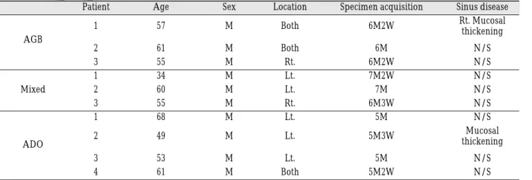
Table 1. Patient’s data.
Patient Age Sex Location Specimen acquisition Sinus disease
1 57 M Both 6M2W Rt. Mucosal
AGB thickening
2 61 M Both 6M N/S
3 55 M Rt. 6M2W N/S
1 34 M Lt. 7M2W N/S
Mixed 2 60 M Lt. 7M N/S
3 55 M Rt. 6M3W N/S
1 68 M Lt. 5M N/S
2 49 M Lt. 5M3W Mucosal
ADO thickening
3 53 M Lt. 5M N/S
4 61 M Both 5M2W N/S
AGB : Autogenous bone graft, Mixed : Autogenous & Deproteinized bovine bone, ADO : Alveolar Distraction Osteogenesis
osteotomized segment was repositioned at its initial position and the surgical access was sutured with 4-0 Vicryl. Antibiotics, non-steroidal analgesics, soft diet, and oral hygiene regimens were followed as the same protocol used in sinus floor elevation procedure. The activation of the distraction device was started after 5-7 days of the latency period and stiches were removed in 2 weeks. The distraction rate of 1mm per day was per- formed until the desired amount of distraction was obtained. The distractor was then maintained in position for 8 weeks to obtain the maturation of the neocallus formed between the basal bone and the distracted seg- ment. After this consolidation period, the distractor was removed. After 4 weeks since the distractor was removed, the implant placement was performed.
Routine radiographic documentations such as panora- ma, CT, tomographs and intraoral radiographs, were obtained, immediately after the operation, at the end of
the distraction procedure, at the time of the implant placement, at the time of prosthetic rehabilitation, and annually thereafter.
2. Harvesting of the specimens
All 10 specimens were harvested at the time of implan- tation about 5 to 7 months after reconstructive proce- dure. All patients consented these procedures with high interests to the bone regenerative capacity of their own, and these all clinical studies were also approved by Institutional Review Board (IRB) of Kangnung National University Dental Hospital. Under local anesthesia, ade- quate incisions were made, a full thickness flap was raised and mobilized for tension-free closure. Bone was inspected, and a trephine bur of 2mm diameter (3i Co., USA) was used to take the specimen from the recon- structed bone (Fig. 1-a, b).
Fig. 1. a. Drawing of harvesting of specimen through crestal approach., b. Intraoperative figure of harvesting of specimen using trephine bur.
ADO
a
b
Sinus graft
3. Histological procedures and assays
Specimens were immediately fixed in 10% NBF (Neutral Buffered Formalin), pH 7.4 and stored for 24 hour at 4℃. Specimens were then decalcified in 9%
formic acid/formalin solution and dehydrated through graded ethanols, cleared in xylene, embedded in paraf- fin, cut into slices of about 4㎛ thickness and stained with hematoxylin and eosin (H&E), Masson Trichrome (MT), and immunohistochemical stainings of MMP-1, -9, -10, TIMP-1, -2, and BMP-2, -4. And the immunodot blot assay was performed for the quantification of the target mRNA especially by computer-assisted analysis of images formed by a microscope.
3-1. Immunohistochemical assay
The endogenous peroxidase was blocked with 3%
hydrogen peroxide for 10 minutes and washed 3 times
with PBS (phosphate buffered saline). Antibodies diluted in DW (distilled water) 1:50 were added to the sections overnight at 4℃ and washed 3 times with PBS (Table 2).
The secondary antibody solution was placed on the sec- tions for 30 minutes and washed 3 times with PBS. Then, horseradish peroxidase-conjugated streptavidin-biotin complex was placed on the sections for 30 minutes and washed 3 times with PBS, followed by a peroxidase reac- tion using 3,3’-diaminobenzidine (DAB) as a chro- mogen. Finally, the sections were washed 3 times with PBS, dehydrated through graded ethanols, and mounted.
Negative control of immunohistochemical study was also performed (Fig. 2-a to 2-c). Each slide was evaluated according to the intensity of positive immunostaining, which were graded as +++, ++, +, +/-, - , which mean strong, moderate, slight, rare and negative respectively.
A ‘rare’grade of +/- was defined to represent a focal or questionable weakly positive signal in the tissue sections.
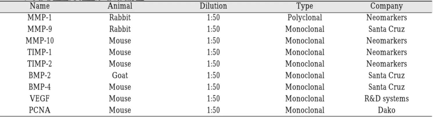
Table 2. Antibodies used in this study.
Name Animal Dilution Type Company
MMP-1 Rabbit 1:50 Polyclonal Neomarkers
MMP-9 Rabbit 1:50 Monoclonal Santa Cruz
MMP-10 Mouse 1:50 Monoclonal Neomarkers
TIMP-1 Mouse 1:50 Monoclonal Neomarkers
TIMP-2 Mouse 1:50 Monoclonal Neomarkers
BMP-2 Goat 1:50 Monoclonal Santa Cruz
BMP-4 Mouse 1:50 Monoclonal Santa Cruz
VEGF Mouse 1:50 Monoclonal R&D systems
PCNA Mouse 1:50 Monoclonal Dako
Fig. 2. Histologic assessement of negative control group (magnification, ×200).
a. H&E staining, b. MT staining, c. Immunohistochemical stainig
3-2. Immunodot blot assay
3-2-1. mRNA extraction
Sections of the bony tissues with 10㎛ thickness were obtained and deparaffinized with xylene, followed by hydration with graded alcohols. After centrifugation, the tissue precipitates were lysed with lysis buffer (300㎕
SDS [sodium dodecyl sulfate] + 6M urea with PMSF [phenylmethylsulfonyl fluoride] and 0.1M DTT [dithio- threitol]). These mixtures were boiled for 3 minutes in 100℃, and then centrifuged (12,000 rpm, 20 minutes).
Supernatants were kept in the - 80℃ freezer until use.
3-2-2. Procedures of immunodot blot
Nitrocellulose transfer membrane (PROTRAN
�, Schleicher&Schuell BioScience Co., Germany) was placed on the Bio-Dot
�apparatus (Bio-Rad Co., USA) using vacuum suction. The experiment was performed, using 6 antibodies such as VEGF, BMP-2, PCNA, MMP- 1, MMP-9, and TIMP-1. A small iliac bone fragment from one of the patients was used as the control group. About 1㎍ of the extracted protein was diluted with 100㎕ PBS and transferred on the membrane. The blotted mem- brane was cut in groups and fixed on the labeled slide glass. After 1 hour’s blocking with skim milk solution in 1× TBST (Tris-buffered saline with 0.1% Tween 20), the membrane was incubated with each antibody for 1 hour using cover glass apparatus. And then membranes were washed with the 1x TBST and incubated in secondary antibody solution. Biotinylated link, with membranes for 30 minutes was used. 1x TBST incubation wass per- formed for 30 minutes, and membranes were incubated with Streptavidine-HRP for 30 minutes, and 1x TBST incubation was done for 30 minutes. The color develop- er, DAB solution activated by H 2 O 2 , was applied to the membrane, and then the membrane was washed and dried out after the color appearance.
3-2-3. Quantification and statistics
Membranes were scanned in 300 dpi (dots per inch) and saved in TIF (tagged image file) after changing to gray shade using Adobe photoshop program (Version 7.0, Adobe Co., USA). Images were analyzed using ImageQuant
�(Version 5.2, Molecular Dynamics Co., USA) and Microsoft Excel (2002, Microsoft Co., USA).
Comparisons in each group were done with ANOVA using SPSS (Version 12.0, SPSS Inc., USA) for Windows and mean values were followed by 95% confidence inter-
vals. Correlation of each factors was analyzed by Pearson’s correlation test (p <0.05).
RESULTS
1. Histomorphologic findings
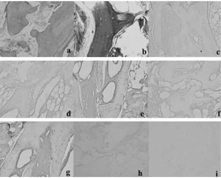
1-1. Sinus floor elevation with autogenous bone graft (AGB group)
In H&E staining, the new regenerated bone is observed and can not be easily distinguished from the natural bone in all cases (Fig. 3-a). In MT staining, the new bone is found on the periphery of old trabecular bone in most cases (Fig. 3-b).
1-2. Sinus floor elevation with mixed bone graft (Mixed group)
In H&E and MT staining, newly formed trabecular bone is predominant as the form of lamellar type with some portions of Haversian canals, but is inferior to the autogenous bone group in the both bony quality and quantity aspects (Fig. 4-a, b).
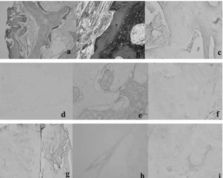
1-3. Alveolar distraction osteogenesis (ADO group) In H&E and MT staining, well-formed new trabecular bone is seen in the distraction gap. Osteophytes, which are layers of osteoid tissue covered with active osteoblasts and typical forms of bony regeneration in ADO, are observed at the outer surface of the trabecular bone (Fig. 5-a, b).
2. Immunohistochemical staining
2-1. Sinus floor elevation with autogenous bone graft (Table 3, Fig. 3-c to 3-i)
Expressions of MMP-1 and MMP-10 are unreliable and invalid, which means that the expression is positive or negative (Fig. 3-c, e), and the positive reaction of MMP-9 is not observed either (Fig. 3-d). Both TIMP-1 and TIMP- 2 are not expressed in immunohistochemical staining, which is negative reaction (Fig. 3-f, g). The expression of BMP-2 is moderately positive, which were observed at the outer surface of the trabecular bone (Fig. 3-h). But the expression of BMP-4 is negative (Fig. 3-i).
2-2. Sinus floor elevation with mixed bone graft (Fig.
4-c to 4-i)
The expression of MMP-1 is unreliable and invalid,
Table 3. Expressions of antibodies in immunohistochemical staining.
MMP-1 MMP-9 MMP-10 TIMP-1 TIMP-2 BMP-2 BMP-4 Negative
Control
AGB +/- - +/- - - ++ - -
Mixed +/- - - - - + - -
ADO +/- - +/- - - ++ - -
- : negative, +/- : rare, + : slight, ++ : moderate, +++ : strong
Fig. 3. Histologic assessement of autogenous bone graft group (magnification, ×200).
a. H&E staining b. MT staining
c. Immunohistochemical stainig, MMP-1 (1:50, polyclonal Ab, Rabbit)
d. Immunohistochemical stainig, MMP-9 (1:50, monoclonal Ab, Rabbit)
e. Immunohistochemical stainig, MMP-10 (1:50, monoclonal Ab, Mouse)
f. Immunohistochemical stainig, TIMP-1 (1:50, monoclonal Ab, Mouse)
g. Immunohistochemical stainig, TIMP-2 (1:50, monoclonal Ab, Mouse)
h. Immunohistochemical stainig, BMP-2 (1:50, monoclonal Ab, Goat)
i. Immunohistochemical stainig, BMP-4 (1:50, monoclonal Ab, Mouse)
which means that the expression is positive or negative (Fig. 4-c), and the positive reactions of MMP-9 and MMP-10 are not observed (Fig. 4-d, e). Both TIMP-1 and TIMP-2 are not expressed in immunohistochemical stain- ing, which is negative reaction (Fig. 4-f, g). The expres- sion of BMP-2 is slightly positive, which were observed at the outer surface of the trabecular bone (Fig. 4-h). But the expression of BMP-4 is negative (Fig. 4-i).
2-3. Alveolar distraction osteogenesis (Fig. 5-c to 5-i) The expressions of MMP-1 and MMP-10 are vague, which means that the expression is positive or negative (Fig. 5-c, e), and the positive reaction of MMP-9 is not observed (Fig. 5-d). Both TIMP-1 and TIMP-2 are not expressed in immunohistochemical staining, which is negative reaction (Fig. 5-f, g). The expression of BMP-2 is moderately positive, which were observed at the outer surface of the trabecular bone (Fig. 5-h). But the expres- sion of BMP-4 is negative (Fig. 5-i).
Fig. 4. Histologic assessement of mixed bone graft group (magnification, ×200).
a. H&E staining b. MT staining
c. Immunohistochemical stainig, MMP-1 (1:50, polyclonal Ab, Rabbit)
d. Immunohistochemical stainig, MMP-9 (1:50, monoclonal Ab, Rabbit)
e. Immunohistochemical stainig, MMP-10 (1:50, monoclonal Ab, Mouse)
f. Immunohistochemical stainig, TIMP-1 (1:50, monoclonal Ab, Mouse)
g. Immunohistochemical stainig, TIMP-2 (1:50, monoclonal Ab, Mouse)
h. Immunohistochemical stainig, BMP-2 (1:50, monoclonal Ab, Goat)
i. Immunohistochemical stainig, BMP-4 (1:50, monoclonal Ab, Mouse)
3. Immunodot blot assay (Fig. 6, 7)
3-1. Comparative quantification of VEGF
ADO group showed high density and followed by AGB group, and then Mixed group. The difference of each group is all statistically significant (p < 0.05).
3-2. Comparative quantification of BMP-2
ADO group showed high density and followed by AGB group, and then Mixed group. The difference between ADO group and AGB group is statistically sig- nificant (p < 0.05), but the difference between AGB group and Mixed group is not significant (p > 0.05).
Fig. 5. Histologic assessement of ADO group (magnification, ×200).
a. H&E staining b. MT staining
c. Immunohistochemical stainig, MMP-1 (1:50, polyclonal Ab, Rabbit)
d. Immunohistochemical stainig, MMP-9 (1:50, monoclonal Ab, Rabbit)
e. Immunohistochemical stainig, MMP-10 (1:50, monoclonal Ab, Mouse)
f. Immunohistochemical stainig, TIMP-1 (1:50, monoclonal Ab, Mouse)
g. Immunohistochemical stainig, TIMP-2 (1:50, monoclonal Ab, Mouse)
h. Immunohistochemical stainig, BMP-2 (1:50, monoclonal Ab, Goat)
i. Immunohistochemical stainig, BMP-4 (1:50, monoclonal Ab, Mouse)
Fig. 7. a. Histogram of immunodot blot assessment.
b. Scatter diagram of correlations.
Fig. 6. a. Natural figure obtained after immunodot blot assay, b. Grayish figure for immunodot blot analysis.
a
a
b
b
AGB Mixed Iliac bone ADO 210
200 190 180 170 160 150
185 180 175 170 165 160 155 150 145
205 200 195 190 185 180 175 170 165 160
210 205 200 195 190 185 180 175 170 165
190 185 180 175 170 165 160 205
200 195 190 185 180 175
AGB Mixed Iliac bone ADO
AGB Mixed Iliac bone ADO AGB Mixed Iliac bone ADO
AGB Mixed Iliac bone ADO AGB Mixed Iliac bone ADO
DensityDensity DensityDensityDensity
Density