1. Introduction
Radiotherapy showed great development in the modern era. From the intensity modulated radiotherapy that can reduce the exposure dose
of the normal organ and enhance the prescription dose of the tumor, to the stereotactic radiotherapy that injects high dose through precision Radiotherapy, the image-guided radiotherapy that verifies the precise position for
A Fusion Study on the Selection of Cyberknife Technique according to the Location of the Pulmonary Tumors
Gab-Jung Kim 1 , Jeong-Ho Kim 2* , Seok-Hwan Bae 3 , Nak-Sang Kim 1 , Sun-Yeol Seo 4
1
Professor, Division of Radiology, Songho University
2
Team member, Department of Radiation Oncology, Konyang University Hospital
3
Professor, Division of Radiological science, Konyang University
4
Team member, Department of Radiology, Daejeon Eulji Medical Center
폐종양의 위치에 따른 사이버나이프 기법의 선택에 관한 융합적 연구
김갑중 1 , 김정호 2* , 배석환 3 , 김낙상 1 , 서선열 4
1
송호대학교 방사선과 교수,
2건양대학교병원 방사선종양학과 팀원,
3
건양대학교 방사선학과 교수,
4대전을지대학교병원 영상의학과 팀원
Abstract Depending on the location of the lung tumor, the choice of treatment technique should be considered when treating the Cyberknife. The 4DCT images of 18 lung cancer patients were analyzed, and location error values were extracted through application program. The evaluation result was lower than the average position error only in the upper and the inner. These results suggest that the Vertebral tracking technique is effective when it is close to the pulmonary attachment or near the vertebral body, and the Synchrony technique is effective at other positions. In the future, we would like to study cyber knife treatment technique according to the position of the tumor as well as the volume of the lung and the respiratory cycle.
Key Words : Cyberknife, Synchrony, Vertebral tracking, Pulmonary tumor, Position Delta Value
요 약 폐 종양의 위치에 따라 사이버나이프 치료 시 치료기법의 선택에 대해 고려해봐야 합니다. 폐암 환자 28 명을 대상으로 18개 지점에 대해 4차원 단층촬영영상을 분석하였고, 응용프로그램을 통해 위치오차값을 추출하였다. 평가 결과 상부와 내측에서만 평균 위치오차값보다 낮았다. 이러한 결과를 통해 폐첨부에 가깝거나 척추체부에 가까운 경우 척추추적기법을 적용하는 것이 효율적이며, 이외의 위치에서는 호흡동조기법이 효율적이다. 이에 본연구를 기반으로 하여 향후 폐종양의 폐내 종양의 위치뿐만 아니라 폐활량에 따른 확장범위 및 환자마다 차이가 발생하는 호흡주기에 따라 사이버나이프 치료기법을 선택할 경우 효율적인 치료기법을 선택할 수 있도록 연구하고자 한다.
주제어 : 사이버나이프, 호흡동조기법, 척추추적, 폐종양, 위치오차값
*Corresponding Author : Jeong-Ho Kim([email protected]) Received June 18, 2019
Accepted July 20, 2019
Revised July 2, 2019
Published July 28, 2019
therapy, and the respiratory-tracking radiotherapy that considers the movement of the tumor from respiration, radiotherapy is applied diversely[1-8].
Among these therapies, the stereotactic radiotherapy was first developed by the Swedish doctor, Dr. Lars Leksell in 1951: narrow beams of radiation are combined in multiple directions to deliver maximum dose to the tumor. Also, minimum dose is applied to the normal organ to prevent any adverse effects, so this therapy replaced the existing multi-fraction therapy of delivering high-dose of radiation for each session. The first therapeutic apparatus applied was the gamma knife that was developed in 1968, and this therapeutic apparatus arranges the radioactive isotope of Co-60 in helmet-type to irradiate the gamma ray in multiple directions in narrow beans through the collimator[9-11].
However, this apparatus has a weakness of limiting the application only on encephalopathy such as the brain tumor and arteriovenous malformation. The therapeutic apparatus that was developed to improve this limitation and to apply the stereotactic radiotherapy on the body is Cyberknife. One comparison with the most widely used medical linear accelerator today is that a small-sized accelerator is mounted on the robot arm composed of 6 joints, so the use of the deflecting electromagnet is no longer necessary[12-14]. The Cyberknife acquired the approval from the US FDA in 2001, and as the body is occurred with the change in position due to respiration and movement of the organs, the photon of kV is used in constant interval to analyze the precise position, and the changes in the position are applied to perform the tumor-tracking therapy by using the cruise-missile-guidance technology of NASA[15-18]. The cruise-missile-guidance technology is based on the discrete time linear movement system, and uses the dynamic system model of the Kalman Filter that assumes the relation of the state vector value according to the change in time. Cyberknife using the
tumor-tracking system is divided in the
therapeutic technique depending on the
therapeutic site such as the head & neck and the
body. Also, the body is divided in therapeutic
technique according to the sites with movements,
and other sites without movements[19]. In other
words, the Cyberknife therapeutic technique can
be divided largely into 3 techniques of Cranial
tracking technique, Vertebral tracking technique
and Synchrony technique. The cranial tracking
technique is limited to the head, and the therapy
is performed by analyzing the morphological
position of the cranium, and the vertebral tracking
technique enables the morphological position
analysis of the vertebrae for the treatment of the
tumor near the vertebrae[20]. As for the
synchrony technique, when the image-guided
radiotherapy is applied on the tumors with
severe change from respiration including the
pulmonary and hepatic tumors, the position
changes each time depending on the rate of
respiration, so the respiration pattern image is
used and radioactive impermeable substance is
inserted near the tumor to be applied in
combination with the image-guided radiotherapy. In
the case of applying the synchrony technique on
the lungs, signifier insert is applied near the
tumor for the location tracking. However, when
there is migration in the signifier insert, or when
the position is inappropriate to have an
overlapped image, the accuracy of the synchrony
technique will be degraded. Especially, when the
radius of positioning is relatively high, the error
will be reflected more greatly. In addition, when
invasive procedure is performed for the signifier
insert, there is a disadvantage of requiring a
long-period of time compared to other
techniques[21-23]. Lungs are the largest organ
among the organs in the chest, and the weight of
the lungs for adults is approximately 500~600g,
with the surface area of about 90m2. The total
volume of the lungs is approximately
5,000~6,000cc, the respiration rate for 1 time is
300~500mL. The segments of the lungs are divided into left and right centrally of the mediastinum. The right lung has 3 lobi with 10 segments, and the left lung has 2 lobi and 8 segments[24]. Also, the movement radius of the lung tissues is different depending on the respiration. In other words, when movement is occurred on the tumor from respiration, the synchrony technique is applied to reduce the error in therapy, but when the movement radius is very low, the synchrony technique is not required. In this study, a standard is to be provided on the selection of the synchrony technique and the vertebral tracking technique according to the interval with the vertebrae.
2. Materials and Methods
2.1 Materials
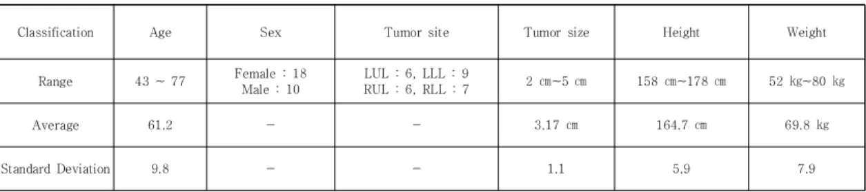
The chest 4DCT images on 36 patients for the pulmonary radiotherapy in hospitals in Daejeon during October, 2015 were used as shown in Table 1. The movements in the point of interest according to the respiratory pattern had to be evaluated. 4 patients with unstable or irregular respiratory patterns, 2 patients with vertebral deformation; in the case of having vertebral deformation on selecting the point of interest in the vertebrae or lung, it becomes a factor on generating big errors on the evaluation, and 2 patients that were not set with the position on the lungs.
2.2 Methods
The measurement points are set to measure the moving distance according to respiration.
However, the measurement points are set in each segment by dividing into upper, middle and lower parts due to the morphologic characteristics of the lung. For the set standard on the upper part, the central point of the No. 2 thoracic vertebrae was set as the standard, and the middle part was No. 6 thoracic vertebrae.
Lastly, the central point of the No. 11 thoracic vertebrae was set on the lower part as the standard. Also, the upper, middle and lower parts were divided into left lung and right lung, and as shown in Fig. 1 to 3, 3 measurement points were set for each segment. The measurement points were set differently for each patient depending on the organ shape and body type. Therefore, in the standard of the central point of the thoracic vertebrae, in the distance increased with 5 cm from the closest distance in contact with the lung tissue, the central point of the angle enabled with the lung tissue was designated as the medial point. Also, in the standard of the central point of the thoracic vertebrae, in the distance decreased 5 cm from the longest distance in contact with the lung tissue, the central point of the angle enabled with the lung tissue was designated as the lateral point. Moreover, the central point of the angle enabled with the lung tissue in the central distance between the medial point distance and lateral point distance was designated as the center point. However, when the central point of angle was not the lung tissue,
Classification Age Sex Tumor site Tumor size Height Weight
Range 43 ~ 77 Female : 18 Male : 10
LUL : 6, LLL : 9
RUL : 6, RLL : 7 2 ㎝~5 ㎝ 158 ㎝~178 ㎝ 52 ㎏~80 ㎏
Average 61.2 - - 3.17 ㎝ 164.7 ㎝ 69.8 ㎏
Standard Deviation 9.8 - - 1.1 5.9 7.9
Table 1. Information of Subject
the point relevant to the lung tissue among the closest angles was set. For the moving distance measurement of each measurement point that was set, the movement according to the respiration pattern on the CT console was replayed repeatedly, and the moving distance of the particular bronchial tube on the relevant point was reflected by indicating the distance through the measure on the console. As shown in Formula (1), the moving distance was calculated through the Vector Calculation on Craniocaudal, Anteriorposterior and Mediolateral.
Fig. 1. Measurement points in T2 level
Fig. 2. Measurement points in T6 level
Fig. 3. Measurement points in T11 level
R
tot=√{(R
vrt)
2+(R
lng)
2+(R
lat)
2} - Eq. (1)
R
tot: Total motion range
R
vrt: Anteriorposterior axis motion range R
lng: Craniocaudal axis motion range R
lat: Mediolateral axis motion range
In the standard of the moving distance according to the respiration pattern for each measurement point, the error value PDVs on the radiotherapy with Cyberknife in Fig. 4 were compared to evaluate the usefulness of the vertebral tracking technique and the synchrony technique for each measurement point.
Fig. 4. The console monitor of cyberknife
3. Result
3.1 The range of motion for each positions
The moving distances of 3 axial directions for each point on total of 28 patients are shown from Fig. 5 to 7. Also, The average moving distance and standard deviation for each axial direction per each point is shown from Table 2.
Fig. 5. Result of motion on T2 level
Fig. 6. Result of motion on T6 level
Fig. 7. Result of motion on T11 level
Vertical Longitudinal Lateral
Right Lung T2
Med. 1.3±0.25 1.2±0.27 1.1±0.23 Cnt. 1.9±0.46 1.4±0.32 2.3±0.48 Lat. 2.3±0.48 2.3±0.48 2.0±0.34
T6
Med. 1.5±0.31 1.8±0.43 1.4±0.21 Cnt. 8.3±1.68 11.3±2.39 8.5±1.63 Lat. 10.7±2.48 14.2±2.72 9.8±2.05
T11
Med. 1.7±0.35 1.4±0.34 1.4±0.3 Cnt. 18.5±4.03 32.1±7.32 8.8±2.27 Lat. 18.2±3.92 22.2±4.79 9.1±2.11
LeftLung T2
Med. 1.4±0.30 1.2±0.24 1.2±0.28 Cnt. 2.0±0.42 1.6±0.28 1.9±0.43 Lat. 1.9±0.43 2.0±0.38 2.1±0.48
T6
Med. 1.5±0.35 1.7±0.35 1.2±0.24 Cnt. 7.7±1.55 11.3±2.53 8.5±1.85 Lat. 10.0±2.15 14.3±3.29 9.5±1.72
T11
Med. 1.7±0.33 1.4±0.26 1.4±0.26 Cnt. 18.5±4.17 9.5±2.19 9.0±1.88 Lat. 18.3±3.56 9.8±2.43 9.3±2.00 Table 2. The data in column Mean value and Standard
deviation of motion length[mm]
At the T2 level, only the left, right, and right lungs were observed within 3 mm from the medial, medial, and lateral sides. In the case of T6 level, only the motion within 2 mm from the inner side was observed but the middle was from 7 mm to 12 mm, unlike the T2 level. In the case of the outer side, the movement was from 9 mm
to 15 mm, and the difference between the left and right was within the error range. In the case of T11 level, we could observe the movement within 2 mm of the medial side and the large movement from 8 to 33 mm in the medial side.
In the case of the outer side, the movement from 9 mm to 23 mm was observed, and it was confirmed that the range of movement was narrower than the intermediate position. And the right side showed a higher range than the left side. This is judged by the effect of the heart.
3.2 Assessment of PDV
The PDV for each node on the Cyberknife therapy is as in Fig. 8, and the range of PDV measuring the moving distance according to each position and direction, and the result of comparison are shown in Fig. 9.
Fig. 8. The graph of PDV
Fig. 9. The graph of measurement value and mean PDV
In the case of PDV, the actual clinical results
show that the tolerance of motion within a
maximum of 4.5 mm is acceptable. Therefore,
the motion of each position was set to an error
limit of 4.5 millimeters. As a result, the validity of
the synchrony technique could not be guaranteed for
all the T2 levels. The T6 and T11 levels were within the error range only from the inside.
4. Discussion
The Cyberknife therapeutic apparatus for stereotactic radiotherapy uses the synchrony technique on the pulmonary tumor. However, for the tumors near the vertebrae, there is no standard provided for selection between the vertebral tracking technique and the synchrony technique, so the change in the lung tissue according to the distance from the vertebrae is evaluated to provide the standard for selecting the therapeutic technique. The upper, middle and lower parts of the lung were divided into left and right, and the movement of the lung tissue was compared on the short, medium and long distances from the vertebrae. The comparison results showed that the points included within the posture error value of PDV during the therapy were only the short distance from the vertebrae for both left and right sides in case of total upper part of the lungs and the middle and lower parts. Also, other points showed very high values than the PDV. The movement of the tissues according to each position was evaluated to provide the standard for selecting the therapeutic technique according to the position of the pulmonary tumor. However, the evaluation positions are very limited, and there may be concerns of having difference for each subject due to the different shapes and functions of the lungs on selecting the position. Therefore, to set the standard on the position of the lungs, classification should be enabled according to the positions and functions of the pulmonary tumors, and the measurement points on the middle and lower parts should be segmentalized more in detail based on the results of this study to be evaluated. This study suggests that it is possible to utilize the existing treatment method as a
basic data to improve the ineffective treatment and to reduce the burden on the patient and the operator and to aim for efficient treatment. In addition, the stability, respiration pattern and stability of patients should be considered in further studies to provide more reliable and detailed standard. Most of the cyber knife treatments for pulmonary tumors used in clinical practice are usually applied with synchrony technique. Therefore, although the data to be compared with the experimental results are insufficient, future studies will examine reliability through continuous data collection.
5. Conclusion
As a result of applying the average moving
distance for each measurement point with 2.1
mm as the error value of the patient of PDV on
the Cyberknife therapy to 28 patients in this
study, all measurement points in the No. 2
thoracic vertebrae height were within the PDV
value, and in the No. 6 thoracic vertebrae and
No. 11 thoracic vertebrae heights, only the
measurement points close to the vertebrae were
shown to be within the PDV value. Also, other
points showed very high values than the PDV
value. As for the synchrony technique, the
exposure amount is higher than that of the
vertebral tracking technique in CT simulation,
and long preparation and therapy time are
required on the cyberknife therapy. Therefore, in
the case of radiotherapy of pulmonary tumor by
using the Cyberknife, the apex of the lung should
use the Vertebral tracking technique, and the
middle and lower parts of the lung should use
the vertebral tracking technique only when the
point is within 5 mm range from the closest
point from the center of the vertebrae. For other
points, the synchrony technique should be used
for therapy.
REFERENCES
[1] Y. B. Kim & C. O. Suh. (2008). Evolution of Radiotherapy: High-precision Radiotherapy, Journal of the Korean Medical Association, 51(7), 604-611.
DOI : 10.5124/jkma.2008.51.7.604
[2] P. Giraud et al. (2011). Centers, Respiratory gating techniques for optimization of lung cancer radiotherapy, Journal of thoracic oncology, 6(12), 2058-2068.
DOI : 10.1097/JTO.0b013e3182307ec2
[3] T. Biswas et al. (2012). SU‐E‐T‐422: Lung SBRT Using Cyberknife: Technique and Treatment Outcome, Medical physics, 39(6Part16), 3801-3802.
DOI : 10.1118/1.4735511|
[4] P. C. Gerszten et al. (2004). CyberKnife frameless stereotactic radiosurgery for spinal lesions: clinical experience in 125 cases, Neurosurgery, 55(1), 89-99.
DOI : 10.1227/01.neu.0000440704.61013.34
[5] S. Y. Seo, M. S. Han, C. G. Kim, M. C. Jeon, Y, K, Kim
& G. J. Kim. (2017). A study on the usefulness of a fusion model designed cloak shield to reduce the radiation exposure of the assistant during CT of severely injured patient. Journal of the Korea Convergence Society, 8(9) 211-216.
DOI : 10.15207/JKCS.2017.8.9.211
[6] Y. J. Jeong & S. H. Kim. (2015). Useful evaluation of 3D target location correction by using Xsight spine tracking system in CyberKnife. Journal of Digital Convergence, 13(1), 331-339.
DOI : 10.14400/JDC.2015.13.1.331
[7] B. H. Han et al. (2014). Evaluation of the Reproducibility of Radiation Output from Diagnostic X-ray Equipment(Standards Based on IEC 60601-2-54).
Journal of Digital Convergence, 12(2), 555-561.
DOI : 10.14400/JDC.2014.12.2.555
[8] K. Y. Lee, B. G. Jung, J. W. Kim, J. S. Park & B. H.
Jeong, (2018). Simulation of the High Frequency Hyperthermia for Tumor Treatment. Journal of the Korea Convergence Society, 9(3), 257-263.
DOI : 10.15207/JKCS.2018.9.3.257
[9] T. R. Mackie et al. (2003). Image guidance for precise conformal radiotherapy, International Journal of Radiation Oncology* Biology* Physics, 56(1), 89-105.
DOI : 10.1016/S0360-3016(03)00090-7
[10] S. Webb. (1991). Optimization by simulated annealing of three-dimensional conformal treatment planning for radiation fields defined by a multileaf collimator, Physics in medicine and biology, 36(9), 1201.
DOI : 10.1088/0031-9155/36/9/004
[11] L. Leksell. (1983). Stereotactic radiosurgery, Journal of Neurology, Neurosurgery & Psychiatry, 46(9), 797-803.
DOI : 10.1136/jnnp.46.9.797
[12] J. Kim, M. Han, S. Yoo, K. Kim & J. H. Cho. (2015).
Improvement of Beam-Quality Evaluation Method for
Medical Linear Accelerator Using Magnetic Field, Journal of Magnetics, 20(2), 120-128.
DOI : 10.4283/JMAG.2015.20.2.120
[13] K. H. Wong, S. Dieterich J. Tang & K. Cleary. (2007).
Quantitative measurement of CyberKnife robotic arm steering, Technology in cancer research & treatment, 6(6), 589-594.
DOI : 10.1177/153303460700600601
[14] M. Sarfaraz. (2007). CyberKnife® robotic arm stereotactic radiosurgery, Journal of the American College of Radiology, 4(8), 563-565.
DOI : 10.1016/j.jacr.2007.05.003
[15] Y. S. Kim. (2008). Cyberknife Robotic Radiosurgery System for Cancer Treatment, Journal of the Korean Medical Association, 51(7), 630-637.
DOI : 10.5124/jkma.2008.51.7.630
[16] T. S. Suh & I. H. Kim. (2008). Physical and Biological Background of Radiosurgery, Journal of the Korean Medical Association, 51(1), 16-26.
DOI : 10.5124/jkma.2008.51.1.16
[17] A. Schweikard, G. Glosser, M. Boddulur, M. J. Murphy
& J. R. Adler. (2000). Robotic motion compensation for respiratory movement during radiosurgery, Computer Aided Surgery, 5(4), 263-277.
DOI : 10.3109/10929080009148894
[18] K. Kitamura et al. (2002). Three-dimensional intrafractional movement of prostate measured during real-time tumor-tracking radiotherapy in supine and prone treatment positions, International Journal of Radiation Oncology* Biology* Physics, 53(5), 1117-1123.
DOI : 10.1016/S0360-3016(02)02882-1
[19] A. Schweikard, H. Shiomi & J. Adler. (2004).
Respiration tracking in radiosurgery, Medical physics, 31(10), 2738-2741.
DOI : 10.1118/1.1774132|
[20] W. Kilby, J. R. Dooley, G. Kuduvalli, S. Sayeh & C. R.
Maurer Jr. (2010). The CyberKnife® robotic radiosurgery system in 2010, Technology in cancer research & treatment, 9(5), 433-452.
DOI : 10.1177/153303461000900502
[21] M. Hoogeman, J. B. Prévost, J. Nuyttens, J. Pöll, P.
Levendag & B. Heijmen. (2009). Clinical accuracy of the respiratory tumor tracking system of the cyberknife: assessment by analysis of log files, International Journal of Radiation Oncology* Biology*
Physics, 74(1), 297-303.
DOI : 10.1016/j.ijrobp.2008.12.041
[22] C. Ozhasoglu et al. (2008). Synchrony–cyberknife respiratory compensation technology, Medical Dosimetry, 33(2), 117-123.
DOI : 10.1016/j.meddos.2008.02.004
[23] R. Shirazi, P. M. Goldfarb, D. B. Fuller & H. Sanati.
(2011). CyberKnife stereotactic body radiation therapy
for palliation and local control in patients with
advanced pancreatic cancer: A retrospective review,
Journal of Clinical Oncology, 29(15_suppl), e14506-e14506.
DOI : 10.1200/jco.2011.29.15_suppl.e14506
[24] J. M. Kuhnigk et al. (2005). New tools for computer assistance in thoracic CT. Part 1. Functional analysis of lungs, lung lobes, and bronchopulmonary segments, Radiographics, 25(2), 525-536.
DOI : 10.1148/rg.252045070
김 갑 중(Gab-Jung Kim) [정회원]
․ 2011년 2월 : 건양대학교 보건학과(보 건학석사)
․ 2015년 2월 : 충북대학교 의용생체공 학과(공학박사 수료)
․ 2017년 3월 ~ 현재 : 송호대학교 방 사선과 교수
․ 관심분야 : 핵의학, 방사선치료학, 의 료영상정보
․ E-Mail : [email protected]
김 정 호(Jeong-Ho Kim) [정회원]
․ 2013년 8월 : 전북대학교 방사선과학 기술학과(이학석사)
․ 2017년 8월 : 전북대학교 방사선과학 기술학과(이학박사)
․ 2011년 9월 ~ 현재 : 건양대학교병원 방사선종양학과 팀원
․ 관심분야 : 방사선물리학, 방사선계측 학, 방사선치료학
․ E-Mail : [email protected]
배 석 환(Seok-Hwan Bae) [정회원]
․ 2005년 2월 : 건양대학교 보건학과 (보건학석사)
․ 2009년 8월 : 건양대학교 보건학과(보 건학박사)
․ 2008년 9월 ~ 현재 : 건양대학교 방사 선학과 교수
․ 관심분야 : 방사선학, 보건의료정책, 의 료영상학
․ E-Mail : [email protected]
김 낙 상(Na-Sang Kim) [정회원]
․ 2000년 2월 : 경산대학교 보건학과 (보건학석사)
․ 2011년 2월 : 대구한의대학교 보건학 과(보건학박사)
․ 2012년 3월 ~ 현재 : 송호대학교 방 사선과 조교수
․ 관심분야 : 전산화단층촬영, 자기공명 영상, 진료영상학
․ E-Mail : [email protected]
서 선 열(Sun-Yeol Seo) [정회원]