치조능확장술과 자가치아골이식술을 이용한 치조능 수평증대술: 증례보고
분당서울대학교병원 치과 구강악안면외과, 치과보철과 김영균․이양진
점차적으로 폭경이 증가하는 나사들을 이용하여 치조능확장술을 시행하고 임프란트를 식립하였다. 임상적으로 양호한 결과를 얻었으며 수술 중 협측 피질골판 골절은 발생하지 않았다. 임프란트 주변 결손부와 협측 피질골판 상방에 자가치아골이식재를 이식함으로써 부가적인 치조능 확장 효과와 동시에 협측 피질골판 흡수를 보상하였 다. 치조능 폭경이 협소한 부위에 임프란트를 식립할 때 나사를 이용한 확장술은 성공적이고 예측 가능한 술식이 며 자가치아골이식재는 치조능증대술과 골유도재생술을 위해 사용될 수 있다.
주요어: 나사, 자가치아골이식재, 치조능확장술 (
구강회복응용과학지 2011:27(1):109~115)
서 론
임프란트 식립 시 치조능의 협설측 폭경이 부 족한 상황에 직면하는 경우가 많다. 이때 해결할 수 있는 방법은 좁은 폭경의 임프란트(narrow implant) 식립, 수평 베니어블록골이식(horizontal veneer block bone graft), 수평골유도재생술 (horizontal guided bony regeneration), 치조능분할 술(ridge splitting procedure) 등이 있다.
1)이 중 치 조능분할술은 골의 높이와 기저골의 폭경은 적 절하지만 치조능 상방부의 폭이 좁은 치조골을 가진 경우 골의 신축성을 이용하여 좁은 치조정 을 넓히는 술식으로 주로 상하악 전치부에 많이 사용되고 있다. 이 술식은 치조골의 순측 형태를 회복시킴으로써 심미적인 효과를 얻을 뿐만 아 니라 협설면의 피질골이 잘 보존될 경우 풍부한
교신저자:
이양진
경기도 성남시 분당구 구미동 300 분당서울대학교병원 치과보철과 부교수
Tel: 82-31-787-7546, Fax: 82-31-787-4068, E-mail: [email protected]
원고접수일: 2011년 01월 30일, 원고수정일: 2011년 02월 17일, 원고채택일: 2011년 03월 25일
혈행 공급으로 임프란트의 골유착을 증진시키는
효과가 있다.
2-4)그러나 수술 도중에 협측골이 파
절되어 분리될 위험성이 있으며 이것을 최소화
하는 것이 매우 중요하다. 치조능분할술은 주로
골절도와 말렛을 이용하여 협측 피질골의 불완
전골절(greenstick fracture)을 야기함으로써 치조
골을 협설측으로 증대시키는 방법이 많이 사용
되었다. 이는 환자의 불편감이 크고 과도하게 큰
힘을 가할 때 협측골의 파절 위험성이 증가하는
문제점이 있다. 따라서 저자 등은 점차적으로 폭
경이 증가하는 나사(screws)들을 이용하여 단계
적으로 치조능을 확장시킨 후 임프란트를 동시
에 식립하고 주변 결손부와 협측에 자가치아골
이식술을 시행함으로써 양호한 결과를 얻은 증
례를 소개하고자 한다.
증례보고
63세 여자 환자가 상악 양측 중절치가 소실된 상태로 내원하였다. 2주전 계단에서 넘어지면서 치아가 소실되었고 #37 치아의 유동성이 매우 심한 상태였다. #37 발치 후 자가치아골이식재로 처리한 후 상악 중절치 부위 임프란트 식립 시 골이식재로 사용하기로 계획하였다. 2009년 12 월 7일 #11-21 부위 치조정 절개를 가하여 피판 을 거상한 결과 치조능의 협설측 폭경이 매우 협 소한 것이 관찰되었다. #11, 21 부위 초기 드릴링 시행 후 SplitMaster expanding screws(SplitMaster
Ⓡ,
Fig. 1. Preoperative intraoral photograph. Both maxillary central incisors were lost 2 weeks ago.
Fig. 2. Preoperative intraoral occlusal view.
Fig. 3. Preoperative panoramic radiograph.
Fig. 4. Mucoperiosteal full-thickness flap was elevated. Buccolingual ridge width is inadequate. Labial concavity is observed.
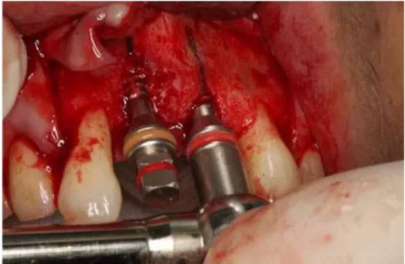
Fig. 5. Ridge was gradually expanded using
Split-Master expander. Alveolar bone
fracture didn’t develop, however, labial
bony dehiscence developed around
right central incisor implant.
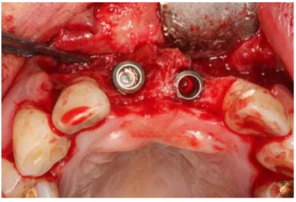
Fig. 6. Implants(ASTRA, 4D/11L) were installed.
Bony dehiscence defects were observed (#11 labial 8mm, mesial 3mm, palatal 2mm, #21 labial 3mm, palatal 1mm).
Fig. 7. Autogenous tooth bone graft material was grafted around the peri-implant defects and over the labial cortical plate.
Mr. Curette, Seoul, Korea)을 이용하여 치조능을 확장시킨 후 임프란트를 식립하였다(ASTRA 4D/11L). 식립 후 임프란트 주변 골결손부와 협 측 피질골 상방에 자가치아골이식재를 이식하고 흡수성 콜라겐막(Ossix plus)을 피개한 후 창상을 일차봉합하였다. 2010년 3월 25일 이차수술을 시 행하였으며 임프란트 주변 골결손부는 모두 신 생골로 잘 충전된 상태였다. 2010년 5월 6일 상 부 보철물이 장착되었다(Fig. 1~13).
Fig. 8. Resorbable collagen membrane(Ossix plus) was covered and primary closure was performed.
Fig. 9. Panoramic radiograph after implant placement.
Fig. 10. Intraoral occlusal view before second
surgery. Three point five months have
passed after implant placement.
Fig. 11. Implants were exposed. Favorable healing of autogenous tooth bone graft is observed.
Fig. 12. Periapical radiograph 3 months after upper prosthetic delivery.
총괄 및 고안
치조능분할술은 1990년 Bruschi 등
5)에 의해 greenstick fracture technique으로 처음 소개된 이 후 ridge widening, split crest procedure, staged ridge splitting등 여러 용어로 문헌에 소개되며 널
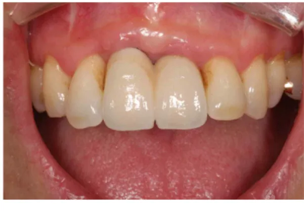
Fig. 13. Intraoral photograph 3 months after upper prosthetic delivery. Oral hygiene is poor and careful maintenance care is necessary.
리 사용되고 있다.
5-8)이 술식은 일반적으로 좁은 폭경의 치조능에 치즐이나 골절도 등으로 greenstick fracture을 일으켜 임프란트 수용부 및 골이식부를 형성하여 부가적인 공여부의 형성 없이 적절한 위치에 넓은 폭경의 임프란트 식립 을 가능하게 한다. 그리고 골재생이 양쪽에서 이 루어 지므로 조직 치유 능력이 좋아 상대적으로 적은 양의 골로 충분히 골유착을 증진시킬 수 있 다. 그리고 임프란트 식립을 위하여 Distraction osteogenesis 는 최소 3개월, GBR은 6개월을 기다 려야 하는 반면, 치조능분할술은 시행 후 1차 식 립 수술이 당일 혹은 1개월 이내 가능하다.
1)분 할술 이후 발생한 중간 공간에 골이식이 필요한 지, 임프란트를 동시에 식립할 경우 골이식재가 골유착을 증진시키는 지에 관해서는 아직 논란 의 대상이 되고 있다.
9)그런데 이 술식은 골의 탄 력성을 이용하는 방법이기 때문에 양측 피질골 사이에 반드시 해면골이 존재할 경우에 시도해 야 한다. 따라서 골유도재생술이나 온레이 블록 골 이식에 비해 적용범위가 제한적일 수 있다.
임프란트의 초기 고정을 얻을 수 있는 기저골의
폭이 매우 협소한 경우나 무치악 치조골의 경사
가 불량한 경우에는 심미적으로 불량한 상부 보
철물이 제작될 수 있으므로 다른 술식을 선택하 는 것이 좋다. 술식의 적응증은 잔존 폭경이 최 소 4mm 이상이어야 하며 폭경이 3mm 이하인 경 우는 다른 골이식술을 부가적으로 시행하고 일 정 치유기간을 가진 후 임프란트를 식립해야 한 다는 보고가 있다.
10)Ridge splitting은 여러 문헌에서 97~97.6%로 높 은 성공율이 보고된 바 있으나 피질골 골절 및 치조정골 흡수, 신경 손상 등의 합병증이 발생할 수 있다. 치조능 분할 후 임프란트를 식립할 때 협순측골이 부분 파절되는 경우가 많으며 이것 은 추후 골개조 과정에서 치조정골 흡수를 유발 할 것이다. 따라서 골절이 발생한 경우엔 부가적 인 골이식과 차단막을 사용하는 것이 흡수를 최 소화할 수 있다. 치조능을 분할한 후 확장시킬 때 깊은 부위에서부터 팽창을 시도해야 한다. 상 방에서 약 3-4mm의 얕은 부분에서 확장을 시도 하면 파절되는 경우가 대부분이다. 파절되지 않 았다 하더라도 임프란트를 식립하는 과정에서 협측 치조골이 파절되는 경우가 많다. 피질골 파 절은 치유과정 중에 흡수가 불가피하게 되면 연 조직 퇴축 등으로 인한 심미적 문제점을 초래할 수 있다.
11-14)임프란트를 식립한 후 협설측 피질 골의 두께가 최소 2mm 이상이어야 치유 기간 중 흡수를 보상할 수 있다. 따라서 치조능 분할 후 협측의 얇은 피질골은 방치할 경우 거의 흡수된 다고 예상하는 것이 좋다. Chiapasco등
3)은 하악 에서 screw type의 기구를 이용하여 4~5일에 걸 쳐 하루에 1mm 씩 점진적 골 확장을 시행한 증 례들을 보고하였으며 피질골 파절을 최소화할 수 있다고 언급하였다.
따라서 저자 등은 점진적으로 폭경이 증가하 는 나사들을 이용하여 치조능의 폭경을 증가시 킨 후 임프란트를 동시에 식립하였다. #11 임프 란트 협측 골열개가 약간 발생하였지만 치조골 파절은 발생하지 않았다. 임프란트 주변 결손부 와 협측 피질골의 흡수를 예상하고 자가치아골 이식재를 이용한 수평골유도재생술을 병행함으 로써 비교적 양호한 결과를 얻을 수 있었다. 그
러나 지나치게 협소한 부위에서 이술식을 적용 할 경우엔 기존의 방법과 마찬가지로 협측 피질 골 파절을 피할 수는 없다. 따라서 기존의 치조 능분할술에 적용되는 적응증을 반드시 준수할 것을 추천한다. 본 연구에서 사용된 screw type 의 기구는 비교적 약한 힘으로 깊은 부위에서 순 차적으로 골을 확장시켜 피질골 골절 방지에 보 다 효과적일 것으로 사료된다. 그리고 통상의 malleting 과정을 최소화 하여 환자의 머리울림 및 그로 인한 두통, 턱관절 손상 등의 부작용이 없는 장점이 있다.
결 론
저자 등이 제시한 술식의 장점은 다음과 같으 며 치조능 폭경이 협소한 증례에서 선택적으로 사용할 경우 좋은 결과를 얻을 수 있다고 사료된 다.
1. 골절도, 말렛 사용으로 인한 불편감을 최소화 하면서 치조능 확장 효과를 얻을 수 있다.
2. 협측 피질골 파절을 최소화할 수 있다.
3. 자가치아골이식재를 이용한 골유도재생술을 병행함으로써 협측 피질골 흡수를 보상하고 임프란트 골유착에 긍정적인 효과를 발휘할 수 있다.
참 고 문 헌
1. Kim YK, Kim SG, Lee BG. Bone graft and implant.
Vol. 2-2. Narae pub, Seoul, Korea. 2007. 435-467.
2. Basa S, Varol A, Turker N. Alternative bone expansion technique for immediate placement of implants in the edentulous posterior mandibular ridges: A clinical report. Int J Oral Maxillofac Implants. 2004; 19: 554-558.
3. Chiapasco M, Ferrini F, Casentini P et al. Dental implants placed in expanded narrow edentulous ridges with the Extension-Crest device: a 1 to 3-year multicenter follow-up study. Clin Oral Implants Res.
2006; 17: 265-272.
4. Engelke WG, Diederchs CG, Jacobs HG, Deckwer I.
Alveolar reconstruction with splitting osteotomy and microfixation of implants. Int J Oral Maxillofac Implants. 1997; 12: 310-318.
5. Bruschi GB, Calesini G, D'Ambrosio F, Scipioni A.
Programization for insertion of osseointegrated implants. Attual Dent. 1990; 6: 10-15.
6. Duncan JM, Westwood M. Ridge widening for the thin maxilla: A clinical report. Int J Oral Maxillofac Implants. 1997; 12: 224-227.
7. Enislidis G, Wittwer G, Ewers R. Preliminary report on a staged ridge splitting technique for implant placement in the mandible: A technical note. Int J Oral Maxillofac Implants. 2006; 21: 445-449.
8. Simon M, Saldoni M, Zaffe D. Jawbone enlargement using immediate implant placement associated with a split-crest technique and guided tissue regeneration.
Int J Periodontics Restorative Dent. 1992; 12:
462-473.
9. Lustmann J, Lewinstein I. Interpositional bone grafting technique to widen narrow maxillary ridge.
Int J Oral Maxillofac Implants. 1995; 10: 568-577.
10. Jensen OT, Cullum DR, Baer D. Marginal bone stability using 3 different flap approaches for alveolar split expansion for dental implants: a 1-year clinical study. J Oral Maxillofac Surg. 2009; 67:1921-1930.
11. Sethi A. Kaus T. Maxillary ridge expansion with stimutaneous implant placement : 5-year results of an ongoing clinical study. Int J Oral Maxillofac Implants. 2000; 15: 491-499.
12. Ferrigno N. Laureti M . Surgical advantages with ITI TE implant placement in conjugation with split crest technique : 18-month results of an ongoing prospective study. Clin Oral Implants Res. 2005; 16:
147-158.
13. Scipioni A, Bruschi GB, Calesini G. The edentulous ridge expansion technique: A five-year study. Int J Periodontics Restorative Dent. 1994; 14: 451-459.
14. Scipioni A, Bruschi GB, Calesini G. The edentulous
ridge expansion technique: A five-year study. Int J
Periodontics Restorative Dent. 1994; 14: 451-459.
Horizontal Ridge Augmentation using Ridge Expansion and Autogenous Tooth Bone Graft: A Case Report
Young-Kyun Kim
1, D.D.S.PhD., Yang-Jin Yi
2, D.D.S.PhD.
1
Department of Oral and Maxillofacial Surgery,
2
Department of Prosthodontics, Section of Dentistry, Seoul National University Bundang Hospital
Implants were placed after performing ridge expansion by inserting screws of gradually increasing thickness. Favorable clinical outcome was obtained. During surgery, buccal cortical plate fracture did not occur. Autogenous tooth bone graft material was grafted around the implant dehiscence defects and over the buccal cortical plate. The method involving the insertion of screws for ridge expansion is a successful and predictable technique for implant placement in narrow alveolar bone. Autogenous tooth bone graft material can be used for ridge augmentation and GBR.
Key words: autogenous tooth bone graft material, ridge expansion, screws
Correspondence to : Yang-Jin Yi, DDS,MSD,PhD
Department of Prosthodontics, Section of Dentistry, Seoul National University Bundang Hospital, 300 Gumi-dong, Bundang-gu, Seongnam-si, Gyeongi-do, 463-707, Korea
Tel: 82-31-787-2780,7546, Fax: 82-31-787-4068, E-mail: [email protected]
Received: January 30, 2011, Last Revision: February 17, 2011, Accepted: March 25, 2011