Copyright ⓒ 2009, The Korean Academy of Oral Biology
97
Journal of Oral Biology
L-trans-pyrrolidine-2,4-dicarboxylate (PDC) induces Excitotoxic and Oxidative Neuronal Death in Cultured Cortical Neurons
Seung Joon Choi
1, Shinae Hwang
1, Do Kyung Kim
2, and Jong-Keun Kim
1*
1
Department of Pharmacology and Center for Biomedical Human Resources (BK 21 Project) Chonnam National University Medical School, Gwangju 501-746, Republic of Korea
2
Department of Oral Physiology, Chosun University School of Dentistry, Gwangju 501-759, Republic of Korea (received May 12, 2009 ; revised May 24, 2009 ; accepted May 29, 2009)
L-trans-pyrrolidine-2,4-dicarboxylate (PDC) is a potent inhibitor of glutamate transporters. In our current study, we investigated whether the neuronal death induced by PDC involves mechanisms other than excitotoxicity in mixed mouse cortical cultures. Cortical cultures at 13-14 days in vitro were used and cell death was assessed by measuring the lactate dehydrogenase efflux into bathing media. Glutamate and PDC both induced neuronal death in a concentration-dependent manner but the neurotoxic effects of glutamate were found to be more potent than those of PDC. Treatment with 10, 100 and 200 µM PDC equally potentiated 50 µM glutamate-induced neuronal death. The neuronal death induced by 75 µM glutamate was almost abolished by treatment with the NMDA antagonists, MK- 801 and AP-5, but was unaffected by NBQX (an AMPA antagonist), trolox (antioxidant), BDNF or ZVAD-FMK (a pan-caspase inhibitor). However, the neuronal death induced by 200 µM PDC was partially but significantly attenuated by single treatments with MK-801, AP-5, trolox, BDNF or ZVAD-FMK but not NBQX. Combined treatments with MK-801 plus trolox, MK-801 plus ZVAD-FMK or MK-801 plus BDNF almost abolished neuronal death, whereas combined treatments with trolox plus ZVAD- FMK, trolox plus BDNF or ZVAD-FMK plus BDNF did not enhance the inhibitory action of any single treatment with these drugs. These results demonstrate that the neuronal death induced by PDC involves not only in the excitotoxicity induced by the accumulation of glutamate but also the
oxidative stress induced by free radical generation. This suggests that apoptotic neuronal death plays a role in PDC- induced oxidative neuronal injury.
Key words: L-trans-pyrrolidine-2,4-dicarboxylate, Excito- toxicity, Oxidative Stress, Apoptosis, Neuronal Cultures
서 론
중추신경계의 대표적 신경전달물질인 glutamate는 정상 상태에서 세포외 액 농도가 1 µM 이하로 유지되어야 신 경세포가 생존할 수 있다 (Schousboe et al, 1997). 세포 외 액 glutamate의 농도는 glutamate 수송체 (transporter) 를 통한 재흡수에 의해 조절된다 (Nicholls과 Attwell, 1990).
세포외 glutamate 농도 증가는 흥분독성 (excitotoxicity) 을 일으키며, 흥분독성은 각종 뇌 질환의 주요 병인으로 작용함이 보고 되어있다. 실제로 glutamate 수송체의 기 능저하와 이에 따른 흥분독성의 유발이 뇌허혈 (Rossi et al, 2000), 척수 손상 (Springer et al, 1997) 및 퇴행성 신경계 질환 (Harris et al, 1996; Lin et al, 1998)의 병 인이 될 수 있음이 보고 되어있다.
L-trans-pyrrolidine-2,4-dicarboxylate(L-PDC)는 현재 알 려져 있는 5 가지 흥분성 아미노산 수송체(Excitatory Amino Acid Transporter, EAAT 1-5) 모두를 강력하게 억제하는 약물로 glutamate 재흡수와 관련된 연구에 최 근까지 많이 사용되고 있다 (Nakagawa et al 2008). 이 약물은 EAAT1-4에는 경쟁적 transportable 억제제로 작 용하고 반면에 EAAT5에는 경쟁적 non-transportable 억 제제로 작용한다 (Wang et al, 1998; Gegelashvili et al,
*Corresponding author: Jong-Keun Kim, M.D Ph.D., Department of Pharmacology, Chonnam National University Medical School, 5 Hak-Dong, Gwangju 501-746, Republic of Korea.
Tel.: +82-62-220-4234, Fax.: +82-62-232-6974
E-mail: [email protected]
2000). 또한 이 약물은 연접 후 glutamate 수용체에는 작 용이 없음이 알려져 있다 (Blitzblau et al, 1996). PDC 의 glutamate 재흡수 억제작용이 배양된 해마신경세포에 서 glutamate에 의한 신경세포사멸을 증강시킨다는 보고 (Robonson et al, 1993) 및 배양된 흰쥐 대뇌 신경세포 에서 흥분독성을 일으킨다는 보고 (Blitzblau et al, 1996;
Velasco et al, 1996; Volterra et al, 1996) 등은 PDC 의 신경세포사멸 작용이 glutamate 재흡수를 억제하여 일 어남을 가리키고 있다. 그러나 in vivo 실험에서 PDC에 의한 세포외 glutamate의 증가가 신경세포사멸과 관계가 없다는 보고 (Massieu et al, 1995), PDC의 지속적인 주 입에 의한 선조체 신경세포사멸이 NMDA 수용체 길항제 인 MK-801로 억제되지 않는다는 보고 (Lievens et al, 1998), 흰쥐 해마와 선조체에 주입한 PDC가 세포 외액의 glutamate의 농도는 약 2-3배 증가시키지만 분명한 신경세 포사멸을 유발시키지 못한다는 보고 (Montiel et al, 2005) 등은 PDC에 의한 신경세포사멸에 glutamate 재흡수 억제 에 의한 흥분독성이외의 요인이 관계함을 시사하고 있다.
본 연구는 배양된 생쥐 대뇌피질세포에서 PDC의 신경 세포사멸 양상을 glutamate에 의한 신경세포사멸과 비교 하여 PDC에 의한 신경세포사멸 기전에 흥분독성 외의 다른 세포사멸기전이 관여하는지를 알아보고자 하였다.
재료와 방법
세포배양
신경세포-교세포의 혼합 배양은 먼저 교세포를 배양하 여 24-well 배양접시에 교세포 층을 만든 후 그 위에 다 시 신경세포를 배양하였다. 교세포 배양은 생후 1~2일된 생쥐 (ICR 계)에서 뇌조직을 적출하여 Ca++과 Mg++가 들어 있지 않은 Hanks' balanced salt solution (HBSS) 에 5 mg/ml glucose, 7 mg/ml sucrose 및 0.35 mg/ml NaHCO3가 첨가된 배양액 (이하 DM)에 넣고 입체현미 경하에서 뇌막을 제거하였다. 대뇌피질을 적출하여 잘게 조각을 내고 0.25% trypsin이 함유된 DM에 넣어 37oC 에서 15분간 처치한 후 1,000 ×g에서 5분간 원심분리하 였다. 그 후 trypsin이 들어있는 용액을 제거하고 분리된 세포를 Eagle's minimal essential medium (MEM)에 2 mM glutamine과 10% fetal bovine serum (FBS) 및 10%
horse serum (HS)이 포함된 배양액 (이하 PM) 1~2 ml 에 넣은 다음 구멍이 좁혀진 pipet으로 약 10회 trituration 하였다. 여기에 epidermal growth factor (EGF)를 10 ng/ml 농도로 첨가한 후 24-well plate (Primaria, Falcon)에 0.5 hemisphere/plate 밀도로 각 well 당 400 µl씩 plating하 여 37oC, 5% CO2와 100% 습도가 유지되는 CO2 배양 기 (Forma사, 미국)에서 배양하였다. 2~4주후 교세포가 충분히 증식하여 배양접시에 완전한 층을 만들면 임신
15~17일된 생쥐를 halothane으로 마취하여 경추전위로 희 생시켜 전신에 70% ethanol을 분무하여 충분히 적신 후 복강을 절개하여 생쥐 태아를 꺼내어 뇌를 적출하였다.
적출된 뇌에서 교세포 배양시와 같은 방법으로 대뇌피질 을 얻어 교세포가 배양된 plate에서 배양액을 완전히 제 거한 후 2.75 hemisphere/plate 밀도로 각 well 당 400 µl 씩 plating하여 37oC, 5% CO2, 100% 습도하에서 배양 하였다. 이때 사용한 PM은 교세포 배양시와 다르게 5%
FBS 및 5% HS로 보충하였다. 교세포의 증식을 억제하 기 위해 혼합배양 3~5일에 cytosine arabinoside를 10 µM 농도로 각 well에 처리하여 약 2일간 작용시켰다. 배양 액 교체는 교세포배양의 경우 2주후부터 1주일에 한번씩, 혼합배양에서는 일주일후부터 일주일에 2번 시행하였다.
약물처리
배양 13~14일 후에 실시하였다. 모든 경우에 세포에 대 한 약물 처리는 처리 전 배양액으로 3번 씻어 주고 나 서 실시하였고, 24-well 배양접시의 한 줄 (4 well)에 같 은 처리를 하였으며, 첫줄은 약물을 투여하지 않은 허위 처리군 (sham wash)으로 하였다. 둘째 줄은 NMDA (500µM)를 처리하여 신경세포만을 모두 죽이는 군 (full kill)으로 하였다. 셋째 줄에서 여섯째 줄까지를 약물 투 여 군으로 사용하였다.
모든 약물은 각 약물에 적절한 용매에 녹여 원액을 만 들어 각 well 당 4~8 µl씩 되게 배양액에 희석하여 배양 액 교환과 함께 투여하였으며, 손상 유발 약물의 억제작 용을 보기 위한 실험에서는 억제약물을 전 처리하거나 두 종류 약물을 동시에 같이 투여하였다.
세포사 측정
위상차 현미경으로 세포 손상 정도를 관찰하였다. 세포 사를 정량화하기 위하여 약물 투여 24 시간 후에 세포막 파괴 시 배양액 내로 유출된 lactate dehydrogenase (LDH) 의 양을 측정하였다. LDH 측정은 96-well plate의 각 well에 배양액 (25 µl)을 넣고 buffer (125 µl)와 0.3 mg/
ml NADH (100µl)를 작용시킨 후 22.7 mM pyruvate (30µl)를 넣어 곧 바로 microplate reader (Molecular Device사, 미국)를 이용하여 340 nm에서 OD값의 변동을 4분간 측정하였다. 이때 standard enzyme으로는 Sigma 사의 표준효소를 사용하였다. 측정치는 sham wash군과 full kill군의 차이를 100으로 하여 각 약물 투여군의 값 과 sham wash군과 차이를 상대적 백분율로 환산하여 평 균 ± 표준오차 (mean ± SEM)로 나타내었다.
사용 시약
Eagle's minimal essential medium, glutamine, Hanks' balanced salt solution은 Gibco사(MD, 미국) 제품을, fetal bovine serum 및 horse serum은 Hyclone사 (Ut, 미국)
제품을 55oC에서 30분간 비활성화 시켜 사용하였다. HEPES (acid), glucose, NaHCO3, NaCl, KCl, MgCl2, CaCl2, NaOH, phenol red와 그 외 trypsin, cytosine arabinoside, epidermal growth factor, sucrose, NMDA, MK-801, NBQX, AP-5, trolox 등은 Sigma-Aldrich사 (MO, 미국) 제품을, z-VAD-FMK는 Enzyme System Products사 (CA, 미국) 제품을, BDNF는 Leinco Technology Inc.(MO, 미 국) 제품을, L-PDC는 Tocris사 (Northpoint, 영국) 제품을 사용하였다.
성적분석
각 군간의 비교는 일측 ANOVA를 시행하여 유의한 경 우에 Student-Neuman- Keuls test로 각 군간의 유의성을 검정하였으며, p < 0.05인 경우 통계학적으로 유의하다고 판정하였다.
실험성적
Glutamate와 PDC의 신경세포사멸작용
Glutamate는 용량의존적인 신경세포사멸작용을 나타냈 다(Fig. 1). 즉 25 µM glutamate 24시간 처리로는 신경세 포사멸이 거의 일어나지 않았으나, 50, 75 µM로 증량함 에 따라 각각 60 ± 5.1% (8예), 80 ± 4.6% (8예)의 신경 세포사멸을 나타냈으며 100 µM처리는 거의 모든 신경세 포를 사멸시켰다(Fig. 1). PDC 역시 용량의존적인 신경 세포사멸을 유도하였으나 그 양상이 glutamate와 약간 차 이가 있었다. 즉 150 µM까지는 20%이하의 경미한 신경
세포사멸을 보이다가 200, 300 µM 처리로 88 ± 4.8%(12 예), 96 ± 6.3% (8예)의 신경세포사멸을 일으켰다(Fig. 1).
300µM 처리로 교세포 손상은 유발되지 않았다.
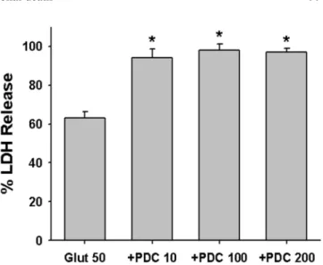
Glutamate에 의한 신경세포사멸에 대한 PDC의 영향 PDC처리가 glutamate에 의한 신경세포사멸에 어떻게 작용하는지를 알아보기 위해 glutamate 50 µM에 의한 신 경세포사멸에 대한 PDC의 영향을 조사하였다. 그 결과 10µM PDC처리에서부터 glutamate에 의한 신경세포사 멸을 유의하게 증가시켰다(Fig. 2). 즉 50 µM glutamate 24시간 처리로 63 ± 3.5% (8예)의 신경세포사멸을 보였던 것이 PDC 10, 100, 200 µM 동시 처리로 각각 94 ± 4.6%
(8예), 98 ± 3.2% (8예), 97 ± 2.1% (8예)로 신경세포사멸 이 증가되었다.
Glutamate에 의한 신경세포사멸에 미치는 각 종 약물 의 영향
Glutamate에 의한 신경세포사멸에 대한 NMDA 수용체 길항제인 MK-801와 AP-5, AMPA와 kainate 수용체 길 항제인 NBQX, 항산화제인 trolox, 신경성장인자인 BDNF 및 caspase 억제제인 ZVAD-FMK의 영향을 조사하였다.
그 결과 NMDA 수용체 길항제인 MK-801 (10 µM)과 AP- 5 (50µM) 처리는 75 µM glutamate에 의한 신경세포사 멸 (16예, 76 ± 4.8%)을 거의 완전히 억제하였으나 나머지 약물은 glutamate에 의한 신경세포사멸에 영향을 미치지 못하였다(Fig. 3).
PDC에 의한 신경세포사멸에 미치는 각 종 약물의 영향 Glutamate에 의한 신경세포사멸이 NMDA 수용체 길항
Fig. 1. Glutamate (
●) and L-trans-pyrrolidine-2,4-dicarboxylate
(PDC,
○) induced concentration-dependent neuronal death in mixed cortical cultures. Each point and bar is the mean ±SEM from 8-12 wells.
Fig. 2. Effect of treatment with 10, 100 or 200 µM L-trans-pyrroli-
dine- 2,4-dicarboxylate (PDC10, PDC100, PDC200) on the 50 µM
glutamate (Glut50)- induced neuronal death at the end of 24 hr
exposure. Mean ± SEM from 8-12 wells. *; Significantly different
from Glut50 control group (p < 0.05).
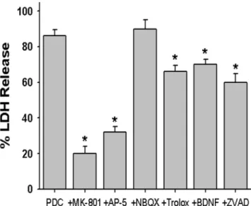
제인 MK-801과 AP-5 처리로만 억제되었으므로 PDC에 의한 신경세포사멸도 같은지를 알기위해 PDC에 의한 신 경세포사멸에 대한 MK-801, AP-5, NBQX, trolox, BDNF 및 ZVAD-FMK의 영향을 조사하였다. 그 결과 AMPA 및 kainate 수용체 길항제인 NBQX를 제외한 다른 약물 (10µM MK-801, 50 µM AP-5, 100 µM trolox, 100 ng/ml BDNF, 100µM ZVAD-FMK) 처리는 200 µM PDC에 의한 신경세포사멸 (24예, 86 ± 3.6%)을 소실시키지는 못 하였으나 유의하게 억제하였다(Fig. 4).
PDC에 의한 신경세포사멸에 대한 각종 약물 병용처 리의 영향
PDC에 의한 신경세포사멸을 각종 약물이 부분적으로 억제하였으므로 이들 약물의 병용처리의 효과를 조사하 였다. 먼저 MK-801과 NBQX의 병용작용을 조사한 결 과 두 약물의 병용처리가 MK-801의 억제작용을 강화하 지 못하였다. 그러나 MK-801과 trolox의 병용처리는 각 각의 약물을 단독 처리하였을 때 보다 현저하게 신경세 포사멸 억제작용이 강화되었다(Fig. 5). 즉 200 µM PDC 에 의해 86 ± 3.6% (24예)의 신경세포사멸이 10 µM MK- 801 및 100 µM trolox 단독 처리로 각각 20 ± 4.1% (12 예) 및 66 ± 3.4% (8예)로 억제되었으며, MK-801과 trolox 를 병용처리하면 2 ± 1.3% (8예)로 거의 소실되었다(Fig. 5).
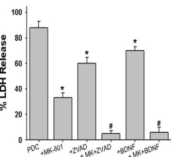
마찬가지로 MK-801과 ZVAD-FMK, MK-801과 BDNF 의 병용처리도 각각의 약물을 단독 처리하였을 때 보다 현저하게 신경세포사멸 억제작용이 강화되었다(Fig. 6). 즉 200µM PDC에 의해 88 ± 4.8% (16예)의 신경세포사멸이 10µM MK-801, 100 µM ZVAD-FMK 및 100 ng/ml BDNF 단독 처리로 각각 33 ± 3.9% (12예), 60 ± .4.8% (8예) 및 70 ± .3.0% (8예)로 억제되었으며, MK-801과 ZVAD- FMK 병용 처리에 의해 5 ± 2.1% (8예)로, MK-801과 BDNF의 병용처리에 의해 6 ± 3.8% (8예)로 PDC에 의 한 신경세포사멸이 거의 소실되었다(Fig. 6).
한편 이와는 다르게 trolox와 ZVAD-FMK, trolox와 BDNF 및 ZVAD-FMK와 BDNF의 병용처리는 단독 처리에 의한 PDC의 신경세포사멸 억제작용과 차이가 없었다(Fig. 7).
Fig. 4. Effect of treatment with 10 µM MK-801, 50 µM AP-5, 10 µM NBQX, 100 µM trolox, 100 ng/ml BDNF or 100 µM ZVAD-FMK on the neuronal death induced by 200 µM L-trans- pyrrolidine- 2,4-dicarboxylate (PDC) at the end of 24 hr exposure.
Mean ± SEM from 8-24 wells. *; Significantly different from PDC control group (p < 0.05).
Fig. 3. Effect of treatment with 10 µM MK-801, 50 µM AP-5, 10 µM NBQX, 100 µM trolox, 100 ng/ml BDNF or 100 µM ZVAD-FMK on the 75 µM glutamate (Glut)- induced neuronal death at the end of 24 hr exposure. Mean ± SEM from 8-16 wells.
*; Significantly different from Glut control group (p < 0.05).
Fig. 5. Effect of single or combined treatment with 10 µM MK-
801, 10 µM NBQX, 100 µM trolox on the neuronal death induced
by 200 µM L-trans-pyrrolidine-2,4-dicarboxylate (PDC) at the end
of 24 hr exposure. Mean ± SEM from 8-24 wells. *; Significantly
different from PDC control group (p < 0.05). #; Significantly dif-
ferent from MK-801-treated group (p < 0.05).
고 찰
본 실험에서 PDC (10, 100, 200 µM) 처리가 glutamate (50µM)에 의한 신경세포사멸을 유의하게 강화시켰으며, 강화 정도는 본 실험에서 사용한 PDC 세 용량 간에 차 이가 없었다. 10 µM PDC는 그 자체가 신경세포독성을
나타내지 않기 때문에 이 성적은 10 µM 농도에서 이미 glutamate 수송체를 억제하고 있음을 가리키고 있다. PDC 의 glutamate-신경독성 강화작용이 수송체의 억제에 의한 재흡수 억제작용뿐만 아니라 PDC가 수송체를 통해 재흡 수되어 glutamate의 유리를 촉진하는 작용도 있다는 보 고(Griffiths et al, 1994; Volterra et al, 1996)로 미루어 본 성적도 수송체 억제와 함께 glutamate 유리의 두 가지 작용이 합해져 나타난 것으로 판단된다.
본 실험에서 glutamate (75 µM)에 의한 신경세포사멸 을 NMDA 수용체 길항제인 MK-801와 AP-5가 거의 완 전히 억제하였고, AMPA 수용체 길항제인 NBQX 처리 는 억제작용을 나타내지 못하였다. 또한 PDC에 의한 신 경세포사멸도 NBQX는 억제하지 못하였으며, MK-801과 의 병용처리도 MK-801의 억제작용을 강화시키지 못하였 다. 이 성적은 본 실험에서 사용하는 생쥐 대뇌피질세포 배양에서 glutamate에 의한 신경세포사멸은 NMDA 수 용체의 활성화를 동반하나 AMPA나 kainate 수용체의 활 성화는 동반하지 않음을 가리키고 있다.
항산화제인 trolox, 신경성장인자인 BDNF 및 caspase 억제제인 ZVAD-FMK처리는 glutamate에 의한 신경세포 사멸에는 영향을 미치지 못하였으나, PDC에 의한 신경 세포사멸은 유의하게 억제하였다. 이 성적은 PDC에 의한 신경세포사멸이 glutamate에 의한 신경세포사멸과 그 기 전이 다름을 가리키고 있다. 또한 PDC에 의한 신경세포 사멸은 MK-801과 trolox, MK-801과 ZVAD-FMK, MK-801과 BDNF의 병용처리에 의해 거의 완전히 억제 되었다. Trolox가 세포막 지질과산화를 억제하는 대표적 항산화제로서 산화성 손상을 억제하는데 가장 광범위하게 사용되는 약물이라는 점 (Chow et al, 1994; Son & Chun, 2007)을 감안하면 본 실험에서 PDC에 의한 신경세포사 멸에 NMDA 수용체를 통한 흥분독성과 함께 산화성 손 상이 유발됨을 시사하고 있다. PDC가 배양된 해마 신경 세포에서 신경세포사멸을 유발하는데 이 때 흥분독성과 함께 산화성 손상이 관여한다는 보고 (Himi et al, 2003)는 본 실험과 일치하는 성적이다.
또한 신경성장인자인 BDNF가 세포자멸사를 억제한다는 보고 (Bamji et al, 1998; Estevez et al, 1998), ZVAD- FMK가 세포자멸사를 실행하는 효소인 caspase를 억제하는 약물 (Ekert et al, 1999; Nicotera et al, 1999; Thornberry, 1999)이라는 점과, 각 종 신경세포의 사멸에 세포자멸사 가 관여한다는 보고 (Huang et al, 1999; Padmanabhan et al, 1999) 등은 PDC의 신경세포사멸에 흥분독성과 함께 세포자멸사 과정이 관여함을 시사한다.
한편 trolox와 ZVAD-FMK, trolox와 BDNF 및 ZVAD- FMK와 BDNF의 병용처리는 단독 처리에 의한 PDC의 신경세포사멸 억제작용과 차이가 없었다. 이 성적은 산화 성 손상과 세포자멸사가 서로 상가적이지 않음을 나타내 는 것으로 이 두 세포사멸의 과정이 같은 기전을 통하여
Fig. 6. Effect of single or combined treatment with 10 µM MK-
801, 100 µM ZVAD-FMK, 100 ng/ml BDNF on the neuronal death induced by 200 µM L-trans-pyrrolidine-2,4-dicarboxylate (PDC) at the end of 24 hr exposure. Mean ± SEM from 8-16 wells.
*; Significantly different from PDC control group (p < 0.05). #;
Significantly different from MK-801-treated group (p < 0.05).
Fig. 7. Effect of single or combined treatment with 100 µM trolox,
100 µM ZVAD-FMK, 100 ng/ml BDNF on the neuronal death
induced by 200 µM L-trans-pyrrolidine-2,4-dicarboxylate (PDC)
at the end of 24 hr exposure. Mean ± SEM from 8-16 wells. *; Sig-
nificantly different from PDC control group (p < 0.05).
나타남을 시사한다. 즉 PDC에 의한 신경세포사멸에 산 화성 손상에 의한 신경세포자멸사가 유발됨을 의미한다.
산화성 손상과 세포자멸사와의 상관관계에는 많은 이견 이 존재하지만 일반적으로 산화성 손상의 강도에 따라 결정된다는 보고가 많다. 즉 손상강도가 크면 괴사가 유 발되고 손상강도가 약하고 오랜 시간동안 진행되면 세포 자멸사가 유발된다는 것이다 (Tan et al, 1998).
이상의 성적은 PDC에 의한 신경세포사멸은 glutamate 에 의한 신경세포사멸과는 달리 배지 내 glutamate 농도 의 증가에 의한 흥분독성 뿐만 아니라 산화성 손상이 관 여함을 가리키며, 이 산화성 손상에 세포자멸사 과정이 관여함을 시사하고 있다.
알 림
이 논문은 2004년도 전남대학교 학술연구비 지원에 의 하여 연구되었음.
참 고 문 헌