Introduction
The development of new implant surfaces and design has enabled a marked reduction of the initial healing period, even to the point of an immediate/
early loading of implants that show high primary sta- bility.1,2 The success of immediate or early loading is dependent on the ability of the clinician to determine the degree of primary stability and changes in stability along with new bone formation and remodeling.3 Pri- mary stability is determined by the bone density, im- plant design, and surgical technique.4,5 The assessment of primary stability is important for the prognosis of implant treatment. Several techniques have been suggested for the determination of implant stability.
Among the various methods, resonance frequency analysis (RFA) is recognized as a useful method for
documentation of clinical implant stability. RFA is a non-invasive intraoral method designed to reflect the bone/implant interface.4-8
The early loading protocol on the maxillary poste- rior single implant is still in doubt, because of defi- ciency in its bone quantity and/or quality. But with the development of a new implant system which has wide diameter and deepened thread (KnifethreadTM, MegaGen Implant Co., Gyeongsan, Korea), it became possible to get excellent initial stability, even at loose bone of the maxillary posterior area. This case report shows a new possibility of early loading at the maxil- lary posterior area with a special implant system. The fixture was evaluated clinically with implant stability quotient (ISQ) value, follow-up checked radiographi- cally for one year.
*Correspondence to: Hyun-Pil Lim, DDS
Department of Prosthodontics, School of Dentistry, Chonnam National University 33 Yongbong-ro, Buk-gu, Gwangju, 500-757, Republic of Korea
Tel: +82-62-530-5577, Fax: +82-62-530-5639, E-mail: [email protected] Received: June 16, 2014/Last Revision: August 14, 2014/Accepted: August 15, 2014
early loading on a maxillary posterior single implant with deepened threads: a case report
Chang-Hoon Han
1, Hyun-Seung Kim
2, Sang-Won Park
2,3, Kwi-Dug Yun
3, Han-Sung Joo
3, Hyun-Pil Lim
3*
1EasyPlant Dental Clinic, Gwangju, Republic of Korea
2RIS Foundation for Advanced Biomaterials, School of Dentistry, Chonnam National University, Gwangju, Republic of Korea
3Department of Prosthodontics, School of Dentistry, Chonnam National University, Gwangju, Republic of Korea
This case report shows an early loading at the maxillary posterior area with the wide diameter implant which has a deepened threads after removal of failed implant. Implant Stability Quotient (ISQ) value has represented favorable result for one year. This clinical report describes the potential of early loading on a maxillary posterior single implant with deepened threads. (J Dent Rehabil Appl Sci 2014;30(3):253-8)
Key words: deepened threads; primary stability; early loading; posterior maxillae
Copyright© 2014 The Korean Academy of Stomatognathic Function and Occlusion.
It is identical to Creative Commons Non-Commercial License.
cc
ISSN 2233-4084
Case Report
A 62-year-old male patient to treat left upper first molar showed root fracture (Fig. 1). For the implant surgery, extraction, CT scan, antibiotic therapy, mouthwash with 0.2% chlorhexidine were performed preoperatively. Local anesthesia was induced by infil- tration of 2% lidocaine with epinephrine 1 : 100,000 (Xylestesin-A; 3M ESPE AG, Seefeld, Germany).
Internal connection type implants 6 mm in diameter and 5.7 mm in length (AnyRidge®, MegaGen Implant Co.) was placed according to the amount of existing host bone (Fig. 2).
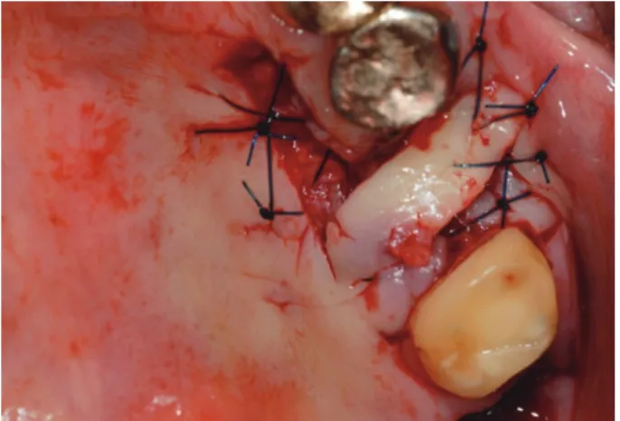
Autogenous bone was grafted into extraction sock- et defect around the implant. And the site was cov- ered with a rotated palatal flap (Fig. 3). Because of uneventful healing, impression for final restoration
was made at 4 months postoperatively and a defini- tive prosthesis (crown) was delivered to the patient.
(Fig. 4, 5) However, the prosthesis showed mobility within 2 weeks, so the fixture was removed (Fig. 6).
Fig. 3. The grafted site was covered with a rotated palatal flap.
Fig. 1. Preoperative standard radiograph (the arrow indicate the root fracture of upper left first molar).
Fig. 2. Immediate placement of fin-type implant fixture with autogenous bone graft.
Fig. 4. The grafted site was covered with a rotated palatal flap.
Fig. 5. Postoperative radiographs at four and half months. (A) Standard view, (B) CT view.
A B
After removal of the failed implant, sinus floor elevation procedure was performed with freeze-dried bone allograft (OraGRAFT, LifeNet Health, Virginia Beach, VA, USA). And then, the implant 7 mm in di- ameter and 8.5 mm in length (AnyRidge®, MegaGen Implant Co.) was placed without further drilling. A speed of 15 rpm with a torque of 50 Ncm was set for insertion of implant (Fig. 7).
After surgery, RFA evaluation is performed using the OsstellTM Mentor (Integration Diagnostics AB, Göteborg, Sweden) according to the manufacturer’s recommendations. A SmartpegTM (Integration Diag- nostics AB) was screwed into the implant with 4 - 5 Ncm. The transducer probe (Osstell MentorTM Probe II) was held so that the probe tip was aimed at the
small magnet on top of the SmartpegTM at a distance of 2 - 3 mm. Double measurements were made for a buccal and palatals sides. The average of ISQ values was recorded and the ISQ value was 70 after the sur- gery.
ISQ value appeared to increase to 71 at the post- operative 1 week, so an impression was taken based by ISQ value. A metal ceramic fixed dental prosthesis was delivered in 2 weeks after surgery (Fig. 8, 9). The ISQ value at that time was 71. This patient was fol- lowed every two weeks with intraoral radiographs until postoperative 1 year, and the socket defect was seen to be filled with regenerated bone. The patient was satisfied with the result (Fig. 10).
Fig. 6. Failed implant fixture.
Fig. 7. Specially deepened implant fixture placement.
Fig. 8. A porcelain-fused-to-metal restoration in 2 weeks after surgery.
Fig. 9. Postoperative radiographs at 2 weeks after re- installation. (A) Standard view, (B) CT view.
A B
Discussion
Norton stated that implants placed in the posterior maxilla were less successful than implants placed in other regions.9 Many clinical documentations also pointed out increased failure risks of implants in these situations: 1. soft bone,10,11 2. occlusal loading (type, magnitude and parafunction),11 3. single tooth replacements11-13 and 4. Implants placed in extraction sockets.9,14
For making a successful immediate/early load- ing protocol at the maxillary posterior area, definite occlusal scheme and proper primary stability were needed. In this case reports, deep threaded ultra wide implants were placed in the maxillary posterior area.
Implant insertion torques of implant was 50 Ncm and final prosthesis were loaded 2 weeks after sur- gery. Good result was showed after 1 year.
The assessment of primary stability is important for the prognosis of implant treatment and espe- cially in immediate loading. Several techniques have been suggested for the determination of implant stability. The use of RFA as a clinical method to measure implant stability and osseointegration was first described by Meredith et al.15 and suggested as a diagnostic instrument to monitor changes in implant stability with repeated measurements over time.16 In one study, RFA yielded a mean ISQ value of 68, a value that was indicative of achievement of high primary implant stability.6 In another study, a cut-off
ISQ value for adequate implant stability of ISQ 47 was proposed.17 Furthermore, ISQ values for suc- cessfully osseointegrated implants varied between 57 and 82, with a mean of 69 after 1 year of loading.18
In this report, ISQ values were measured at the in- sertion time and checked weekly. The conditions for early loading on maxillary posterior single implants were set as 50 Ncm and 70 or more ISQ value/ The initial decrease in the ISQ values within the first 3 weeks has been reported in many studies.19,20 How- ever, ISQ values at the time of surgery and 1 or 2 weeks after surgery were not decreased but stable in this case.
This case report is showing a new possibility of loading protocol at the maxillary posterior area with a deep threaded wide implant. It is concluded that early loading can be used even in case of maxillary posterior single implant with special implant system.
To safe load implant early, it would seem reasonable to check the implant stability with RFA prior to load should be confirmed.
References
1. Attard NJ, Zarb GA. Immediate and early implant loading protocols: a literature review of clinical studies. J Prosthet Dent 2005;94:242-58.
2. Ostman PO. Immediate/early loading of dental im- plants. Clinical documentation and presentation of a treatment concept. Periodontol 2000 2008;47:90- 112.
3. Sennerby L, Meredith N. Implant stability measure- ments using resonance frequency analysis: biologi- cal and biomechanical aspects and clinical implica- tion. Periodontol 2000 2008;47:51-66.
4. Chee W, Jivraj S. Efficiency of immediately loaded mandibular full-arch implant restorations. Clin Im- plant Dent Relat Res 2003;5:52-6.
5. Sennerby L, Meredith N. Resonance frequency analysis: measuring implant stability and osseointe- gration. Compend Contin Educ Dent 1998;19:493- 8.
6. Glauser R, Sennerby L, Meredith N, Rée A, Lun- dgren A, Gottlow J, Hämmerle CH. Resonance frequency analysis of implants subjected to im- Fig. 10. Postoperative radiographs at 13 months after
re-installation. (A) Standard view, (B) CT view.
A B
mediate or early functional occlusal loading. Suc- cessful vs. failing implants. Clin Oral Implants Res 2004;15:428-34.
7. Ostman PO, Hellman M, Wendelhag I, Sennerby L. Resonance frequency analysis measurements of implants at placement surgery. Int J Prosthodont 2006;19:77-83.
8. Ostman PO, Hellman M, Sennerby L. Immediate occlusal loading of implants in the partially eden- tate mandible: a prospective 1-year radiographic and 4-year clinical study. Int J Oral Maxillofac Im- plants 2008;23:315-22.
9. Norton MR. A short-term clinical evaluation of immediately restored maxillary TiOblast single- tooth implants. Int J Oral Maxillofac Implants 2004;19:274-81.
10. Glauser R, Rée A, Lundgren A, Gottlow J, Häm- merle CH, Schärer P. Immediate occlusal loading of Brånemark implants applied in various jaw bone regions: a prospective, 1-year clinical study. Clin Implant Dent Relat Res 2001;3:204-13.
11. Achilli A, Tura F, Euwe E. Immediate/early func- tion with tapered implants supporting maxillary and mandibular posterior fixed partial dentures:
preliminary results of a prospective multicenter study. J Prosthet Dent 2007;97:S52-8.
12. Rao W, Benzi R. Single mandibular first molar im- plants with flapless guided surgery and immediate function: preliminary clinical and radiographic re- sults of a prospective study. J Prosthet Dent 2007;
97:S3-S14.
13. Rocci A, Martignoni M, Gottlow J. Immediate load- ing of Brånemark system TiUnite and machined- surface implants in the posterior mandible: a
randomized open-ended clinical trial. Clin Implant Dent Relat Res 2003;5 Suppl 1:57-63.
14. Ericsson I, Nilson H, Lindh T, Nilner K, Randow K. Immediate functional loading of Brånemark sin- gle tooth implants. An 18 months’ clinical pilot fol- low-up study. Clin Oral Implants Res 2000;11:26- 33.
15. Meredith N, Alleyne D, Cawley P. Quantitative de- termination of the stability of the implant-tissue interface using resonance frequency analysis. Clin Oral Implants Res 1996;7:261-7.
16. Sim CP, Lang NP. Factors influencing resonance frequency analysis assessed by Osstell mentor dur- ing implant tissue integration: I. Instrument posi- tioning, bone structure, implant length. Clin Oral Implants Res 2010;21:598-604.
17. Nedir R, Bischof M, Szmukler-Moncler S, Bernard JP, Samson J. Predicting osseointegration by means of implant primary stability. Clin Oral Implants Res 2004;15:520-8.
18. Ersanli S, Karabuda C, Beck F, Leblebicioglu B.
Resonance frequency analysis of one-stage dental implant stability during the osseointegration period.
J Periodontol 2005;76:1066-71.
19. Barewal RM, Oates TW, Meredith N, Cochran DL.
Resonance frequency measurement of implant stability in vivo on implants with a sandblasted and acid-etched surface. Int J Oral Maxillofac Implants 2003;18:641-51.
20. Crismani AG, Bernhart T, Schwarz K, Celar AG, Bantleon HP, Watzek G. Ninety percent success in palatal implants loaded 1 week after placement: a clinical evaluation by resonance frequency analysis.
Clin Oral Implants Res 2006;17:445-50.
*교신저자: 임현필
(500-757) 광주광역시 북구 용봉로 33 전남대학교 치의학전문대학원 보철학교실 Tel: 062-530-5577|Fax: 062-530-5639|E-mail: [email protected]
깊은 나사선을 갖는 임플란트를 이용한 상악 구치부 조기 하중: 증례보고
한창훈
1, 김현승
2, 박상원
2,3, 윤귀덕
3, 주한성
3, 임현필
3*
1이지플란트 치과의원, 2전남대학교 미래형생체부품소재 RIS 사업단, 3전남대학교 치의학전문대학원 보철학교실
본 증례는 상악 구치부에서 실패한 임플란트 제거 후 깊은 나사선을 갖는 직경이 큰 임플란트를 식립 후 조기하중을 가 한 증례이다. 1년 경과 후, 임플란트 안정성 지수에서 안정적인 결과를 보였다. 본 임상증례는 상악 구치부에서 깊은 나 사선을 갖는 임플란트를 이용한 조기하중의 가능성을 보여준다.
(구강회복응용과학지 2014;30(3):253-8)
주요어: 깊은 나사선; 초기 안정성; 조기하중; 상악 구치부