Yeungnam Univ J Med 2018;35(2):165-170
Effects of small tidal volume and positive end-expiratory pressure on oxygenation in pressure-controlled ventilation-volume guaranteed mode
during one-lung ventilation
Sung Hye Byun, So Young Lee, Jin Yong Jung
Department of Anesthesiology and Pain Medicine, Catholic University of Daegu School of Medicine, Daegu, Korea Background: The purpose of this study was to investigate whether tidal volume (TV) of 8 mL/kg without positive end-expiratory pressure (PEEP) and TV of 6 mL/kg with or without PEEP in pressure-controlled ventilation-volume guaranteed (PCV-VG) mode can maintain arterial oxygenation and decrease inspiratory airway pressure effectively during one-lung ventilation (OLV).
Methods: The study enrolled 27 patients undergoing thoracic surgery. All patients were ventilated with PCV- VG mode. During OLV, patients were initially ventilated with TV 8 mL/kg (group TV8) without PEEP. Ventila- tion was subsequently changed to TV 6 mL/kg with PEEP (5 cmH2O; group TV6+PEEP) or without (group TV6) in random sequence. Peak inspiratory pressure (Ppeak), mean airway pressure (Pmean), and arterial blood gas analysis were measured 30 min after changing ventilator settings. Ventilation was then changed once more to add or eliminate PEEP (5 cmH2O), while maintaining TV 6 mL/kg. Thirty min after changing ventilator settings, the same parameters were measured once more.
Results: The Ppeak was significantly lower in group TV6 (19.3±3.3 cmH2O) than in group TV8 (21.8±3.1 cmH2O) and group TV6+PEEP (20.1±3.4 cmH2O). PaO2 was significantly higher in group TV8 (242.5±111.4 mmHg) than in group TV6 (202.1±101.3 mmHg) (p=0.044). There was no significant difference in PaO2 between group TV8 and group TV6+PEEP (226.8±121.1 mmHg). However, three patients in group TV6 were dropped from the study because PaO2 was lower than 80 mmHg after ventilation.
Conclusion: It is postulated that TV 8 mL/kg without PEEP or TV 6 mL/kg with 5 cmH2O PEEP in PCV-VG mode during OLV can safely maintain adequate oxygenation.
Keywords: Airway pressure; Arterial oxygenation; One-lung ventilation; Pressure-controlled ventilation-volume guaranteed
Copyright ©2018 Yeungnam University College of Medicine
This is an Open Access article distributed under the terms of the Creative Commons Attribution Non-Commercial License (http://creative- commons.org/licenses/by-nc/4.0/) which permits unrestricted non-commercial use, distribution, and reproduction in any medium, provided the original work is properly cited.
Received: April 11, 2018, Revised: May 28, 2018 Accepted: May 31, 2018
Corresponding Author: Jin Yong Jung, Department of Anesthesiology and Pain Medicine, Catholic University of Daegu School of Medicine, 33, Duryugongwon-ro 17-gil, Nam-gu, Daegu 42472, Korea
Tel: +82-53-650-4505, Fax: +82-53-650-4517 E-mail: [email protected]
INTRODUCTION
Development of one-lung ventilation (OLV) in anesthesio- logy has greatly influenced the development of lung surgery.
During OLV, respiration is achieved only with a dependent lung, and the anesthesiologist must prevent hypoxemia due to increased shunting, as well as lung injury due to increased airway pressure, in contrast with two-lung ventilation [1-4].
According to previous studies comparing oxygenation rates between different ventilator modes during OLV, improve- ment in oxygenation in volume-controlled ventilation (VCV) and pressure-controlled ventilation (PCV) showed moderate differences in accordance with pulmonary function. Patients who were expected to have decreased pulmonary function based on preoperative pulmonary function tests showed bet-
ter oxygenation with PCV, compared to that with VCV [5].
In patients with normal pulmonary function, there was no difference between VCV and PCV [6]. In studies comparing VCV with pressure-controlled ventilation-volume-guaran- teed (PCV-VG), PCV-VG showed better oxygenation during OLV [7,8].
In order to prevent lung injury during OLV, a protective ventilation strategy has been widely used, i.e., by lowering tidal volume and applying positive end-expiratory pressure (PEEP), which reduces lung injury while maintaining oxygen- ation [1,2,9].
The primary goal of this study was to investigate whether a decrease in tidal volume of 6 mL/kg with or without 5 cmH2O PEEP would be enough to prevent lung barotrauma by redu- cing inspiratory airway pressure, while maintaining appro- priate oxygenation during OLV using PCV-VG, which has the merits of PCV and the tidal volume-designating characteristic of VCV.
MATERIALS AND METHODS
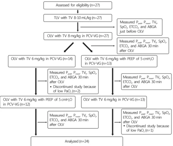
The study proceeded with approval from the institutional review board of our hospital. The study enrolled 27 subjects who were classified as American Society of Anesthesiologists physical status 1 or 2 and scheduled for pulmonary surgery under OLV (Table 1). All subjects provided written informed consent before surgery. Patients scheduled for pneumonec- tomy were excluded. All patients received pulmonary function tests preoperatively. Subjects with forced vital capacity (FVC) lower than 70% of predicted value or those with forced ex- piratory volume at 1 second (FEV1) lower than 70% of pre- dicted value were excluded. Patients with previous lung sur- gery or heart disease were also excluded.
All subjects were given an intramuscular injection of glyco- pyrrolate 0.2 mg before entering the operating room as pre- medication. In the operating room, the electrocardiogram, pulse oximeter, non-invasive blood pressure, and bispectral index were monitored. Anesthesia was induced with 4 μg/mL of propofol and 4 ng/mL of remifentanil, using target cont- rolled infusion (TCI). For intubation, 100% oxygen was admi- nistrated via mask. After loss of consciousness, 1 mg/kg rocu- ronium was injected for muscle relaxation. After checking mu- scle relaxation, patients were intubated with a left-side dou- ble-lumen endotracheal tube (DLT, Silbroncho®, Fuji System
Corp, Tokyo, Japan). The position of the DLT was checked by auscultation, followed by fiberoptic bronchoscopy (FOB) for confirmation. After these procedures, a 20-G catheter was installed for perioperative blood pressure monitoring and blood collection for arterial blood gas analysis (ABGA). A central venous catheter was inserted in the subclavian vein, on the ipsilateral side of the operation site. Anesthesia maintenance was achieved using 6 L/min 100% oxygen, propofol, and re- mifentanil TCI. One hour after induction, vecuronium 0.08 mg/kg/h was additionally injected for muscle relaxation.
The location of the DLT was examined again with FOB after the patient was placed in lateral decubitus position. All patients underwent surgery using the same anesthetic equip- ment (Avance, GE Healthcare, Wauwatosa, WI, USA), using PCV-VG as ventilation mode. After induction, tidal volumes were 8-10 mL/kg during two-lung ventilation. During OLV, patients were initially placed on TV 8 mL/kg (group TV8) without PEEP. Ventilation was subsequently changed to TV 6 mL/kg with PEEP (5 cmH2O; group TV6+PEEP) or with- out (group TV6), in random sequence. Peak airway pressure (Ppeak), mean airway pressure (Pmean), exhaled tidal volume (TVE), peripheral oxygen saturation (SpO2), end-tidal CO2
(ETCO2), and ABGA were measured and examined just be- fore OLV and 30 min after each mode of OLV was applied.
Ventilation was changed once more to add or eliminate PEEP (5 cmH2O), while maintaining TV 6 mL/kg. Thirty min after changing ventilator settings, the same parameters were meas- ured once more (Fig. 1). PCV-VG mode was used under 40 cmH2O of maximum airway pressure (Pmax), I:E ratio of 1:2, and the minimal time to reach target airway pressure was set as 5 s. Respiratory rates during anesthesia were set equal- ly for all patients at 12 times per min. When SpO2 levels were below 90%, or when arterial partial pressure of oxygen (PaO2) was below 80 mmHg in ABGA, subjects were discon- tinued from this study and hypoxia was corrected by tidal volume elevation, relocation of DLT, or two-lung ventilation, if needed.
Sample size was calculated as 24, with 80% power and 0.05 statistical significance, using the average and standard deviation of established values [5,9], according to all three ventilation methods. Assuming a 10% drop-out rate, 27 sub- jects were included in the study. Each mode of ventilation was statistically assessed using SPSS for Windows, version 17.0 (SPSS, Chicago, IL, USA) and repeated measures analysis
Fig. 1. Consort diagram. TLV, supine two lung ventilation; TV, tidal volume; OLV, one-lung ventilation;
Ppeak, peak inspiratory pressure; Pmean, mean inspiratory pressure; TVE, exhaled tidal volume; SpO2, peri- pheral oxygen saturation; EtCO2, end-tidal CO2; ABGA, arterial blood gas analysis; OLV, one-lung ventila- tion; PCV-VG, pressure-controlled ventilation-volume-guaranteed; PEEP, positive end expiratory pressure;
PaO2, arterial oxygen pressure.
of variance. All measured values were presented as averages and standard deviations, and p-values <0.05 were considered to indicate statistical significance.
RESULTS
Of the 27 patients, three dropped out and the study was conducted statistically on the remaining 24 subjects. In these three patients, arterial oxygen partial pressure after 30 min of respiration with tidal volume of 6 mL/kg on ABGA was 60.8, 70.2, and 71.8 mmHg, respectively. Although the pe- ripheral oxygen saturation did not fall below 90%, tidal vol- ume was increased after interrupting the study.
Among 24 patients, 16 (males, n=16/18) had a past smok- ing history and two (n=2/16) had both smoking and tubercu- losis histories. No female patients (n=6) had a history of smo-
king or tuberculosis (Table 1).
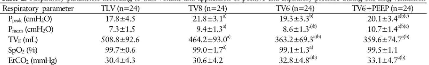
The Ppeak values were significantly higher in the TV8 group (21.8±3.1 cmH2O) compared to the TV6 (19.3±3.3 cmH2O) and TV6+PEEP (20.1±3.4 cmH2O) groups (p<0.002). The Pmean values were significantly higher in the TV6+PEEP group (10.7±1.4 cmH2O) compared to those in the other groups (p=0.000). The TVE values were significantly higher in the TV8 group (464.2±93.0 mL), but there was no statistically significant difference between TV6 (363.2±69.3 mL) and TV6+PEEP (359.6±74.7 mL). The ETCO2 values were sig- nificantly lower in the TV8 group (30.6±4.2 mmHg) com- pared to those in the other groups (p=0.003), but there were no significant differences between TV6 (32.8±4.8 mmHg) and TV6+PEEP (33.1±4.7 mmHg) (p>0.05) (Table 2).
The arterial oxygen partial pressure was significantly high- er in TV8 group (242.5±111.4 mmHg) than in TV6 group
Table 1. Demographic variables
Age (yr) 66.0±9.9
Sex (male/female) 18/6
Height (cm) 162.5±9.1
Weight (kg) 63.7±10.0
BMI (kg/m2) 24.1±3.0
Smoking history (male/female) 16/0
Tuberculosis history (male/female) 2/0 Duration of operation (min) 261.6±68.1 Preoperative FVC (% of predictive) 95.4±10.7 Preoperative FEV1 (% of predictive) 96.6±12.5 Diagnosis
Lung cancer 20
Empyema 2
Secondary pneumothorax 2
Operation site (left/right) 11/13
Surgical approach
VATS 18
Open thoracotomy 6
BMI, body mass index; VATS, video-assisted thoracoscopic surgery.
Table 2. Respiratory parameters according to tidal volume and application of positive end-expiratory pressure during one-lung ventilation
Respiratory parameter TLV (n=24) TV8 (n=24) TV6 (n=24) TV6+PEEP (n=24)
Ppeak(cmH2O) 17.8±4.5 21.8±3.1a) 19.3±3.3b) 20.1±3.4a)b)c)
Pmean(cmH2O) 7.3±1.5 9.4±1.3a) 8.6±1.3a)b) 10.7±1.4a)b)c)
TVE(mL) 508.8±92.6 464.2±93.0a) 363.2±69.3a)b) 359.6±74.7a)b)
SpO2(%) 99.7±0.6 99.0±1.7a) 99.1±1.3a) 99.5±1.1
EtCO2(mmHg) 30.4±4.3 30.6±4.2 32.8±4.8a)b) 33.1±4.7a)b)
Data are presented as mean±standard deviation.
Ppeak, peak inspiratory pressure; Pmean, mean inspiratory pressure; TVE, exhaled tidal volume; SpO2, peripheral oxygen saturation;
TLV, supine two-lung ventilation; TV8, one-lung ventilation with tidal volume 8 mL/kg; TV6, one-lung ventilation with tidal volume 6 mL/kg; PEEP, 5 cmH2O positive end expiratory pressure.
a)p<0.05 compared with TLV, b)p<0.05 compared with TV8, c)p<0.05 compared with TV6.
Table 3. Arterial blood gas values according to tidal volume and application of positive end-expiratory pressure during one-lung ventilation
Arterial blood gas value TLV (n=24) TV8 (n=24) TV6 (n=24) TV6+PEEP (n=24)
PaO2(mmHg) 444.7±115.9 242.5±111.4a) 202.1±101.3a)b) 226.8±121.1a)
PaCO2(mmHg) 38.5±4.0 39.3±5.7 42.6±6.8a)b) 43.4±7.5a)b)
SaO2(%) 99.8±0.1 98.7±1.8a) 98.9±0.9a) 98.9±1.2a)
Hematocrit (%) 34.0±4.0 34.6±3.8 34.3±3.8 34.4±4.0
Data are presented as mean±standard deviation.
PaO2, arterial oxygen pressure; PaCO2, arterial carbon dioxide pressure; SaO2, arterial oxygen saturation; TLV, supine two lung ventilation; TV8, one-lung ventilation with tidal volume 8 mL/kg; TV6, one-lung ventilation with tidal volume 6 mL/kg; PEEP, 5 cmH2O positive end expiratory pressure.
a)p<0.05 compared with TLV, b)p<0.05 compared with TV8, c)p<0.05 compared with TV6.
(202.1±101.3 mmHg) (p=0.044). There was no significant di- fference between TV8 and TV6+PEEP groups (226.8±121.1
mmHg) (p>0.05). The arterial carbon dioxide partial pressure was significantly higher in both TV6 (42.6±6.8 mmHg) and TV6+PEEP (43.4±7.5 mmHg) groups compared to that in the TV8 (39.3±5.7 mmHg) group (p=0.003, 0.002) (Table 3).
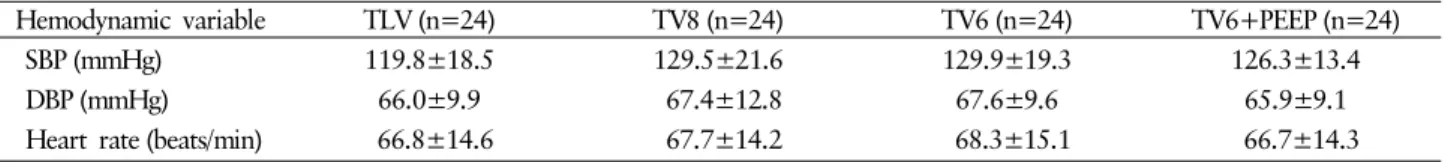
There was no difference in arterial oxygen saturation, he- matocrit levels, and hemodynamic variables among the three groups (p>0.05) (Table 3, 4).
DISCUSSION
During OLV with PCV-VG, a tidal volume of 8 mL/kg and a tidal volume of 6 mL/kg with PEEP of 5 cmH2O could safely be applied for appropriate oxygenation. Tidal volume of 6 mL/kg without PEEP showed the lowest maximum and aver- age inspiratory pressure. However, about 11% of our subjects (3/27) showed hypoxemia with arterial oxygen partial pres- sures under 80 mmHg at a tidal volume of 6 mL/kg without PEEP. Therefore, the authors believe that single application of tidal volume 6 mL/kg with PCV-VG without PEEP creates a high risk of intraoperative hypoxemia.
PCV-VG is the latest addition to respiration methods used in anesthetic machines; it calculates the patient’s lung com-
Table 4. Hemodynamic variables according to tidal volume and application of positive end-expiratory pressure during one-lung ventilation
Hemodynamic variable TLV (n=24) TV8 (n=24) TV6 (n=24) TV6+PEEP (n=24)
SBP (mmHg) 119.8±18.5 129.5±21.6 129.9±19.3 126.3±13.4
DBP (mmHg) 66.0±9.9 67.4±12.8 67.6±9.6 65.9±9.1
Heart rate (beats/min) 66.8±14.6 67.7±14.2 68.3±15.1 66.7±14.3
Data are presented as mean±standard deviation. No significant differences were noted among the groups.
SBP, systolic blood pressure; DBP, diastolic blood pressure; TLV, supine two lung ventilation; TV8, one-lung ventilation with tidal volume 8 mL/kg; TV6, one-lung ventilation with tidal volume 6 mL/kg; PEEP, 5 cmH2O positive end expiratory pressure.
pliance by initial respiration with VCV mode, followed by programmed control of tidal volumes, which are previously set [10]. Song et al. [11] reported that using PCV-VG mode incre- ased expiratory volume compared to that using VCV mode.
In PCV mode, the tidal volume can change depending on lung compliance, leading to hypercarbia caused by decreased minute volumes. In PCV-VG mode, when compared to VCV mode, expiratory volume is increased and can be maintained, keeping the minute volume at appropriate levels. Theoreti- cally, PCV-VG can lower airway pressure during inspiration, thus improving oxygenation by matching the V/Q ratio of the dependent lung. Bouls and Ghobrial [7] previously re- ported that using tidal volumes of 8-10 mL/kg can improve oxygenation during OLV in PCV-VG mode, compared to that in VCV mode. Moreover, Pu et al. [8] reported that using tidal volumes of 8-10 mL/kg can improve oxygenation during OLV in PCV-VG mode. However, Song et al. [11] reported that tidal volume of 8 mL/kg reduces peak airway pressure, but does not lead to statistically significant differences in ar- terial oxygen partial pressure between PCV-VG and VCV.
According to previous studies, application of PCV-VG in an- esthesiology is desirable. Therefore, the authors investigated the optimal tidal volume for use of PCV-VG mode, which can successfully lower the airway pressure while also improv- ing oxygenation.
In OLV using VCV mode, a minimum tidal volume of 8 mL/kg maintains oxygenation while not causing atelectasis [12]. Moreover, tidal volume of 9 mL/kg or lower has not been reported to cause lung injury [13]. Kim et al. [14] repor- ted that the probability of hypoxemia is 70% when tidal vol- ume of 6 mL/kg is used during OLV with VCV mode and 65% when 5 cmH2O of PEEP is added to tidal volume of 6 mL/kg. These data show a much higher hypoxemia inci- dence compared with 5% hypoxemia incidence when tidal volume 10 mL/kg was used for OLV with VCV. In addition,
at tidal volume of 10 mL/kg, there were no cases showing expansion pressure of up to 37 cmH2O, leading to the con- clusion that there was no pulmonary damage. Therefore, in patients with normal lung function, high tidal volume such as 10 mL/kg increases the arterial oxygen partial pressure, and OLV does not cause complications such as lung injury. In the present study, using tidal volume of 6 mL/kg in PCV-VG mode caused hypoxemia in 11% of patients. Hypoxemia did not occur when using tidal volume of 8 mL/kg or 6 mL/kg with 5 cmH2O of PEEP in PCV-VG mode, thus leading to the conclusion that both methods can be applied safely while using PCV-VG mode. In addition, it is thought that there was low risk of barotrauma in PCV-VG mode, since peak inspiratory pressures were measured at about 20 cmH2O in all three groups.
The limitation of this study is the consistent application of ventilator modes, i.e., tidal volume of 8 mL/kg, followed by 6 mL/kg with or without 5 cmH2O of PEEP. In addition, the shunt ratios during lung surgery may have changed due to pulmonary artery ligation, or handling of the surgical site.
As a result, the surgery itself may have influenced the arterial oxygen partial pressure. Since the authors conducted the study in the routine order noted above, the early period of OLV may have maintained better arterial oxygenation. Another limitation is that ideal body weight was not used in calculat- ing the tidal volume. In overweight patients, the tidal volumes may have been overestimated, resulting in possible high air- way pressure and causing negative influence on oxygenation.
However, since our subjects’ mean body mass index was ap- proximately 24 kg/m2, its effect on the study was thought to be minimal. In addition, further study on the clinical implica- tion of low tidal volume in PCV-VG is needed because the postoperative pulmonary status was not measured in this study.
In conclusion, while using PCV-VG mode during OLV, a tidal volume of 8 mL/kg or 6 mL/kg with added PEEP of
5 cmH2O is thought to be a safe method of artificial ventila- tion, and can reduce airway pressure while maintaining appro- priate oxygenation.
CONFLICT OF INTEREST
No potential conflict of interest relevant to this article were re ported.
ORCID
Sung Hye Byun, https://orcid.org/0000-0002-9287-5087 Jin Yong Jung, https://orcid.org/0000-0003-2662-3540
REFERENCES
1. Yun du G, Han JI, Kim DY, Kim JH, Kim YJ, Chung RK. Is small tidal volume with low positive end expiratory pressure during one-lung ventilation an effective ventilation method for endoscopic thoracic surgery? Korean J Anesthesiol 2014;67:
329-33.
2. Kim H. Protective strategies for one-lung ventilation. Korean J Anesthesiol 2014;67:233-4.
3. Jung JD, Kim SH, Yu BS, Kim HJ. Effects of a preemptive alveolar recruitment strategy on arterial oxygenation during one-lung ventilation with different tidal volumes in patients with normal pulmonary function test. Korean J Anesthesiol 2014;67:96-102.
4. Lee W, Lee JY, Choi DN, Shin CM, Cho K, Kim MH, et al.
Airway dimensions and margin of safety with the left-sided
double-lumen tube in patients of a short stature. Anesth Pain Med 2015;10:110-7.
5. Tuğrul M, Camci E, Karadeniz H, Sentürk M, Pembeci K, Akpir K. Comparison of volume controlled with pressure con- trolled ventilation during one-lung anaesthesia. Br J Anaesth 1997;79:306-10.
6. Unzueta MC, Casas JI, Moral MV. Pressure-controlled versus volume-controlled ventilation during one-lung ventilation for thoracic surgery. Anesth Analg 2007;104:1029-33.
7. Boules NS, Ghobrial HZ. Efficiency of the newly introduced ventilatory mode “pressure controlled ventilation-volume gua- ranteed” in thoracic surgery with one lung ventilation. Egypt J Anaesth 2011;27:113-9.
8. Pu J, Liu Z, Yang L, Wang Y, Jiang J. Applications of pressure control ventilation volume guaranteed during one-lung venti- lation in thoracic surgery. Int J Clin Exp Med 2014;7:1094-8.
9. Végh T, Juhász M, Szatmári S, Enyedi A, Sessler DI, Szegedi LL, et al. Effects of different tidal volumes for one-lung venti- lation on oxygenation with open chest condition and surgical manipulation: a randomised cross-over trial. Minerva Anes- tesiol 2013;79:24-32.
10. Keszler M. Volume-targeted ventilation. Early Hum Dev 2006;
82:811-8.
11. Song SY, Jung JY, Cho MS, Kim JH, Ryu TH, Kim BI. Volu- me-controlled versus pressure-controlled ventilation-volume guaranteed mode during one-lung ventilation. Korean J Anes- thesiol 2014;67:258-63.
12. Gal TJ. Con: low tidal volumes are indicated during one-lung ventilation. Anesth Analg 2006;103:271-3.
13. Fernández-Pérez ER, Sprung J, Afessa B, Warner DO, Vachon CM, Schroeder DR, et al. Intraoperative ventilator settings and acute lung injury after elective surgery: a nested case con- trol study. Thorax 2009;64:121-7.
14. Kim SH, Jung KT, An TH. Effects of tidal volume and PEEP on arterial blood gases and pulmonary mechanics during one- lung ventilation. J Anesth 2012;26:568-73.