Isolation and Genetic Characterization of Protease-Producing Halophilic Bacteria from Fermenting Anchovy
Jin-Ho Lee*
*Department of Food Science & Biotechnology, Kyungsung University, Busan 608-736, Korea
Received November 14, 2011 /Revised December 26, 2011 /Accepted December 27, 2011Three protease-producing halophilic bacteria were isolated from fermenting anchovy. Isolated FAM 10, FAM 114, and FAM 115 were found to grow optimally at salt concentrations of 2-4%, 10%, and 6%, respectively, and could grow in salinity of up to 18-22%. The salinity conditions for optimum protease production were 6% in FAM 10 and 10% in FAM 114 and FAM 115. The protease activity of FAM 10 was gradually inhibited by the addition of NaCl up to 10%, and was not evident at 14%, whereas FAM 114 and FAM 115 displayed protease activity at 14% NaCl and could not be measured at 18%.
These results demonstrated that the three isolated strains belong to protease-producing, moderately halophilic bacteria. Strain FAM 10, FAM 114, and FAM 115 were identified as Salinivibrio sp., Halobacillus sp., and Halobacillus sp. respectively, based on comparative analyses of the 16S rRNA gene and the 16S-23S intergenic space sequence (IGS), biochemical testing, and Gram staining. Salinivibrio sp. FAM 10 had two 16S rDNAs containing different sequences at position 191 and four IGSs that harbored no tRNA gene and tRNA genes for isoleucine, alanine, glutamate, lysine, and/or valine.
Halobacillus sp. FAM 114 and FAM 115 had completely identical 16S rRNA gene sequences and showed 99% identity to the sequences of various Halobacillus strains. The three IGSs found in the ge- nome of both strains displayed 99% sequence identity with Halobacillus aidingensis and Halobacillus sp.
JM-Hb, and had IGS
0with no tRNA gene and IGS
IAwith tRNA genes for isoleucine and alanine.
Key words : Fermenting anchovy, halophilic bacteria, protease, Salinivibrio, Halobacillus
*Corresponding author
*Tel:+82-51-633-4716, Fax:+82-51-622-4986
*E-mail : [email protected]
서 론
멸치젓갈은 멸치와 소금을 혼합하여 상온에서 6~12개월 보 관하면서 발효, 숙성시킨 우리나라의 대표적인 전통 발효식품 중의 하나이다[11,13]. 젓갈은 발효 과정에서 증식하는 미생물 에 의해 분비되는 단백질 분해효소와 멸치 내에 존재하는 자 가소화효소의 공동작용에 의해서 멸치조직이 액화와 분해가 진행되면서 발효 및 숙성이 이루어지게 된다. 이러한 과정을 통해 많은 유리 아미노산, 저분자의 펩티드, 생화학반응에 의 해 유도되는 여러 화합물질들이 젓갈액에 축적되어, 멸치젓갈 특유의 풍미와 영양학적 측면에서 양질의 단백질을 제공하게 된다[13].
발효과정중에 20~25%의 소금을 함유한 멸치조직이 용해되 기 위해서는 단백질 분해효소를 분비하는 호염성 미생물들 (halophilic microorganisms)이 필요하다. 대부분의 단백질 분 해효소들의 활성은 고농도의 NaCl이 존재하는 조건에서 그 활성이 감소하기 때문에 멸치액젓을 제조하는데는 매우 긴 시간이 소요된다[3,7]. 또한, 최근 소비자의 식습관의 변화로 인해서 저염도의 젓갈의 요구성이 증대되고 있지만, 식품 안 전성과 위생측면에서의 품질관리의 어려움 때문에 고 품질의
저염도 젓갈액을 제조하는데 많은 문제를 내포하고 있다 [17,20].
지금까지 젓갈 발효에 관련된 많은 미생물들이 분리, 동정 되었는데, 그 대표적인 균은 유산균, Bacillus sp. Pseudomonas sp. Micrococcus sp, Brevibacterium sp. Microbacterium sp., Halobacterium sp., Halobacillus sp. 등이다[3,9,11,12,15]. 그러 나, 이러한 미생물에 대한 분류, 생리적 특징, 효소학적 특징등 에 대한 상세한 사항은 아직 잘 규명되지 않고 있다[3,7]. 그 중에서 특히, 16S rRNA 유전자를 이용하여 그 서열의 차이를 이용하여 멸치젓갈의 특정 속(genus)의 한 종(species)을 정확 히 구분하는 것은, 종 간에 16S rDNA 서열의 차이가 거의 없는 문제점 때문에 많은 어려움을 내포하고 있다[8]. 이러한 문제점을 해결하기 위해 최근에는 16S-23S ribosomal RNA 유전자사이 부위(intergenic spacer region, IGS)의 염기서열의 변화를 이용하여 속, 종, 균주(strain)를 동정하는데 많이 사용 되고 있다[2,8,16]. 따라서, 멸치젓갈의 발효에 관련된 미생물 에 관한 체계적이고 과학적인 연구를 위해서는 16S rDNA와 함께 16S-23S ribosomal RNA 유전자 사이 부위의 변형에 대 한 분석연구가 병행되어야 한다.
본 연구에서는 멸치젓갈 발효과정에서 분리된 protease를
분비하는 호염성세균을 분리하고, 단백질 분해효소의 활성
을 확인하였으며, 16S rDNA와 16S-23S 유전자 사이의 부위
에 대한 염기서열을 비교 분석하여 미생물을 동정하여 보고 한다. 특히, 동정된 Salinivibrio와 Halobacillus는 16S-23S IGS 내에 다양한 형태의 tRNA 유전자가 존재하는 특징을 확인 하였다.
재료 및 방법
배지
호염성 세균을 분리하고 배양하기 위해 marine 한천배지 (KisanBiotech, Korea)를 사용하였다. 필요할 경우, 80 g/l의 염화나트륨과 10 g/l의 전지분유 (skim milk)를 첨가하여 사 용하였다.
세포성장 및 단백질분해효소 생산
세포성장과 단백질분해효소를 생산할 목적으로, 분리된 균 을 30 ml의 marine 배지를 함유한 250 ml 플라스크에서 30℃, 24시간 배양하였다. 이때, 염화나트륨을 각각 0, 4, 8, 12, 16, 20, 24% (w/v) 첨가하였다. 세포성장은 분광광도계(Shimadzu UV mini-1240)를 사용하여 600 nm에서 흡광도를 측정하여 관찰하였다.
단백질분해효소를 분비하는 호염성 세균의 분리
4 kg의 멸치와 1 kg의 소금을 혼합하고 밀봉한 다음 15~20
℃에서 보관하면서 3, 6, 12주 후에 각각 주기적으로 표본을 취하였다. 샘플은 10% 식염수에 희석한 후 10% NaCl과 1%전 지분유를 함유한 marine 한천배지에 도말하여 30℃에서 2~4 일간 배양하였다. 흰 한천배지에서 콜로니 주변에 큰 환을 형 성하는 균를 예비적으로 선별하였으며, 분리된 균주들은 2~3 회 순수분리 후 20% 글리세롤에 보관하면서 사용하였다.
호염성세균의 동정
세포의 형태는 그람염색(Difco Gram stain kit 사용) 후 광 학현미경(Model Nikon Eclipse E200)으로 관찰하였다. 생화 학적 실험은 API kit (20 NE, 50CHB)를 사용하였다.
16S rDNA 증폭과 염기서열 분석
16S rDNA는 16S-27F (5’-AGAGTTTGATCCTGGCTCAG- 3’)와 16S-1492R (5’-GGTTACCTTGTTACGACTT-3’) 프라이 머를 사용하여 중합효소연쇄반응(polymerase chain reaction, PCR)을 이용하여 증폭하였다[4]. 증폭된 DNA 단편은 솔젠트
㈜의 plasmid T-blunt와 접합(ligation)하여 대장균 TOP10에 형질전환하였다. 10개의 콜로니로부터 플라스미드 DNA를 추 출하여 M13-F (5’-GTAAAACGACGGCCAGT-3’), M13-R (5’- GGAAACAGCTATGACCATG-3’), 16S-785F (5’-GGATTAG ATACCCTGGTA-3’), 그리고 16S-805R (5’-GACTACCAGGG TATCTAATC-3’) 프라이머를 이용하여 염기서열을 분석하였다.
16S-23S ribosomal RNA 유전자사이 공간(intergenic spacer, IGS)의 증폭과 염기서열분석
16S-23S IGS는 16/23S-1525F (5’-GCTGGATCACCTCCT TTCT-3’)와 16/23S-40R (5’-CTGGTGCCAAGGCATCCA-3’) 프라이머를 이용하여 PCR로 증폭하였다[2]. 아가로즈 젤에서 200~800 염기쌍(base pair, bp) 사이의 주요 단편을 정제하여 T-blunt plasmid에 각각 클로닝하고, 그로부터 얻은 10개의 콜로니를 이용하여 M13-F 와 M13-R 프라이머를 사용하여 염 기서열을 분석하였다.
염기서열의 분석
16S rDNA와 16S-23S IGS의 염기서열은 National Center for Biotechnology Information (NCBI; http://www.ncbi.nlm.
nih.gov/)의 BLAST server를 사용하여 분석하였다. 다중서열 배열 (multiple sequence alignment)은 CLUSTAL W 프로그 램을 사용하여 분석하였다[23]. 계통발생도(phylogenetic tree) 는 MEGA5 프로그램을 이용하여[14], 통계적 방법은 neigh- bor-joining 법[21]으로 계통발생론 평가는 Bootstrap법을 이 용하여 구축하였다. 16S-23S IGS에 존재하는 tRNA의 분석은 transfer RNA database server (http://trnadb.bioinf.uni- leipzig.de/)를 사용하였다.
재조합 DNA 기술
모든 재조합 DNA 기술은 분자클로닝(molecular cloning) 에 기술된 방법에 준하여 실시하였다[22]. 세포내 염색체 DNA는 염색체 DNA 정제키트(Qiagen, Dutch)를 사용하여 분리하였다. DNA 증폭은 Pfu-X DNA polymerase (솔젠트, 한국)를 사용하였다. PCR 정제와 겔 추출키트는 Gene All Biotechnology (한국) 제품을 사용하였다. PCR로 얻은 모든 클론은 DNA염기서열을 분석하여 확인하였다.
염기서열 가입번호(accession number)
16S rDNA와 16S-23S IGS 부위 DNA 염기서열의 GenBank 가입번호는 JN381953~JN382964이다.
단백질분해효소 분석법
단백질분해효소의 효소활성도는 Chung 등이 기술한 방법
을 변형하여 측정하였다[6]. 2% 카제인을 함유한 2.5 ml의 100
mM Tris-HCl buffer (pH 8.0)와 0.2 ml의 세포배양액을 혼합
한 다음 37℃에서 30분간 반응하였다. 반응은 30% trichloro-
acetic acid 를 1 ml 첨가하여 정지한 다음, 13,000 rpm에서
10분간 원심분리 후, 280 nm에서 흡광도를 측정하였다. 단백
질분해효소 활성도 1 Unit는 30분간 1 흡광도 증가를 촉매하
는 효소의 양으로 정의하였다. 효소 활성도와 세포성장 흡광
도는 5회 반복하여 측정하여 평균치를 표시하였다.
결과 및 고찰
단백질분해효소 생산 호염세균의 분리 및 특징
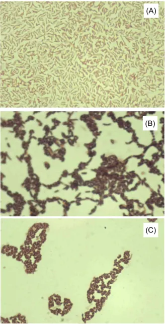
발효가 진행중인 멸치젓갈을 10% NaCl과 전지분유를 함유 한 marine 한천배지에 도말하여 배양하여 단백질분해 환을 형성하는 콜로니를 분리하여 단백질분해효소 생산 호염균을 선별하였다. 큰 환을 형성하는 24개의 콜로니를 1차 선별하고, 몇 단계의 순수분리 작업을 거쳐서 최종적으로 3개의 균주를 선택하였다(Fig. 1). 균주 FAM 10은 크림색깔을 나타내었으 며, FAM 114와 FMA 115는 황색을 보였다. 또한 선별된 균주 가 protease를 분비하는 호염성균인 것을 확인하기 위해 액체 배지에 NaCl을 2~26% 첨가하면서 각 균주의 세포 성장과 단 백질분해효소 활성을 관찰하였다(Fig. 2, 3). FAM 10, FMA 114, 그리고 FAM 115는 각각 NaCl 2~4%, 10%, 그리고 6%
조건에서 최적 성장조건을 나타내었다(Fig. 2). 또한, 모든 균 이 14%까지는 세포성장이 이루어지다가 18%부터 성장이 급 격히 저하되기 시작하였으며, 26%에서는 모든 균의 세포 성장 이 억제되었다. 이때 배양액 상등액을 이용하여 단백질 분해 효소 활성을 측정하여 본 결과(Fig. 3), FAM 10은 NaCl 6%
조건에서 가장 높은 활성을 보였으며, 14% 첨가조건에서는 단백질분해효소 활성을 관찰할 수 없었다. FMA 114와 FAM 115는 모두 10%에서 단백질분해효소 활성이 가장 높았으며, 14% 조건에서는 활성이 급격히 저하되다가 18%부터는 효소 활성이 없었다. 효소활성도 측면에서는 FAM 115가 FAM 10 과 FAM 114보다 더 우수한 활성도를 나타내었다. 따라서, NaCl 첨가조건에서 세포 성장과 효소활성도를 기반으로, 본 연구에서 분리한 FAM 10, 114, 115는 모두 단백질분해효소를 분비하는 중간 정도의 호염세균(moderately halophilic bac- teria)임을 알 수 있었다.
(A)
(B) (C)
Fig. 1. Screening of protease-producing halophilic bacteria from fermenting anchovy on marine agar plate with 10% NaCl and 1% skim milk. (A) FAM 10, (B) FAM 114, and (C) FAM 115.
단백질분해효소 생산 호염세균의 생화학적 특성
그람염색 후에 현미경으로 관찰한 결과 FAM 10은 휜 모양 (curved-shaped)의 그람음성 세균이였다(Fig. 4A). API 20 NE 키트를 사용하여 FAM 10에 대한 생화학적 특성을 조사하였 다. glucose acidification, arginine dihydrolase, β-glucosidase (esculine 가수분해), protease (젤라틴 가수분해), cytochrome oxidase 는 양성반응을 보였으며, nitrate reduction, 인돌 생 산, urease, 그리고 β-galactosidase 실험은 음성을 나타내었다.
여러 탄소원에 대한 자화도를 테스트한 결과, glucose, man- nose, mannitol, N-acetylglucosamine, maltose, gluconate,
0 1 2 3 4 5 6
0 5 10 15 20 25
NaCl (%)
Growth
FAM10 FAM114 FAM115
Fig. 2. Effect of NaCl on cell growth of FAM 10, FAM 114, and FAM 115. Cells were grown in 250 ml flask containing 30 ml of marine medium supplemented with a gradient of salts at 30℃ for 24 hr.
0 2 4 6 8 10 12
0 5 10 15 20 25
NaCl (%)
Protease activity (U/ml)
FAM10 FAM114 FAM115
Fig. 3. Effect of NaCl on protease production of FAM 10, FAM 114, and FAM 115. After growth on marine medium with 10% NaCl cell supernatants were used for measurement of protease activity.
(A)
(B)
(C)
Fig. 4. Light micrograph of isolated three strains after Gram staining (x1,000). (A) FAM 10, (B) FAM 114, and (C) FAM 115.
caprate, 그리고 citrate는 자화할 수 있었으나, arabinose, adi- pate, malate, 그리고 phenyl-acetate는 자화할 수 없었다 (Table 1)[1,10,19]. FAM 114와 115 균을 현미경으로 관찰한 결과, 모두 막대형(rod-sahped), 그람양성 세균임이 확인되었 으며(Fig. 4B, 4C), 그 생화학적 특성은 Table 2에 나타내었다.
FAM 114는 D-arabitol을 이용하지 못한 반면, FAM 115는 탄 소원으로 D-arabitol을 사용할 수 있었으며, 그 외의 모든 생화 학적인 특성은 동일하였다[5,24,25].
균주 FMA 10의 16S-rDNA와 16S-23S 유전자 사이 부위의 서열분석
균주를 동정하기 위해 FAM 10의 16S rDNA 서열을 결정하
Table 1. Phenotyphic characteristics of the
Salinivibrio
sp. FAM 10Characteristics Results
Shape Curved rod
Gram stain -
Colony color cream
Nitrate reduction -
Indole production -
Glucose acidification +
Citrate +
Arginine dihydrolase +
Urease -
β -glucosidase (esculin hydrolysis) + Protease (gelatin hydrolysis) +
β -galactosidase -
Cytochrome oxidase +
Utilization of carbon source
a)Glucose +
Arabinose -
Mannose +
Mannitol +
N-acetyl-glucosamine +
Maltose +
Gluconate +
Caprate +
Adipate -
Malate -
Phenyl-acetate -
a)API 20NE kit was used.
였다. FAM 10은 191번째부터 염기서열이 각각 “AC”와
“TAGTTCATTACGAGCTG”을 갖는 2가지 다른 형태의 16S rDNA (GenBank Accession Numbers JN381953과 JN381954) 가 존재하였다. 대부분의 미생물은 염색체에 16S rRNA gene 이 여러 복사본이 존재하는데[18], FAM 10은 염색체내에 적어 도 2개의 다른 copy의 16S rRNA gene들이 존재함을 의미한다.
BLAST를 이용하여 상동성 분석을 한 결과, 많은 Salinivibrio sp. (AJ640132.1, GU212631.1, EU308487.1, HQ641751.1, EU308488.1, X95527.1, X95531.1, EU308484.1, X95530.1, X74699.1, 그리고 AF057016.1)의 16S rDNA서열들과 98~99%
의 서열 동일성(sequence identity)을 확인하였다[1,10,19]. Fig.
5는 FAM 10의 계통발생도를 표현한 것으로, Salinivibrio에 속 하며, 특히 Salinivibrio costicola와 가장 유사한 것으로 확인이 되었다. 16S rRNA 유전자 데이터베이스에 따르면, Salinivibrio 는 균주에 따라서 191번째(JN381953 서열번호기준)부터 변화 된 서열을 보이는 균들이 많았다. 예를 들어서 Salinivibrio sp.
TA09_2, TA09_1, TA012_1, TA012_2, TA012_3, NTU-05,
Salinivibrio costicola strain SI3, GSP020, GSP12, Salinivibrio bu-
daii strain PT, 그리고 Salinivibrio costicola subsp. vallismortis
Table 2. Phenotyphic characteristics of the
Halobacillus
sp. FAM 114 and FAM 115Characteristics Results Characteristics Results
Shape rod Arbutine -
Gram stain + Esculine -
Colony color yellow Salicine -
Utilization of carbon source
a)Cellobiose -
Glycerol + Maltose +
Erythritol - Lactose -
D-arabinose - Melibiose +
L-arabinose + Saccharose +
Ribose + Trehalose +
D-Xylose + Inuline -
L-Xylose - Melezitose -
Adonitol - D-Raffinose -
β -Methyl-xyloside - Amidon +
Galactose + Glycogene +
D-Glucose + Xylitol -
D-Fructose + β -Gentiobiose -
D-Mannose - D-Turanose -
L-Sorbose - D-Lyxose -
Rhamnose - D-Tagatose -
Dulcitol - D-Fucose +
Inositol - L-Fucose -
Mannitol + D-Arabitol -/+
b)Sorbitol - L-Arabitol -
α -Methyl-D-mannoside - Gluconate -
α -Methyl-D-glucoside - 2-ceto-gluconate -
N-acetyl glucosamine + 5-ceto-gluconate -
Amygdaline -
a)API 50CHB kit was used.
b)Showing (-) reaction in FAM 114; showing (+) reaction in FAM 115.
Salinivibrio sp. 18AG (AJ640132)
Salinivibrio sp. TA09 1 (EU308487) Salinivibrio sp. TA09 2 (EU308488)
Salinivibrio sp. H3B27 (GU212631) Salinivibrio costicola strain E-367 (X95529)
Salinivibrio costicola strain cepa6 (X95531) Salinivibrio costicola strain V-15 (X95530) Salinivibrio costicola 16S strain NCIMB 701-T (X95527)
Salinivibrio costicola strain SI3 (HQ641751) Salinivibrio costicola strain GSP020 (DQ157161)
Salinivibrio costicola strain GSP12 (AY505534)
Salinivibrio costicola subsp. vallismortis strain DV (AF057016) FAM 10 (JN381953)
FAM 10 (JN381954) Salinivibrio sp. 18AG (AJ640132)
Salinivibrio sp. TA09 1 (EU308487) Salinivibrio sp. TA09 2 (EU308488)
Salinivibrio sp. H3B27 (GU212631) Salinivibrio costicola strain E-367 (X95529)
Salinivibrio costicola strain cepa6 (X95531) Salinivibrio costicola strain V-15 (X95530) Salinivibrio costicola 16S strain NCIMB 701-T (X95527)
Salinivibrio costicola strain SI3 (HQ641751) Salinivibrio costicola strain GSP020 (DQ157161)
Salinivibrio costicola strain GSP12 (AY505534)
Salinivibrio costicola subsp. vallismortis strain DV (AF057016) FAM 10 (JN381953)
FAM 10 (JN381954)
Fig. 5. Phylogenetic tree of strain FAM 10. The 16S rRNA sequence was analyzed and compared with nucleotide sequence database using BLAST program. The tree was created by neighbor-joining method. The numbers on the tree indicate the percentages of bootstrap based on 1,000 replication. The scale bar indicates 0.001 change per nucleotide.
Fig. 7. Alignment of 16S-23S IGS sequences of
Salinivibrio
sp. FAM 10. Sources of sequence are as follows: 1, 310 bp of IGSO; 2, 534 bp of IGSIA-1; 3, 568 bp of IGSEKV; 4, 633 bp of IGSIA-2. The symbols [ ] and □ indicate tRNA gene for a specific amino acid and anticodon sequence corresponding to amino acid, respectively. The anticodon sequences for isoleucine, alanine, glutamic acid, lysine, and valine are GAT, TGC, TTC, TTT, and TAC, respectively. The symbol * shows the consensus sequence in 4 IGSs.S (1) (2) (3)
Fig. 6. Amplification of 16S-23S IGS from isolated strains FAM 10, FAM 114, and FAM 115. S, 1Kb DNA ladder, (1) FAM 10: 4 bands (about 750, 650, 600, 380 bp), (2) FAM 114:
2 bands (about 500, 230 bp), and (3) FAM 115: 2 bands (about 500, 230 bp).
strain DV는 JN381953과 동일한 “AC” 서열을 가지며, S.
costicola srain NCIMB 701-T, strain E-367, strain H-178는 JN381954와 동일한 “TAGTTCATTACGAGCTG” 서열을 갖 는 것으로 확인이 되었다. 그러나, Salinivibrio sp. 18AG, H3B27, cepa 6, 그리고 V-15 균주들은 동일한 위치에 또 다 른 공통서열인 “TGATTCGTTAGAGTCA”를 나타내었다.
따라서, 여러 Salinivibrio 균주들 사이에서 이 위치에 많은 변화가 존재함을 확인할 수 있었다. 그러나, 지금까지 보고 된 여러 종류의 Salinivibrio 균주들 모두 동일한 균주내에 2 가지 다른 형태의 16S rDNA 염기서열을 갖는 균주는 아직 보고된 봐 없다. 따라서, 본 연구에서 분리된 FAM 10은 기 존에 보고된 균주의 16S rDNA와는 다른 특징을 갖는 균주 임을 확인할 수 있었다.
rRNA 오페론(rrn operon)의 유전적 배열을 더 확인하기
위하여, 16S-23S IGS 서열을 분석하였다[2,8]. FAM 10의
Halobacillus trueperi strain HLSB (FJ999554) Halobacillus trueperi strain LS5 (FJ937876) Halobacillus trueperi strain whb45 (FJ444991)
Halobacillus trueperi strain S6-16 (EU624440)
Halobacillus trueperi strain DSM 10404T (AJ310149) Halobacillus trueperi strain XJSL8-9 (GQ903458)
Halobacillus sp. CNJ955 PL04 (DQ448804) FAM 114 and 115 (JN381955)
Halobacillus litoralis strain XJSL8-10 (GQ903459) Halobacillus litoralis strain SL-4 (NR029304)
Halobacillus sp. CNJ915 (DQ448762) Halobacillus profundi (AB189298) Halobacillus dabanensis strain XJSL8-5 (GQ903454)
Halobacillus dabanensis strain XJSL8-45 (GQ903453)
Halobacillus karajensis strain I122 (DQ089675) Marinococcus albus strain I18 (DQ093354)
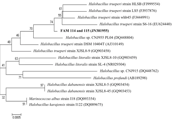
Fig. 8. Phylogenetic tree of strain FAM 114 and 115
.
The 16S rRNA sequence was analyzed and compared with nucleotide sequence database using BLAST program. The tree was created by neighbor-joining method. The numbers on the tree indicate the percentages of bootstrap based on 1,000 replication. The scale bar indicates 0.0005 change per nucleotide.16S-23S IGS 부위를 PCR로 증폭한 결과, 300에서 800 bp 사이 에 특징적인 4가지 단편을 얻을 수 있었다(Fig. 6). 그 단편들 의 염기서열을 확인하고 다중배열하여 분석하였다(Fig. 7). 염 기서열, 서열길이, 그리고 존재하는 tRNA 형태에 따라서 분 류한 결과, FAM 10에는 4가지 다른 유형의 IGS들이 존재하였 다. 즉, tRNA가 존재하지 않는 310 bp의 IGS (IGS
O라 명명), 각각 이소루이신과 알라닌을 운반하는 tRNA가 존재하는 534 bp의 IGS (IGS
IA라 명명), 각각 이소루이신과 알라닌을 운반하 는 tRNA가 존재하는 633 bp의 IGS (IGS
IA라 명명), 각각 글루 탐산, 라이신, 그리고 발린을 운반하는 tRNA가 존재하는 568 bp의 IGS (IGS
EKV라 명명) (JN381956, JN381957, JN381958, 그 리고 JN381959, Table 3)가 확인되었다. FAM10내 존재하는 tRNA 유전자를 제외한 IGS들의 염기서열은 Salinivibrio cos- ticola ATCC 33508 에 존재하는 5가지 유형의 IGS (2 가지 IGS
O, 2가지 IGS
E, 그리고 1가지 IGS
I) 들과 99% 서열 동일성 을 나타내었다. 그러나, FAM 10에 IGS내의 tRNA 유형과 유 전자 배열은 기존에 보고된 Salinivibrio costicola ATCC 33508 와[10] 많은 Vibrio species [16]에 존재하는 형태와는 다른 유 형과 유전적 배열을 나타내었다. 따라서, 계통발생도와 16S-23S IGS 서열과 tRNA 유전자 배열, 생화학적인 특징을 비교하여 보면, 본 연구에서 분리된 FAM 10은 기존에 보고된 Salinivibrio 균주와는 그 특징이 다른 새로운 Salinivibrio sp.
임을 확인하였다.
균주 FMA 114와 FAM 115의 16S-rDNA와 16S-23S 유전자 사이 부위의 서열분석
염기서열분석 결과, FAM 114와 FAM 115는 완전히 동일한 16S rRNA 유전자 서열을 가졌으며(JN381955), Halobacillus trueperi whb45, HLSB3, LS5, S6-16, GSP38, XJSL8-8, XJSL8-9, 그리고 Halobacillus sp. CNJ955 (FJ444991.1, FJ999554.1, FJ937876.1, EU624440.1, AY505522.1, GQ903457.1, GQ903458.1, and DQ448804.1)의 16S rDNA서열과 99% 동일성을 보여주었 다. 계통발생도 분석결과, FAM 114와 FAM 115는 Halobacillus 에 속하며, 특히 Halobacillus trueperi와 가장 유사한 것으로 확 인이 되었다(Fig. 8)[3,4,23,24].
FAM 114와 115의 16S-23S IGS 부위를 PCR로 증폭한 결
과, 젤 상에서 약 230 bp와 500 bp에 해당되는 2개의 단편을
확인하고 그 염기서열을 분석하였다(Fig. 6). FAM 114는
tRNA 유전자가 존재하지 않는 2가지 종류의 170 bp의 IGS
(IGS
0라 명명)와 각각 이소루이신과 알라닌을 운반하는
tRNA 유전자를 함유하는 412 bp의 IGS (IGS
IA라 명명)가 존
재함을 확인하였다(JN381960, JN381961, 그리고 JN381962,
Table 3). FAM 115에는 1개의 170 bp IGS
0와 1개의 412 bp
IGS
IA(JN381963와 JN381964)가 존재함을 확인하였다. 두 균
주에 존재하는 3개의 IGS
0와 2개의 IGS
IA는 각각 1~2 염기만
을 제외하고는 모두 같은 염기서열을 가지며, tRNA 유전자
역시 같은 배열을 나타내었다(Fig. 9). 또한, FAM 114와 FAM
115의 tRNA를 제외한 IGS 서열은 Halobacillus aidingensis와
Fig. 9. Alignment of 16S-23S IGS sequences of
Halobacillus
sp. FAM 114 and FAM 115. Sources of sequence are as follows: 1, 170 bp of IGSO-1 in FAM 114; 2, 170 bp of IGSO-2 in FAM 114; 3, 170 bp of IGSO in FAM 115; 4, 412 bp of IGSIAin FAM 114; 5, 412 bp of IGSIAin FAM 115. The symbols [ ] and □ indicate tRNA gene for a specific amino acid and anticodon sequence corresponding to amino acid, respectively. The anticodon sequences for isoleucine and alanine are GAT and TGC, respectively. The symbol * shows the consensus sequence in 5 IGSs.Table 3. Profile and characteristics of the 16S-23S IGSs in FAM 10, FAM 114, and FAM 115.
Strain IGS
designation IGS length (bp)
Salinivibrio
sp. FAM 10Halobacillus
sp. FAM 114Halobacillus
sp. FAM 115IGSOa IGSIA -1b IGSIA -2 IGSEKVc IGSO -1 IGSO -2 IGSIA IGSO IGSIA
310 534 633 568 170 170 412 170 412
a IGSO without tRNA gene.
bIGSIAwith tRNA genes for isoleucine and alanine, respectively.
cIGSEKVwith tRNA genes for glutamic acid, lysine, and valine, respectively.
Halobacillus sp. JM-Hb과 99% 동일성을 보여주었다. 따라서, 계통발생도와 16S-23S IGS분석, 생화학적인 특성에 기초하 여, 분리된 FAM 114와 FAM 115를 각각 Halobacillus sp.
FAM 114와 FAM115로 명명하였다.
중국 염전에서 분리한 H. trueperi whb7과 H. trueperi whb45의 경우[4], 각각 병원성 곰팡이인 Candida albicans와 간암세포 Bel 7402의 증식억제효과를 나타내는 활성이 보고 되었다. 향후 건강 기능성 멸치액젓을 개발하기 위한 목적 으로, 본 연구에서 분리된 FAM 114와 FAM 115 균주에 대 한 항진균 활성과 암세포 제해 활성을 관찰 할 필요성이 있 다고 사료된다.
감사의 글
본 논문은 2011학년도 경성대학교 학술연구비지원에 의하
여 연구되었습니다.
References
1. Amoozegar, M. A., P. Schumann, M. Hajighasemi, A. Z.
Fatemi, and H. R. K. Heidari. 2008.
Salinivibrio proteolyticus
sp. nov., a moderately halophilic and proteolytic species from a hypersaline lake in Iran.Int. J. Syst. Evol. Microbiol.
58, 1159-1163.
2. Bourget, N. L., H. Philippe, I. Mangin, and B. Decaris. 1996.
16S rRNA and 16S to 23S internal transcribed spacer se- quence analyses reveal inter- and intraspecific
Bifidobacterium
phylogeny.Int. J. Syst. Bacteriol.
46, 102-111.3. Chaiyanan, S., S. Chaiyanan, T. Maugel, A. Huq, F. T. Robb, and R. R. Colwell. 1999. Polyphasic taxonomy of a novel
Halobacillus
,Halobacillus thailandensis
sp. nov. isolated from fish sauce.Syst. Appl. Microbiol.
22, 360-365.4. Chen, L., G. Wang, T. Bu, Y. Zhang, Y. Wang, M. Liu, and X. Lin. 2010. Phylogenetic analysis and screening of anti- microbial and cytotoxic activities of moderately halophilic bacteria isolated from the Weihai solar saltern.
World J.
Microbiol. Biotechnol.
26, 879-888.5. Chen, Y. G., Z. X. Liu, Y. Q. Zhang, Y. X. Zhang, S. K. Tang, E. Borrathybay, W. J. Li, and X. L. Cui. 2009.
Halobacillus naozhouensis
sp. nov., a moderately halophilic bacterium iso- lated from a sea anemone.Antonie van Leeuwenhoek
. 96, 99-107.6. Chung, S. S., Y. U. Shin, H. J. Kim, G. H. Jin, H. M. Rho, and H. H. Lee. 2000. Isolation of high yielding alkaline pro- tease mutants of
Vibrio metschnikovii
strain RH530 and de- tergency properties of enzyme.J. Microbiol. Biotechnol.
10, 349-354.7. Essghaier, B., M. Bejji, H. Jijakli, A. Boudabous, and N. S.
Zouaoui. 2009. High salt-tolerant protease from a potential biocontrol agent
Bacillus pumilus
M3-16.Ann. Microbiol.
59, 553-558.8. Gűrtle, V. and V. A. Stanisich. 1996. New approaches to typ- ing and identification of bacteria using the 16S-23S rDNA spacer region.
Microbiol.
142, 3-16.9. Hong, Y., J. H. Kim, B. H. Ahn, and S. K. Cha. 2000. The effects of low temperature storage and aging of
Jeot-kal
on the microbial counts and microflora.Korean J. Food Sci.
Technol.
32, 1341-1349.10. Huang, C. Y., J. L. Garcia, B. K. C. Patel, J. L. Cayol, L.
Baresi, and R. A. Mah. 2000.
Salinivibrio costicola
subsp.val- lismortis
subsp. nov., a halotolerant facultative anaerobe from Death valley, and emended description ofSalinivibrio costicola
.Int. J. Syst. Evol. Microbiol.
50, 615-622.11. Hur, S. H. 1996. Critical review on the microbiological stand- ardization of salt-fermented fish product.
J. Korean Soc. Food Sci. Nutr.
25, 885-891.12. Kim, M. S., E. J. Park, M. J. Jung, S. W. Roh, and J. W.
Bae. 2009. Analysis of prokaryote communities in Korean traditional fermented food, Jeotgal, using culture-dependent method and isolation of a novel strain.
Kor. J. Microbiol.
30, 26-31.13. Kim, Y. M. 2008. Present status and prospect of fermented seafood industry in Korea
. Food Sci. Industry
41, 16-33.14. Kumar, S., R. Jansen, F. Sasse, and G. Hofle. 2004. MEGA3:
integrated software for molecular evolutionary genetics analysis and sequence alignment.
Brief Bioinform.
5, 150-163.15. Lim, H. T., S. K. Chung, G. N. Kim, J. U. Ha, and H. D.
Paik. 2002. Characterization of protease produced by
Bacillus amyloliquefaciens
HTP-8 isolated from Korea fer- mented anchovy sauce.Kor. J. Microbiol. Biotechnol.
30, 26-32.16. Lee, S. K. Y., H. Z. Wang, S. H. W. Law, R. S. S. Wu, and R. Y. C. Kong. 2002. Analysis of the 16S-23S rDNA inter- genic spacers (IGSs) of marine vibrios for species-specific signature DNA sequences.
Mar. Pol. Bul.
44, 412-420.17. Lee, S. M., J. M. Lim, K. H. Kim, S. Y. Cho, K. S. Park, Y. M. Sin, C. Y. Cheung, J. I. Cho, H. J. You, K. H. Kim, D. H. Cho, C. J. Lim, and O. H. Kim. 2008. Microbiological study using monitoring of microorganism in salt-fermented fishery products.
J. Fd. Hyg. Safety
23, 198-205.18. Martín, J. F., C. Barreiro, E. G. Lavado, and B. M. Barriuso.
2003. Ribosomal RNA and ribosomal proteins in corynebacteria.
J. Biotechnol.
104, 41-53.19. Mellado, E., E. R. B. Moore, J. J. Nieto, and A. Ventosa. 1996.
Analysis of 16S rRNA gene sqquences of
Vibrio costicola
strains: description ofSalinivibrio costicola
gen. nov., comb.nov.
Int. J. System Bacteriol.
46, 817-821.20. Park, C. K., T. J. Kang, and K. O. Cho. 2002. Studies on the processing of rapid- and low salt-fermented liquefaction of anchovy (I).
Korean J. Dietary Culture
17, 197-213.21. Saitou, N. and M. Nei. 1987. The neighbor-joining method:
a new method for constructing phylogenetic trees.
Mol. Biol.
Evol.
4, 406-425.22. Sambrook, J. and D. W. Russell. 2001. Molecular cloning:
a laboratory manual, 3rded. Cold Spring Harbor Laboratory Press, Cold Spring Harbor, N.Y.
23. Thompson, J. D., D. G. Higgins, and T. J. Gibson. 1994.
CLUSTAL W: improving the sensitivity of progressive mul- tiple sequence alignment through sequence weighting, posi- tions-specific gap penalties and weight matrix choice.
Nucleic Acids Res.
22, 4673-4680.24. Yoon, J. H., S. J. Kang, C. H. Lee, H. W. Oh, and T. K.
Oh. 2005.
Halobacillus yeomjeoni
sp. nov., isolated from a ma- rine solar saltern in Korea.Int. J. Syst. Evol. Microbiol.
55, 2413-2417.25. Yoon, J. H., S. J. Kang, and T. K. Oh. 2008.