Sulforaphane-Induced Apoptosis was Regulated by p53 and Caspase-3 Dependent Pathway in Human Chondrosarcoma, HTB-94
Won Kil Lee and Song Ja Kim*
Department of Biological Sciences, College of Natural Sciences, Kongju National University, Korea Received April 1, 2011 /Accepted May 26, 2011
Sulforaphane (SFN) is an isothiocyanate, isolated from glucoraphanin in broccoli and other crucifer- aous vegetables. Recent studies have revealed that SFN induces anti-proliferation and apoptosis by cell cycle arrest in various cancer cells. In this study, we investigated the effect of SFN induced apop- tosis in chondrosarcoma HTB-94 cells. SFN caused suppression of proliferation and apoptosis in a dose-dependent manner as determined by cell phenotype, MTT assay and FACS analysis in HTB-94 cells. Treatment of SFN led to caspase-3 activation and p53 accumulation as determined by Western blot analysis. Also, SFN significantly induced DNA fragmentation and nuclear degradation though ac- tivation of caspase-3, as detected by DNA electrophoresis and immunostaining, respectively. Our re- sults indicate that SFN-induced apoptosis was regulated by p53 and caspase-3 dependent pathways.
Furthermore, SFN may act as a potent anti-proliferation agent, and as a promising candidate for mo- lecular-targeting chemotherapy against human chondrosarcoma cells.
Key words : Chondrosarcoma, sulforaphane, apoptosis, caspase-3
*Corresponding author
*Tel:+82-41-850-8507, Fax:+82-41-850-0927
*E-mail : [email protected]
서 론
연골육종(Chondrosarcoma)은 골육종에 이어 두 번째로 흔 한 원발성 악성종양으로, 대개 연골세포에서 발생하며 서서히 자라고 천천히 전이하는 특징이 있다. 연골육종은 원발성 악 성 골종양의 약 10-20%를 차지하며, 발병 연령은 30~60세 이 후로 골육종(Osteosarcoma)보다 발병 시기가 늦으며 20대 이 하에게서는 드물게 발생한다. 연골육종 중 약 75% 정도가 중 심성 연골육종이며 골반골에 가장 많이 발생하며 대퇴골, 상 완골, 늑골, 두개골 등에 발생한다. 연골육종은 항암 약물치료 와 방사선치료가 잘 듣지 않기 때문에 수술이 가장 중요한 치료방법으로 선택되고 있다. 그러므로, 연골육종의 성장을 억제하거나 세포사멸(apoptosis)을 일으킬 수 있는 약물을 찾 는 것이 매우 중요하다.
Sulforaphane (SFN)은 양배추, 브로콜리, 콜리플라워, 케일, 겨자 등, 십자화과 채소에 많이 들어있는 것으로 알려져 있다 [8]. SFN은 이소시아네이트(isothiocyanate) 구조를 갖는 물질 로서 강력한 항산화, 항염증, 그리고 항암효과가 있는 것으로 알려져 있으며, glutathione S-transferase (GST) enzyme ac- tivity의 활성을 증가시켜 줌으로서 많은 발암성 물질을 억제 하는 것으로 알려져 있다[13-16]. 또한, Phase Ⅱ detoxification enzymes로 알려져 있는 quinone reductase [NAD(P)H:
(quinone-acceptor) oxidoreductase]의 활성을 증가시켜 발암
물질에 의한 암세포의 성장을 억제하기도 한다[3,16]. 그리고, 이소시아네이트에 의한 항암효과가 동물실험에서도 확인되 었으며[5,7], 다양한 인간 암세포를 이용한 in vitro 및 in vivo 실험에서 증식억제 효과가 있는 것으로도 알려져 있다 [6,8,14,17,18,25-27]. 또한, SFN은 인간 대장암세포에서 p53 의 존적인 caspase-2-JNK 경로를 통하여 세포사멸을 조절한다고 도 알려져 있다[23].
세포사멸은 병리적인 상태뿐만 아니라 정상적인 상태의 조 직에서도 확인된다. 세포사멸은 세포괴사(necrosis)와 함께 세 포죽음(cell death)의 한 종류이지만 일시적인 세포손상으로 발생하는 세포괴사와는 구별된다. 세포사멸이 일어나는 세포 는 탈수에 의한 세포수축, 세포 내 칼슘이온 농도 증가, DNA 분절, 염색질 응축, phosphatidylserine이 세포막 밖으로 노출 되는 등 다양한 생리적 현상이 동반되며 최종적으로 apoptotic body를 형성하며 죽게 된다. 일반적으로 많은 암세포에서 다 양한 항암제를 이용하여 세포사멸에 대한 연구가 in vitro 및 in vivo에서 많이 이루어지고 있다.
여러 암세포 유형에서 SFN이 유도하는 세포사멸에 대한
연구는 그동안 많이 이루어져 왔으나[14,19,20], 인간 연골암세
포에서 SFN을 처리하여 유도되는 세포사멸에 대한 분자적 기
전연구는 거의 알려져 있지 않다. 이에, 본 연구에서는 인간
연골암세포주인 HTB-94 세포주에 SFN을 처리하여 유도되어
지는 세포사멸 조절기전에 관한 연구를 수행하였으며, 그 결
과, SFN에 의한 연골암세포사멸은 p53 및 caspase-3 의존적인
경로를 통하여 일어난다는 것을 알 수 있었다.
재료 및 방법
Cell culture
실험에 사용된 인간 연골암 세포주 HTB-94 cell (American Type Culture Collection, Manassas, VA, U.S.A.)는 10%의 FBS (fetal bovine serum), 50 μg/ml의 streptomycin, 50 units/ml의 penicillin이 함유된 Dulbecco's modified Eagle's medium (DMEM, Gibco-BRL, Gaithersburg, MD, U.S.A.)에 5% CO
2, 37℃ incubator에서 배양하였다. 계대배양은 배양접 시에 1×10
6cells/dish의 밀도로 배양하였다. 배지는 2~3일마 다 교환하였으며 배양접시의 세포밀도가 거의 80%가 되었을 때 본 연구에 사용된 시약을 처리하였다.
Western Blot Analysis
단백질의 정량분석을 위하여 세포를 50 mM Tris-HCl pH 7.4, 150 mM NaCl, 1% Nonidet P-40, 0.1% SDS가 함유된 cell lysis buffer에 단백질 분해효소 억제제[10 μg/ml leupeptin, 10 μg/ml pepstatin A, 10 μg/ml aprotinin, 1 mM 4-(2-aminoethyl) benzensulfonyl fluoride]를 첨가하여 세포 단백질을 추출하였다. 추출한 단백질은 SDS-polyacrylamide gel에 전기영동하여 단백질을 크기 별로 분리한 후 nitro- cellulose (NC) 막으로 이동시켰다. 단백질 발현분석을 위해 1차 항체인 p53, pro-caspase 3, actin (Santa Cruz Biotechnology Inc., Santa Cruz, CA, U.S.A.)을 2시간 동안 단 백질이 부착된 NC와 반응시킨 후, 2차 항체를 1시간 동안 반 응시켜 chemiluminescence 시스템을 이용하여 분석하였다.
MTT (3-(4,5-dimethylthiazol-2yl)-2,5-diphenyl-2H- tetrazolium bromide) assay
인간 연골암 세포주 HTB-94 cell를 96 well plate에 세포를 2.5×10
4cells/well로 200 μl/well 분주하여 incubator에 배양 한다. 세포밀도가 80% 이상 되었을 때 시약을 처리하였으며 시약을 처리한 후 24시간 동안 배양하였다. 배양한 후 MTT reagent Ⅰ 을 최종 농도 10%로 첨가하고 4시간 동안 배양한 후, 다시 solubilization sulution인 MTT reagent Ⅱ (10% SDS in 0.01 M HCl, DMSO)를 최종 부피의 2배 첨가하여 formazan 결정을 12시간 동안 용해한 후 ELISA (Enzyme-Linked Immuno Sorbent Assay) reader를 사용하여 흡광도(595 nm) 값을 측정한다.
DNA fragmentation
배양된 HTB-94는 1X PBS로 2회 세척 후 lysis buffer (100 mM Tris-HCl, pH 7.4, 0.5 mM EDTA, 0.2% SDS, 200 mM NaCl)를 사용하여 세포를 용해시켰다. 용해된 세포액은 13,000 rpm에서 10분 동안 원심분리하여 상등액을 수집하였 으며, 이 상등액에 20 μg RNase A (Sigma, St. Louis, MO,
U.S.A.)를 37℃에서 2시간 동안 반응하였으며, 이후 0.01 mg/ml의 proteinase K (Sigma, St. Louis, MO, U.S.A.)를 37℃
에서 2시간 반응시켰다. 반응이 끝난 용액에 PCI [Phenol:Chloroform:Isoamyl alcohol=25:24:1]를 사용하여 DNA를 정제하였으며, 3 M sodium acetate와 에탄올을 사용 하여 DNA를 침전하였다. 침전된 DNA는 70% 에탄올로 세척 하여 상온에서 건조 후 멸균된 3차 증류수를 사용하여 DNA를 녹였다. 용해된 DNA는 2% agarose gel에 전기영동하여 DNA 절편을 분리하였다.
Fluorescence Activated Cell Sorter (FACS)
1X PBS로 세척 후 Trypsin-EDTA를 사용하여 부착된 세포 를 부유시켜 상등액을 1,000 rpm에서 10분 동안 모아 70% 에 탄올로 4℃에서 24시간 고정시켰다. Propidium Iodide (BD bioscience, San Diego, CA, U.S.A.) 염색을 위해 50 μg/ml Propidium Iodide, 0.1% Nonident P-40, 100 μg/ml RNase A 1X PBS에 녹여 20분 동안 37℃에서 암반응을 시켰다. 염색 한 시료는 Fluorescence Activated Cell Sorter (FACS)를 이용 하여 nylon mesh filter를 통해 부유하는 것의 세포주기를 측 정하였고 data 분석은 Cellquest anlalysis program (Becton Dickinson, Mountain View, CA, USA)을 이용하였다.
Immunofluorescence staining
연골암 세포주 HTB-94에서 SFN에 의한 핵과 세포골격의 변화를 관찰하기 위하여 immunofluorescence를 수행하였다.
배양된 HTB-94는 3.5% paraformaldehyde로 10분 동안 상온 에서 고정시켰으며, 0.1% Triton X-100을 이용하여 세포막 투 과성을 높여주었다. 고정된 세포는 DAPI [4',6-dia- midino-2-phenylindole dihydrochloride]와 phalloidin을 사 용하여 염색하였으며, 형광현미경으로 관찰하였다.
Statistics analysis
본 실험의 결과는 여러 차례의 실험 중 평균적인 결과를 사용하였다. 통계적 검증은 student-t test를 사용하였고, 신뢰 구간은 5%로 하여 p값이 0.05보다 낮은 경우를 유의하다 판단 하였다.
결 과
Sulforaphane은 인간 연골암 세포주 HTB 94에서 세포 사멸을 유도한다
Sulforaphane (SFN)은 여러 종양세포에서 항암효과와 염증
유발인자의 활성저해 및 세포사멸에 관여하는 것으로 알려져
있다. 그러나 연골암세포에서 SFN에 의한 세포사멸에 대한
연구는 미비하여, 본 연구에서는 인간 연골암 세포주 HTB-94
세포에 SFN을 처리하여 세포사멸에 미치는 영향 및 이의 분자
Fig. 1. SFN induces apoptosis and inhibits cell proliferation in HTB-94 cells. HTB-94 cells were treated with the indicated concen- trations of SFN for 24 hr. Cells photographs were taken using phase contrast microscope (magnification, X200) (A). Apoptotic cells were determined by MTT assay (B). The data present a typical experiment (A) and as mean values with standard deviation (B) (n=4). * p <0.05, ** p <0.001 compared with untreated cells.
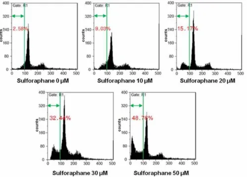
Fig. 2. SFN stimulates cell death in HTB-94 cells. HTB-94 cells were untreated or treated with the various concentrations of SFN for 24 hr and harvested at the indicated times for FACS analysis of cell cycle disrubution. The cells were fixed with 80%
ethanol in PBS, stained with propidium iodide (PI). Data are presented as results of a typical experiment.
적 기전을 알아보았다. HTB-94 세포에 SFN을 24시간 처리하 여 세포형태를 위상차 현미경을 이용하여 확인한 결과 세포의 부피 감소와 더불어 SFN의 처리농도가 높아질수록 세포사멸 정도가 증가함을 알 수 있었다(Fig. 1A). 이에 세포사멸정도를 정량화 하고자 MTT assay를 한 결과 SFN의 처리농도가 증가 할수록 세포생존률이 감소함을 확인할 수 있었다(Fig. 1B). 유 세포분석기(Fluorescence Activated Cell Sorter, FACS)를 사
용하여 세포사멸 정도를 확인해 본 결과 세포사멸을 나타내는
Sub G1기 세포수가 SFN 10 μM에서 9.03%, 20 μM에서
15.17%, 30 μM에서 32.3% 정도 증가하였으며, 50 μM에서는
48.76% 정도로 거의 반 정도의 세포사멸이 일어난 것으로 확
인되었다(Fig. 2). 이는 연골암세포의 사멸정도가 SFN 처리농
도 의존적으로 일어남을 확인할 수 있었다. 본 연구결과를 종
합해 볼 때, SFN은 인간 연골암 세포주인 HTB-94에서 처리농
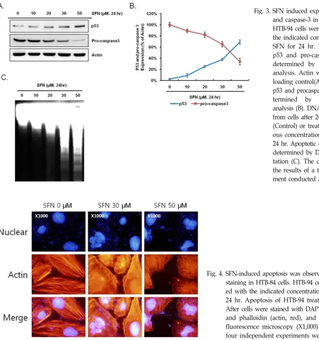
Fig. 3. SFN induced expression of p53 and caspase-3 in HTB-94 cells.
HTB-94 cells were treated with the indicated concentrations of SFN for 24 hr. Expression of p53 and pro-caspase 3 were determined by Western blot analysis. Actin was used as a loading control(A). Amount of p53 and procaspase-3 were de- termined by densitometric analysis (B). DNA was isolated from cells after 24 hr untreated (Control) or treatment of vari- ous concentrations of SFN for 24 hr. Apoptotic cell death was determined by DNA fragmen- tation (C). The data represent the results of a typical experi- ment conducted at three times.
Fig. 4. SFN-induced apoptosis was observed by nuclear staining in HTB-84 cells. HTB-94 cells were treat- ed with the indicated concentrations of SFN for 24 hr. Apoptosis of HTB-94 treated with SFN.
After cells were stained with DAPI (nuclei, blue) and phalloidin (actin, red), and visualized by fluorescence microscopy (X1,000) (C). At lease four independent experiments were conducted.
도 의존적으로 세포사멸을 유도하는 것을 확인할 수 있다.
Sulforaphane은 HTB-94에서 p53과 caspase-3 활성 의존경로를 통하여 세포사멸을 유도한다
위의 실험결과를 토대로 연골암 세포주 HTB-94에서 SFN 처리 시 일어나는 세포사멸에 관련된 분자적기전을 알아보고 자 세포사멸 주요 관련단백질로 알려진 p53 단백질의 발현정 도를 Western blot analysis를 통해 알아본 결과 SFN 처리농도 의존적으로 발현량이 증가함을 확인 할 수 있었다. 본 실험에 사용되어진 HTB-94 세포는 mutant p53 genotype을 가지는 세포주로 알려져 있어[28] 세포사멸 신호전달계에는 p53 단백 질이 관여되지 않을 것으로 예상하였으나, SFN 처리 시 급격 한 발현량의 증가가 관찰되지는 않았지만 농도의존적인 양상
으로 p53 단백질의 축적을 확인할 수 있었다. 이는 SFN 처리
에 의한 세포사멸에 p53 단백질이 관여함을 말해주는 결과라
하겠다. 반면, DNA의 분해를 통해 세포사멸을 일으키는 것으
로 알려진 pro-caspase 3의 발현량을 확인해 본 결과 SFN 처리
농도의존적으로 감소되어짐을 알 수 있었다(Fig. 3A). 이는
pro-caspase 3의 절단을 통한 caspase-3의 활성을 의미하는 것
으로 DNA 분해를 통한 세포사멸을 의미하는 것이라 할 수
있다. Fig. 3B는 loading control로 사용된 actin 단백질과 p53,
pro-caspase 3의 발현량을 densitometric analysis를 통해 정량
화한 것이다. 세포사멸의 결과로서 caspase-3의 활성에 의해
세포 내 DNA가 180~200 bp의 길이로 잘려지는 특징을 이용
한 DNA Fragmentation 실험을 행한 결과 기대한 것처럼 SFN
처리농도가 높아짐에 따라 DNA의 단편정도가 증가함을 관찰
할 수 있었다(Fig. 3C). 이에 세포 내 핵의 분해를 확인하고자 핵 염색액인 DAPI를 이용하여 핵 염색을 실시한 결과, 30 μM SFN 처리군에서 핵 분해단편을 확인할 수 있었으며, 50 μM SFN 처리 시 핵분해 단편을 가진 더 많은 세포를 확인할 수 있었다(Fig. 4). 연골암 세포주에 SFN 처리 시 세포형태 변화가 관찰되어 세포골격 단백질인 actin을 염색하는 phalloidin staining을 한 결과 대조군에서는 선명하고 길게 뻗은 actin fiber를 관찰할 수 있었으나, SFN이 처리된 세포군에서는 ac- tin fiber의 응축 및 세포부피 감소 등을 확인할 수 있었다(Fig.
4). 위의 결과를 종합해 볼 때, SFN에 의한 연골암 세포주 HTB-94에서의 세포사멸은 p53 및 caspase-3 의존적인 활성기 전을 통해 이루어짐을 알 수 있었다.
고 찰
세포사멸의 주된 조절기전으로서 p53 단백질에 의한 cas- pase-3의 활성은 매우 중요하다. p53 단백질은 전사인자로서 세포사멸을 유도하는 신호전달경로에서 작용하는 주요 단백 질로 DNA 손상수복 시에 세포주기를 중지시키는 작용을 한 다고 알려져 있다[1,2]. 이전 본 연구실의 실험결과, 연골세포 사멸에서 p53 단백질의 발현은 MAPK 중 한가지인 p38 kin- ase 신호전달계에 의해 조절되어 지는 것을 확인하였다[12].
이렇게 조절되어진 p53 단백질은 pro-caspase 3의 활성을 자 극하고 활성화된 caspase-3는 핵을 분해하여 세포사멸 과정을 진행한다. 또한, p53 단백질은 p21과 같은 세포주기에 관련된 단백질을 발현시켜 세포증식에 관여하는 것으로 알려져 있다.
이전의 많은 연구에서, 다양한 암세포에서의 세포사멸과 세포 주기를 조절하는 p53 단백질의 발현을 sulforaphane이 증가시 키는 것이 연구되어 졌다[23].
SFN은 십자화과 채소에 많이 들어있는 이소시아네이트 구 조를 갖는 물질로 알려져 있다. 이전의 연구에서는 다양한 동 물 실험이나 인간 암세포에서 SFN을 처리하면 세포주기를 억 제하고, 세포사멸을 유도하는 것으로 알려져 있고, 이렇게 SFN이 유도하는 세포사멸은 caspase-dependent pathway로 조절된다고 알려져 있다[4,17,24]. 그 중, SFN이 식도선암의 성장을 억제한다는 것이 in vivo와 in vitro 실험에서 확인되었 고, 췌장암 세포에서는 SFN이 FOXO의 전사활성을 유도하여 세포사멸을 유도한다고 알려져 있다[21,22]. 일반적으로 세포 주기의 억제는 암세포의 증식억제와 세포사멸을 유도한다고 알려져 있고 그에 따라 암세포와 세포주기의 억제에 관련된 실험은 많은 연구가 되어져 있다. 그 중, G2/M 세포주기의 억제는 cdc2, cyclin B, p53, p21의 발현이 증가하는 것으로 알려져 있다. SFN은 구강암세포와 전립선암세포에서 p21 단 백질을 활성화 하여 G2/M 세포주기를 억제함으로써 세포의 증식을 억제하는 것을 확인하였다[9,10].
이처럼 SFN은 다양한 암세포에서 세포의 증식을 억제하거
나 세포사멸을 유도하는 것을 확인하였다. 그러나 SFN에 의한 인간 연골암세포에서의 세포사멸에 관련된 연구결과가 거의 없어 본 연구를 수행하였다. 그 결과, SFN은 연골암세포주 HTB-94에서 p53단백질의 발현을 증가시키고, caspase-3의 활 성을 증가하여 세포 내 DNA에 손상을 입혀 세포사멸을 유도 하는 것을 확인하였다. HTB94에 작용하는 SFN의 세포사멸 효과에 관련된 세포 내 조절인자는 매우 다양하며 복잡하기 때문에 본 연구를 바탕으로 더 명확하고 구체적인 신호전달경 로를 파악하는 것이 필요하다. 또한, 본 연구실에서는 토끼관 절 연골세포에서의 세포형태 변화는 세포사멸 및 염증반응, 분화에 깊이 관여한다는 것을 보고한 바 있다[11]. 연골암 세포 주 HTB 94에 SFN을 처리 시 actin fiber의 응축 및 세포형태의 변화를 관찰한 바(Fig. 4), 이러한 세포골격단백질의 변화는 세포사멸 및 염증반응, 분화에 관련이 있을 것으로 예상된다.
이에, 이전의 연구결과를 토대로 SFN에 의한 연골암세포의 세포사멸기작이 세포형태 변화와 관련이 있는지에 대한 연구 가 수행되어야 할 것이다. 본 연구결과는 향후 연골암세포의 치료에 SFN이 치료약제로서 도움이 될 것으로 기대한다.
감사의 글
이 논문은 정부(교육과학기술부)의 재원으로 한국연구재단 의 지원을 받아 수행된 기초연구사업임(No. 2009-0084569 &
2010-0003239).
References