Open Access
The Prognostic Impact of the Number of Metastatic Lymph Nodes and
a New Prognostic Scoring System for Recurrence in Early-Stage Cervical Cancer with High Risk Factors: A Multicenter Cohort Study (KROG 15-04)
Original Article
Purpose
We aimed to assess prognostic value of metastatic pelvic lymph node (mPLN) in early-stage cervical cancer treated with radical surgery followed by postoperative chemoradiotherapy.
Also, we sought to define a high-risk group using prognosticators for recurrence.
Materials and Methods
A multicenter retrospective study was conducted using the data from 13 Korean institutions from 2000 to 2010. A total of 249 IB-IIA patients with high-risk factors were included. We evaluated distant metastasis-free survival (DMFS) and disease-free survival (DFS) in relation to clinicopathologic factors including pN stage, number of mPLN, lymph node (LN) ratio (num- ber of positive LN/number of harvested LN), and log odds of mPLNs (log(number of positive LN+0.5/number of negative LN+0.5)).
Results
In univariate analysis, histology (squamous cell carcinoma [SqCC] vs. others), lymphovas- cular invasion (LVI), number of mPLNs ( 3 vs. > 3), LN ratio ( 17% vs. > 17%), and log odds of mPLNs ( 0.58 vs. > 0.58) were significant prognosticators for DMFS and DFS.
Resection margin involvement only affected DFS. No significant survival difference was ob- served between pN0 patients and patients with 1-3 mPLNs. Multivariate analysis revealed that mPLN > 3, LVI, and non-SqCC were unfavorable index for both DMFS (p < 0.001, p=0.020, and p=0.031, respectively) and DFS (p < 0.001, p=0.017, and p=0.001, respec- tively). A scoring system using these three factors predicts risk of recurrence with relatively high concordance index (DMFS, 0.69; DFS, 0.71).
Conclusion
mPLN > 3 in early-stage cervical cancer affects DMFS and DFS. A scoring system using mPLNs > 3, LVI, and non-SqCC could stratify risk groups of recurrence in surgically resected early-stage cervix cancer with high-risk factors.
Key words
Uterine cervical neoplasms, Adjuvant treatment,
Combined modality therapy, Lymphatic metastasis, Scoring system
Jeanny Kwon,
MD, PhD1Keun-Young Eom,
MD, PhD2Young Seok Kim,
MD, PhD3Won Park,
MD, PhD4Mison Chun,
MD, PhD5Jihae Lee,
MD6Yong Bae Kim,
MD, PhD7Won Sup Yoon,
MD, PhD8Jin Hee Kim,
MD, PhD9Jin Hwa Choi,
MD10Sei Kyung Chang,
MD11Bae Kwon Jeong,
MD, PhD12Seok Ho Lee,
MD, PhD13Jihye Cha,
MD14+ + + + + + + + + + + + + + + + + + + + + + + + + + + + + + + + + + + + + + + + + + + + + + + + + + + + + + + + + + + + + + + + + + + + + + + + + + + + + + + + + + + + + + + + + + + + + + + + + + + + + + + + + + + + + + + + + + + + + + + + + + + + + + + + + + + + + + + + + + + + + + + + + + + + + + + + + + + + + + + + + + + + + + + + + + + + + + + + + + + + + + + + + + + + + + + + + + + + + + + + + + + + + + + + + + + + + + + + + + + + + + + + + + + + + + + + + + + + + + + + + + + + + + + + + + + + + + + + + + + + + + + + + + + + + + + + + + + + + + + +
Correspondence: Keun-Young Eom, MD, PhD Department of Radiation Oncology, Seoul National University Bundang Hospital, 82 Gumi-ro 173beon-gil, Bundang-gu, Seongnam 13620, Korea
Tel: 82-31-787-7653 Fax: 82-31-787-4019
E-mail: [email protected]
Received July 20, 2017 Accepted October 20, 2017 Published Online October 24, 2017
*A list author’s aliations appears at the end of the paper.
Introduction
Cervical cancer is newly diagnosed in 3,500 patients in the Korea in 2014 [1]. Despite a declining trend over the last 40 years, the incidence and mortality rate among young women increased [2]. Since cancer screening using Pap smear has been widely used, about three quarters of newly diagnosed cases are the International Federation of Gynecology and Obstetrics (FIGO) stage I or IIA [3]. These early cervical can- cer are subject to radical surgery and has a relatively favor- able prognosis with a 5-year overall survival (OS) rate of 90%
and a disease-free survival (DFS) rate of 82% [4]. However, pathologic findings including positive resection margin (RM), parametrial (PM) involvement, and lymph node (LN) metastasis are considered as high risk factors for recurrence and patients with those factors are candidate for postopera- tive chemoradiotherapy (CRT) [5]. Pelvic LN metastasis is found in nearly half of cases, even in early stage cervical can- cer [6] and in this setting, the 5-year relative survival rate was 57.4% [7]. In a Gynecologic Oncology Group (GOG) 49 study including FIGO IB patients, the 5-year DFS of patients with metastatic pelvic LN (mPLN) was 10% lower than those without [8]. In pooled analysis of GOG 24/56/59, LN metas- tasis was statistically significant variable for both DFS (haz- ard ratio [HR], 1.9) and OS (HR, 2.0) [9].
Several researchers have assessed prognostic meaning of mPLN in various ways. The simplest is counting the number of mPLN, which is intuitive and widely used in other cancers [10,11]. The LN ratio (LNR) which has been developed to reflect the extent of LN resection and the log odds of positive LN (LODDs) which has the advantage of reflecting the num- ber of negative LNs are suggested as prognostic variables related to LN status [12]. However, those studies were lim- ited in that most of them are single institutional retrospective series and also, included heterogeneous population in terms of tumor stages and treatment characteristics. Until now, it is unclear that among several parameters related to LN sta- tus, which one serves as a best prognostic factor.
We therefore aimed to assess prognostic value of the num- ber of and related parameters of mPLN in patients with early-stage cervical cancer treated with radical surgery fol- lowed by postoperative CRT due to pathologic high risk fac- tors. Also, we sought to find a high risk group of recurrence in these patients.
Materials and Methods
1. Patient cohort
We conducted a multicenter retrospective study (Korean Radiation Oncology Group [KROG] 15-04) at affiliated 13 institutions in South Korea. After the approval of Institu- tional Review Board of each institution, medical records of patients who were newly diagnosed of FIGO IB-IIA cervical cancer between 2000 and 2010, and treated with radical sur- gery followed by postoperative CRT due to pathologic high risk features including positive RM, PM invasion, and LN metastasis were reviewed. We intended to enroll homoge- neous group of patients without clinically involved LN on preoperative positron emission tomography/computed tomography (PET/CT) or magnetic resonance imaging (MRI). This is because definitive CRT or radiotherapy (RT) is often recommended in those with gross mPLNs which are LNs with increased metabolism than the background (PET/CT) or those with a short diameter greater than 1 cm (MRI). Additionally, patients who received preoperative chemotherapy (CTx), who did not receive radical surgery, who have insufficient information on the extent of surgery or pathologic report, who did not complete adjuvant CRT, or who have with history of other malignancies were excluded. Finally, a total of 259 patients were candidates of this study, and each institutional review board’s approval was achieved. The patient selection process is depicted in S1 Fig.
2. Treatment
All patients underwent radical hysterectomy with pelvic LN dissection according to our inclusion criteria. Para-aortic LN sampling or dissection was carried out in 120 patients (46.3%). Postoperative RT was delivered to whole pelvis in daily fraction of 1.8-2 Gy with median dose of 50.4 Gy (range, 41.4 to 60.4 Gy). Brachytherapy was performed in 36 patients (18%) with median dose of 18 Gy (range, 5 to 28 Gy) follow- ing whole pelvic RT. Nineteen patients (7.3%) underwent para-aortic nodal irradiation with a median dose of 45 Gy (range, 45 to 60 Gy), of whom nine patients received RT pro- phylactically. Most of all patients (97.3%) received platinum- based CTx during CRT. The most common CTx regimen was weekly cisplatin (50.2%), and doublet of 5-fluorouracil and cisplatin was the second (24.5%).
3. Variables and statistical analysis
All patients were staged using 2009 FIGO staging system [13]. As FIGO staging system does not describe presence of
LN metastases, pathologic nodal status was described sepa- rately as pN0 or pN1 based on seventh edition of American Joint Committee on Cancer staging system [14]. Additionally, we evaluated parameters related to LN status such as the number of mPLNs, LNR, and LODDs. LNR was calculated by dividing the number of mPLNs by the number of har- vested LNs (Eq. 1).
LNR= No. of mPLNs No. of harvested LNs (1)
LODDs was defined as the logarithm of odds ratio of neg- ative LNs over positive LNs (Eq. 2) [12].
LODDs=log No. of mPLNs+0.5 No. of negative LN+0.5 (2)
In this Eq. (2), 0.5 is added constant to avoid an infinite number. Cutoff value of these parameters was determined by maximal chi-square method.
Distant metastasis-free survival (DMFS) was defined as an interval between the surgery and the detection of distant metastasis or last follow-up. DFS was calculated from the surgery to the detection of any recurrence or last follow-up.
In-pelvic recurrences are regarded as locoregional failures while para-aortic LN failures as distant. Survival curves were estimated by Kaplan-Meier method and survival differences were evaluated using log-rank test. Statistically significant variables in univariate analyses were included in a step-wise Cox proportional hazard regression model for multivariate analyses. In the modeling process, variables with p < 0.20 were put into and those with p > 0.10 were eliminated from the regression model. A p-values < 0.05 was considered sta- tistically significant. All statistical analyses were performed by R ver. 3.2.4 (R Foundation for Statistical Computing, Vienna, Austria, http://www.r-project.org).
4. Ethical statement
The study was approved by the Institutional Review Board of Seoul National University Bundang Hospital (IRB No.
B-1511/324-109) and performed in accordance with the prin- ciples of the Declaration of Helsinki. The informed consent was waived.
Results
1. Patient demographics
The median age of patients was 47 years (range, 25 to 74 years) and about three-quarters of patients was FIGO stage IB. The most common histology was squamous cell carci- noma (SqCC; 71.8%) followed by adenocarcinoma (ADC;
18.5%). Deep stromal invasion greater than 1/2 thickness, bulky tumor more than 4 cm, and lymphovascular invasion (LVI) were found in 88.6%, 33.2%, and 78.9% of patients, respectively. Patients with PM extension or positive RM were 45.6% and 14.3%, respectively. Two hundred and seven patients (79.9%) were pN1. The median number of harvested LNs and mPLNs were 26 (range, 4 to 85) and 1 (range, 0 to 19), respectively. Of 120 patients who underwent para-aortic LN sampling or dissection, 10 patients (3.9%) had metastatic para-aortic LNs. Thirty-six patients (13.9%) received a con- solidative CTx following adjuvant CRT at the discretion of treating physician. Patient demographics are summarized in Table 1.
2. Survival analysis
As the presence of para-aortic LN metastases is an obvious poor prognostic factor for DMFS and DFS, 10 patients with para-aortic LN metastases were excluded from subsequent survival analyses to avoid possible confounding effect. Con- sequently, 249 patients were included for survival analyses of DMFS and DFS. Median follow-up duration was 70 months (range, 2.5 to 468.5 months). Five-year DMFS and DFS of 249 patients were 83.5% and 78.1%, respectively. Dis- tant failure occurred in 42 patients and crude distant failure rate was 16.9%. The lungs were the most common site of fail- ure (33.3%) and the second most common was para-aortic LN (28.6%). Locoregional failure occurred in 23 patients (9.2%) and there were 11 patients who experienced both locoregional and distant failure.
Determined cutoff value for the number of mPLNs, LNR and LODDs for DMFS were 3, 17%, and 0.58, respectively (S2 Fig.). The proportion of patients with mPLNs 3, LNR
17%, and LODDs 0.58 were 81.1%, 86.0%, and 87.6%, respectively.
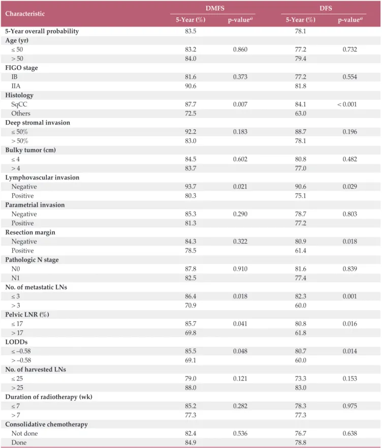
In univariate analysis, non-SqCC (p=0.007), the presence of LVI (p=0.021), mPLNs > 3 (p=0.018), pelvic LNR > 17%
(p=0.041), and LODDs > 0.58 (p=0.048) were significant poor predictors for DMFS. However, no survival difference was observed according to the number of harvested LNs (p=0.121), duration of RT (p=0.282), or consolidative CTx (p=0.536). As for DFS, the survival rate was significantly dif- ferent according to following factors: non-SqCC (p < 0.001),
presence of LVI (p=0.029), mPLNs > 3 (p=0.001), pelvic LNR
> 17% (p=0.016), and LODDs > 0.58 (p=0.014), and RM pos- itivity (p=0.018), respectively. DFS was not influenced by the number of harvested LNs (p=0.153), duration of RT (p=0.975), or consolidative CTx (p=0.638). These results are presented in Table 2.
Interestingly, pathologic N stage itself was independently associated with neither DMFS (p=0.910) nor DFS (p=0.839) (Fig. 1A). As the cutoff value of mPLN determined by max- imal chi-square test was 3, we compared survival differences between three groups of patients: patients with pN0, patients with 1-3 mPLNs, and patients with mPLNs more than 3. Sur- vivals of patients with 1-3 mPLNs were significantly higher (5-year DMFS, 86.0%; 5-year DFS, 82.7%) than those with
> 3 mPLNs (5-year DMFS, 70.9%, p=0.017; 5-year DFS, 60.0%, p=0.001). However, survival differences between patients with pN0 and 3 mPLNs were not statistically significant (5-year DMFS, 87.7%, p=0.616; 5-year DFS, 81.6%, p=0.535).
Fig. 1B depicts survival curves according to these three groups.
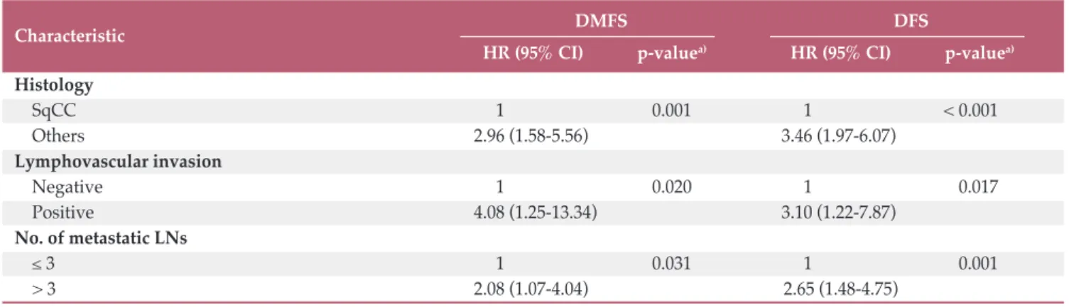
In multivariate analysis (Table 3), both DMFS and DFS were significantly affected by three factors: non-SqCC histol- ogy (DMFS: p=0.001; HR, 2.96 and DFS: p < 0.001; HR, 3.46), LVI (DMFS: p=0.020; HR, 4.08 and DFS: p=0.017; HR, 3.10), and mPLNs > 3 (DMFS: p=0.031; HR, 2.08 and DFS: p=0.001;
HR, 2.65). S3 Fig. shows survival curves according to these factors. Other LN-related parameters (pN stage, LNR, or LODDs) lost their significance after adjusting interaction between variables.
3. A scoring system for prediction of DMFS and DFS
A scoring system was built based on the results of multi- variate analysis (Table 4). Non-SqCC histology, presence of LVI, and mPLNs > 3 were counted independently as 1 point with total score ranging from 0 to 3. As a result, patients were stratified into four groups as follows: 28 patients (12.1%) with score 0, 125 patients (53.9%) with score 1, 69 patients (29.7%) with score 2, and 10 patients (4.3%) with score 3, respectively.
The 5-year DMFS and DFS were 100% in patients with score Table 1. Patient and tumor characteristics
Characteristic No. (%)
Age at diagnosis, median (range, yr) 47 (25-74) FIGO stage
IB 202 (78.0)
IIA 57 (22.0)
Histology
Squamous cell carcinoma 186 (71.8)
Adenocarcinoma 48 (18.5)
Adenosquamous carcinoma 11 (4.3)
Mucinous adenocarcinoma 4 (1.5)
Small cell carcinoma 3 (1.2)
Others 7 (2.7)
Deep stromal invasion (%)a)
50 28 (11.4)
> 50 218 (88.6)
Bulky tumor (cm)a)
4 163 (66.8)
> 4 81 (33.2)
Lymphovascular invasiona)
Negative 51 (21.1)
Positive 191 (78.9)
Parametrial extension
Negative 141 (54.4)
Positive 118 (45.6)
Resection margin
Negative 222 (85.7)
Positive 37 (14.3)
Pathologic N stage
N0 52 (20.1)
N1 207 (79.9)
No. of harvested pelvic LNs
Median (range) 26 (4-85)
25 125 (48.3)
> 25 134 (51.7)
No. of metastatic pelvic LNs
Median (range) 1 (0-19)
0 52 (20.1)
1 88 (34.0)
2 44 (17.0)
3 75 (29.0)
Para-aortic LN dissection
Not done 139 (53.7)
Done 120 (46.3)
Para-aortic LN metastasis
Absent 110 (91.7)
Present 10 (8.3)
Duration of radiotherapy (wk)
7 199 (76.8)
> 7 60 (23.2)
Consolidative chemotherapy
Not done 223 (86.1)
Done 36 (13.9)
Table 1. Continued
FIGO, International Federation of Gynecology and Obstet- rics; LN, lymph node. a)Available cases analysis.
Characteristic No. (%)
High-risk factor
1 168 (64.9)
2 79 (30.5)
3 12 (4.6)
(Continued)
Characteristic DMFS DFS
5-Year (%) p-valuea) 5-Year (%) p-valuea)
5-Year overall probability 83.5 78.1
Age (yr)
50 83.2 0.860 77.2 0.732
> 50 84.0 79.4
FIGO stage
IB 81.6 0.373 77.2 0.554
IIA 90.6 81.8
Histology
SqCC 87.7 0.007 84.1 < 0.001
Others 72.5 63.0
Deep stromal invasion
50% 92.2 0.183 88.7 0.196
> 50% 83.0 78.1
Bulky tumor (cm)
4 84.5 0.602 80.8 0.482
> 4 83.7 77.0
Lymphovascular invasion
Negative 93.7 0.021 90.6 0.029
Positive 80.3 75.1
Parametrial invasion
Negative 85.3 0.290 78.7 0.803
Positive 81.3 77.2
Resection margin
Negative 84.3 0.322 80.9 0.018
Positive 78.5 61.4
Pathologic N stage
N0 87.8 0.910 81.6 0.839
N1 82.5 77.4
No. of metastatic LNs
3 86.4 0.018 82.3 0.001
> 3 70.9 60.0
Pelvic LNR (%)
17 85.7 0.041 80.8 0.016
> 17 69.8 61.8
LODDs
–0.58 85.5 0.048 80.7 0.014
> –0.58 69.1 60.0
No. of harvested LNs
25 79.0 0.121 73.3 0.153
> 25 88.0 83.0
Duration of radiotherapy (wk)
7 85.2 0.282 78.3 0.975
> 7 77.3 77.3
Consolidative chemotherapy
Not done 82.4 0.536 76.7 0.638
Done 84.9 78.8
Table 2. Univariate survival analysis for DMFS and DFS
DMFS, distant metastasis-free survival; DFS, disease-free survival; FIGO, International Federation of Gynecology and Obstetrics; SqCC, squamous cell carcinoma; LN, lymph node; LNR, lymph node ratio; LODDs, log odds of positive lymph nodes. a)p-value by log-rank test.
0. In contrast, 5-year DMFS and DFS were 50% and 40% in patients with score 3. In patients with score 1 and 2, 5-year DMFS and DFS were 88.6%/71.2% and 85.9%/62.1%, respec- tively (Fig. 2). The Harrell's C index of the scoring system for DMFS and DFS were 0.69 and 0.71.
Additionally, we developed prognostic nomogram for DFS using above three risk factors to specifically estimate indi- vidual patient’s prognosis (Fig. 3A, Supplementary Method).
The nomogram suggested that non-SqCC histology had the largest impact on prognosis. The concordance index of our nomogram predicting 5-year survival probability was 0.69.
The calibration plot depicted a good correspondence between the nomogram-predicted survival and actuarial sur- vival rate at 5 years (Fig. 3B).
Discussion
The present study enrolled homogenous groups of patients diagnosed of FIGO IB-IIA cervical cancer without clinically Fig. 1. Distant metastasis-free survival (DMFS) and disease-free survival (DFS) curves according to pN stage (A), and number of metastatic pelvic lymph nodes (LNs) (B).
DMFS
1.0
0 0.2 0.4 0.6 0.8
0
Time (mo)
48 60
36 24
12 72 84
pN0pN1 pN0
pN1
DFS
1.0
0 0.2 0.4 0.6 0.8
0
Time (mo)
48 60
36 24
12 72 84
DMFS
1.0
0 0.2 0.4 0.6 0.8
0
Time (mo)
48 60
36 24
12 72 84
DFS
1.0
0 0.2 0.4 0.6 0.8
0
Time (mo)
48 60
36 24
12 72 84
pN0
1-3 LN metastasis
> 3 LN metastasis pN0
1-3 LN metastasis
> 3 LN metastasis
A
B
involved LNs, treated by radical surgery and adjuvant CRT owing to postoperative high risk features. This study demon- strates that the number of mPLN shows best prognostic per- formance among parameters related to LN status. DMFS and DFS of patients with mPLN 3 did not differ from those with pN0. In contrast, the risk of recurrence in patients with mPLNs > 3 remained high even though postoperative CRT was performed. Additionally, we proposed a scoring system which can discriminate poor prognostic group of patients using three factors including tumor histology, LVI, and the number of mPLN. It is simple and correlates well with DMFS and DFS.
Characteristic DMFS DFS
HR (95% CI) p-valuea) HR (95% CI) p-valuea) Histology
SqCC 1 ( 0.001 1 ( < 0.001
Others 2.96 (1.58-5.56) 3.46 (1.97-6.07)
Lymphovascular invasion
Negative 1 ( 0.020 1 ( 0.017
Positive 4.08 (1.25-13.34) 3.10 (1.22-7.87)
No. of metastatic LNs
3 1 ( 0.031 1 ( 0.001
> 3 2.08 (1.07-4.04) 2.65 (1.48-4.75)
Table 3. Multivariate analysis for DMFS and DFS
DMFS, distant metastasis-free survival; DFS, disease-free survival; HR, hazard ratio; CI, confidence interval; SqCC, squamous cell carcinoma; LN, lymph node. a)p-value by Cox proportional hazard model with backward stepwise regression.
Fig. 2. Distant metastasis-free survival (DMFS) (A) and disease-free survival (DFS) (B) curves according to the scoring system.
DMFS
1.0
0 0.2 0.4 0.6 0.8
0
Time (mo)
48 60
36 24
12 72 84
Score 0 Score 1 Score 2 Score 3
A
DFS
1.0
0 0.2 0.4 0.6 0.8
0
Time (mo)
48 60
36 24
12 72 84
Score 0 Score 1 Score 2 Score 3
B
Table 4. A scoring system for prediction of distant failureSqCC, squamous cell carcinoma; LN, lymph node.
Characteristic 0 1
Histology SqCC Others
Lymphovascular invasion Negative Positive
No. of metastatic LNs 3 > 3
Some of well-known prognosticators such as bulky tumor size, deep stromal invasion, positive RM, and PM involve- ment for early cervical cancers did not affect survivals in the present study. As those factors are mainly derived from sur- gical series to establish indications of adjuvant treatment [8,15,16], proper prognostic factors in early cervical cancers with high risk features are currently unknown when stan- dard treatment including radical surgery and postoperative CRT were performed. Our findings suggest that prognosti- cators for survivals in this specific setting would be different from those in surgical series and should be defined sepa- rately.
The LN status affects prognosis of patients and several parameters associated with LN status were suggested in the previous studies. It is agreed that pN1 disease is a heteroge- neous group in terms of the location and the tumor burden of involved LN. Nodal staging system based on the number of metastatic LNs is widely used in other cancers of breast, stomach, and rectum [14]. In studies of patients with early stage cervical cancer treated with radical surgery followed by RT, some authors showed that the survival difference of patients between with one mPLN and 2 was statistically significant [7,15]. KROG1303 study showed that the hazard of death increases continuously with the number of mPLNs (up to 5) [11]. On the other hand, Tsai et al. [10] reported that patients with only one mPLN had achieved similar outcomes compared to those with pN0 (5-year DFS, 84% vs. 87%;
p=0.48), and patients with 2 mPLNs had lower survival rates (87% vs. 61%, p < 0.001) than those with pN0. In the present study, there was no significant difference in survival
between patients with pN0 and those with 1-3 mPLNs which is higher cut-off value than Tsai et al. [10]. The main differ- ence between two is that whether concomitant CTx was administered or not. Possible explanation of the observed difference in the number of significant mPLN will be that the risk of recurrence, especially distant failure, might be atten- uated by concomitant CTx in patients with 2-3 mPLNs.
The number of mPLNs directly depends on the extent of LN dissection. Inadequate LN dissection results in underes- timation of LN metastasis. The prognostic advantage of LNR as a surrogate for LN status, which reflects the number of mPLN as well as the degree of LN dissection, has been rec- ognized. Fleming et al. [17] reported decreased survivals in patients with high LNR (6.6% for progression-free survival, and 7.6% for OS) compared to those with low LNR in early stage cervix cancer. Li et al. [18] and Polterauer et al. [19]
showed that the prognosis of patients with high LNR greater than 10% was poor. The present study suggests a cutoff value of 17% for LNR. As the median number of harvested LNs was 26 in the current study, which is not much different from the previous studies (Fleming et al. [17], median 19; Chen et al. [20], median 26), relatively high LNR cutoff in our study may be attributed to the improved survival of patients with pN1 in our study. The 5-year OS of pN1 patients in the pres- ent study was 84.9% which is higher than 22.4% (OS) in Chen et al. [20]. Additionally, Fleming et al. [17] reported a 5-year PFS of around 67% in patients with > LNR 6.6%, which is similar to those with LNR > 16% in our study (68.3%). More recently, Li et al. [18] and Zhou et al. [21] suggested cutoff values of LNR as 20% and 17% which is consistent with our Fig. 3. Nomogram predicting disease-free survival (DFS) (A) and calibration plot (B). SqCC, squamous cell carcinoma.
0 10 20 30 40 50 60 70 80 90 100 Points
Histology Lymphovascular invasion Pelvic lymph node metastasis Total points 5-Year DFS probability
SqCC
Non-SqCC
Negative
Positive
0 20 40 60 80 120 160 200 240 280
0.9 0.8 0.7 0.6 0.5 0.4 0.3
≤ 3
> 3
Actual survival probability at 5 yr
1.0
0 0.2 0.4 0.6 0.8
0
Predicted survival probability at 5 yr 0.8 0.6 0.4
0.2 1.0
A B
results.
LODDs reflects not only the extent of LN dissection, but also negative LNs. It has the advantage of further discrimi- nating patients with LNR close to zero or one [22]. For exam- ple, if the number of mPLN is zero, the LNR will be zero regardless of the dissection extent, but the LODDs will vary depending on the number of negative LNs (S4 Fig.). It has been studied actively in rectal and gastric cancers, but not in cervical cancers. Kwon et al. [12] reported the prognostic superiority of LODDs to the number of mPLNs and LNR in a patient group similar to our study, but it was limited due to small number of patients. In the present study, LODDs was a significant prognostic factor in univariate analysis, but lost its significance in multivariate analysis. In order for LODDs to acquire superior predictability, the number of neg- ative LNs should be different among patients with mPLNs
3 with or without recurrences, but there was no significant difference (t test, p=0.458 for distant recurrence, p=0.2488 for overall recurrence).
In addition to the number of mPLN, our study revealed that LVI and histologic type were meaningful factors for pre- dicting DFS as well as DMFS. The prognostic significance of LVI has been reported in many studies including GOG 49 [8]
and is still an important prognosticator even after adjuvant RT [23]. In addition, LVI has been reported to be significantly associated with pelvic LN metastasis in stage IB cervical can- cer [24]. Similarly, LVI was associated with mPLN (chi- square test, p=0.002) in our study, but it was an independent prognostic factor for both DMFS and DFS after adjusting interaction between two variables. These are in line with pre- vious studies.
ADC in cervix cancer has been considered to have biolog- ical aggressiveness and to be more resistant to adjuvant treat- ment [25,26]. In the present study, ADC histology carried a poor prognosis with 71.8% of DMFS and 61.8% of DFS at 5 years compared to SqCC with 87.7% of DMFS and 84.1%
of DFS at 5 years, respectively. The significantly lower sur- vivals of ADC are consistent with the findings of Hosaka et al. [25] and Galic et al. [27]. In the study of Intaraphet et al.
[28], the risk of cancer-related death was significantly higher in patients with small cell neuroendocrine carcinoma or ADC than those with SqCC. Of note, an unfavorable prognosis of ADC was observed only in advanced stages while there was no significant survival difference in early stages [28]. More recently, Winer et al. [29] also reported that equivalent sur- vival outcomes regardless of histologic type were achieved in patients with early stage cervix cancer. The discrepancies between studies may be explained by the difference of patient characteristics: all patients in our study have early stages, but belong to the high-risk group. It could be sup- ported by the study of Mabuchi et al. [30] showing that ADC was an independent poor prognostic factor for survivals in
intermediate- and high-risk group, but not in low-risk group of patients with early cervical cancer.
Our study is limited inherently by its retrospective nature:
the data were collected from 13 institutions in a retrospective manner. It resulted in the following weakness: data on the performance status of patients, pretreatment anemia, trans- fusion, and treatment-related toxicities were missing and unable to analyze. In addition, favorable treatment outcomes of our study may suggest a potential selection bias. However, to the best of our knowledge, the present study is the largest study focusing on early cervical cancer patients without clin- ical LN involvement carrying high-risk factors who under- went radical surgery followed by adjuvant CRT. For this specific patient population, our study suggested three prog- nostic factors including histologic type, LVI, and the number of mPLN for DMFS and DFS, and provides simple scoring system for predicting DMFS and DFS. It is worthwhile to identify patient groups at risk of treatment failure despite adjuvant CRT.
In conclusion, the risk of recurrence, especially distant fail- ure, remains high when adjuvant CRT is performed in patients with mPLNs > 3. New prognostic scoring system consisted of mPLNs > 3, presence of LVI and non-SqCC is suggested, and it could stratify patients according to risk of recurrence in early cervix cancer with high-risk factors. Fur- ther treatment options such as consolidative CTx after adju- vant CRT should be considered to improve treatment outcomes in patients having poor prognostic factors and prospective trials are warranted.
Electronic Supplementary Material
Supplementary materials are available at Cancer Research and Treatment website (http://www.e-crt.org).
Conflicts of Interest
Conflict of interest relevant to this article was not reported.
Author Details
1Department of Radiation Oncology, Chungnam National Univer- sity College of Medicine, Daejeon, 2Department of Radiation Oncol- ogy, Seoul National University Bundang Hospital, Seongnam,
3Department of Radiation Oncology, Asan Medical Center, Univer- sity of Ulsan College of Medicine, Seoul, 4Department of Radiation Oncology, Samsung Medical Center, Sungkyunkwan University School of Medicine, Seoul, 5Department of Radiation Oncology, Ajou University School of Medicine, Suwon, 6Department of Radi- ation Oncology, Ewha Womans University Mokdong Hospital, Ewha Womans University School of Medicine, Seoul, 7Department of Radiation Oncology, Yonsei Cancer Center, Yonsei University
College of Medicine, Seoul, 8Department of Radiation Oncology, Korea University Ansan Hospital, Ansan, 9Department of Radiation Oncology, Keimyung University Dongsan Medical Center, Keimyung University School of Medicine, Daegu, 10Department of Radiation Oncology, Chung-Ang University Hospital, Seoul, 11Department of Radiation Oncology, CHA Bundang Medicial Center, CHA Univer-
sity School of Medicine, Seongnam, 12Department of Radiation Oncology, Gyeongsang National University Hospital, Jinju,
13Department of Radiation Oncology, Gachon University Gil Med- ical Center, Gachon University of Medicine and Science, Incheon,
14Department of Radiation Oncology, Wonju Severance Christian Hospital, Wonju, Korea
1. Jung KW, Won YJ, Oh CM, Kong HJ, Lee DH, Lee KH, et al.
Cancer statistics in Korea: incidence, mortality, survival, and prevalence in 2014. Cancer Res Treat. 2017;49:292-305.
2. Moon EK, Oh CM, Won YJ, Lee JK, Jung KW, Cho H, et al.
Trends and age-period-cohort effects on the incidence and mortality rate of cervical cancer in Korea. Cancer Res Treat.
2017;49:526-33.
3. Nowakowski A, Cybulski M, Buda I, Janosz I, Olszak-Wasik K, Bodzek P, et al. Cervical cancer histology, staging and sur- vival before and after implementation of organised cervical screening programme in Poland. PLoS One. 2016;11:e0155849.
4. Landoni F, Maneo A, Colombo A, Placa F, Milani R, Perego P, et al. Randomised study of radical surgery versus radiother- apy for stage Ib-IIa cervical cancer. Lancet. 1997;350:535-40.
5. Peters WA 3rd, Liu PY, Barrett RJ 2nd, Stock RJ, Monk BJ, Berek JS, et al. Concurrent chemotherapy and pelvic radiation therapy compared with pelvic radiation therapy alone as adjuvant therapy after radical surgery in high-risk early-stage cancer of the cervix. J Clin Oncol. 2000;18:1606-13.
6. Kidd EA, Siegel BA, Dehdashti F, Rader JS, Mutch DG, Powell MA, et al. Lymph node staging by positron emission tomog- raphy in cervical cancer: relationship to prognosis. J Clin Oncol. 2010;28:2108-13.
7. Howlader N, Noone AM, Krapcho M, Miller D, Bishop K, Altekruse SF, et al. SEER cancer statistics review 1975-2013 [Internet]. Bethesda, MD: National Cancer Institute; 2016 [cited 2016 Dec 26]. Available from: https://seer.cancer.gov/csr/
1975_2013/.
8. Delgado G, Bundy B, Zaino R, Sevin BU, Creasman WT, Major F. Prospective surgical-pathological study of disease-free interval in patients with stage IB squamous cell carcinoma of the cervix: a Gynecologic Oncology Group study. Gynecol Oncol. 1990;38:352-7.
9. Stehman FB, Bundy BN, DiSaia PJ, Keys HM, Larson JE, Fowler WC. Carcinoma of the cervix treated with radiation therapy. I. A multi-variate analysis of prognostic variables in the Gynecologic Oncology Group. Cancer. 1991;67:2776-85.
10. Tsai CS, Lai CH, Wang CC, Chang JT, Chang TC, Tseng CJ, et al. The prognostic factors for patients with early cervical can- cer treated by radical hysterectomy and postoperative radio- therapy. Gynecol Oncol. 1999;75:328-33.
11. Lee HJ, Han S, Kim YS, Nam JH, Kim HJ, Kim JW, et al. Indi- vidualized prediction of overall survival after postoperative
radiation therapy in patients with early-stage cervical cancer:
a Korean Radiation Oncology Group study (KROG 13-03). Int J Radiat Oncol Biol Phys. 2013;87:659-64.
12. Kwon J, Eom KY, Kim IA, Kim JS, Kim YB, No JH, et al. Prog- nostic value of log odds of positive lymph nodes after radical surgery followed by adjuvant treatment in high-risk cervical cancer. Cancer Res Treat. 2016;48:632-40.
13. Pecorelli S, Zigliani L, Odicino F. Revised FIGO staging for carcinoma of the cervix. Int J Gynaecol Obstet. 2009;105:107-8.
14. Compton CC, Byrd DR, Garcia-Aguilar J, Kurtzman SH, Olawaiye A, Washington MK. AJCC cancer staging atlas: a companion to the seventh editions of the AJCC cancer staging manual and handbook. New York: Springer; 2012.
15. Monk BJ, Wang J, Im S, Stock RJ, Peters WA 3rd, Liu PY, et al.
Rethinking the use of radiation and chemotherapy after radi- cal hysterectomy: a clinical-pathologic analysis of a Gyneco- logic Oncology Group/Southwest Oncology Group/Radiation Therapy Oncology Group trial. Gynecol Oncol. 2005;96:
721-8.
16. Sedlis A, Bundy BN, Rotman MZ, Lentz SS, Muderspach LI, Zaino RJ. A randomized trial of pelvic radiation therapy ver- sus no further therapy in selected patients with stage IB carci- noma of the cervix after radical hysterectomy and pelvic lymphadenectomy: a Gynecologic Oncology Group Study.
Gynecol Oncol. 1999;73:177-83.
17. Fleming ND, Frumovitz M, Schmeler KM, dos Reis R, Munsell MF, Eifel PJ, et al. Significance of lymph node ratio in defining risk category in node-positive early stage cervical cancer.
Gynecol Oncol. 2015;136:48-53.
18. Li C, Liu W, Cheng Y. Prognostic significance of metastatic lymph node ratio in squamous cell carcinoma of the cervix.
Onco Targets Ther. 2016;9:3791-7.
19. Polterauer S, Hefler L, Seebacher V, Rahhal J, Tempfer C, Hor- vat R, et al. The impact of lymph node density on survival of cervical cancer patients. Br J Cancer. 2010;103:613-6.
20. Chen Y, Zhang L, Tian J, Fu X, Ren X, Hao Q. Significance of the absolute number and ratio of metastatic lymph nodes in predicting postoperative survival for the International Feder- ation of Gynecology and Obstetrics stage IA2 to IIA cervical cancer. Int J Gynecol Cancer. 2013;23:157-63.
21. Zhou J, Sun JY, Chen SY, Li FY, Lin HX, Wu SG, et al. Prog- nostic value of lymph node ratio in patients with small-cell carcinoma of the cervix based on data from a large national
References
registry. Onco Targets Ther 2015;9:67-73.
22. Aurello P, Petrucciani N, Nigri GR, La Torre M, Magistri P, Tierno S, et al. Log odds of positive lymph nodes (LODDS):
what are their role in the prognostic assessment of gastric ade- nocarcinoma? J Gastrointest Surg. 2014;18:1254-60.
23. Morice P, Piovesan P, Rey A, Atallah D, Haie-Meder C, Pautier P, et al. Prognostic value of lymphovascular space invasion determined with hematoxylin-eosin staining in early stage cer- vical carcinoma: results of a multivariate analysis. Ann Oncol.
2003;14:1511-7.
24. Chandacham A, Charoenkwan K, Siriaunkgul S, Srisomboon J, Suprasert P, Phongnarisorn C, et al. Extent of lymphovascu- lar space invasion and risk of pelvic lymph node metastases in stage IB1 cervical cancer. J Med Assoc Thai. 2005;88 Suppl 2:S31-6.
25. Hosaka M, Watari H, Mitamura T, Konno Y, Odagiri T, Kato T, et al. Survival and prognosticators of node-positive cervical cancer patients treated with radical hysterectomy and system- atic lymphadenectomy. Int J Clin Oncol. 2011;16:33-8.
26. Kim HJ, Rhee WJ, Choi SH, Nam EJ, Kim SW, Kim S, et al.
Clinical outcomes of adjuvant radiation therapy and prognos- tic factors in early stage uterine cervical cancer. Radiat Oncol J. 2015;33:126-33.
27. Galic V, Herzog TJ, Lewin SN, Neugut AI, Burke WM, Lu YS, et al. Prognostic significance of adenocarcinoma histology in women with cervical cancer. Gynecol Oncol. 2012;125:287-91.
28. Intaraphet S, Kasatpibal N, Siriaunkgul S, Sogaard M, Patu- manond J, Khunamornpong S, et al. Prognostic impact of his- tology in patients with cervical squamous cell carcinoma, adenocarcinoma and small cell neuroendocrine carcinoma.
Asian Pac J Cancer Prev. 2013;14:5355-60.
29. Winer I, Alvarado-Cabrero I, Hassan O, Ahmed QF, Alosh B, Bandyopadhyay S, et al. The prognostic significance of histo- logic type in early stage cervical cancer: a multi-institutional study. Gynecol Oncol. 2015;137:474-8.
30. Mabuchi S, Okazawa M, Matsuo K, Kawano M, Suzuki O, Miyatake T, et al. Impact of histological subtype on survival of patients with surgically-treated stage IA2-IIB cervical can- cer: adenocarcinoma versus squamous cell carcinoma.
Gynecol Oncol. 2012;127:114-20.