신규항암제인 Heptaplatin 의 인체 흑색종세포 (SK-MEL-28) 에 대한 세포생존률 및 유세포 분석
최수라 • 명평근 #
충남대학교 약학대학 임상생화학실 (Received October 6 , 2003; Revised November 5 , 2003)
Cell Viability and Flow Cytometry Analysis of a Novel Antitumor Agent, Heptaplatin in Human Melanoma Cell Line, SK-MEL-28
Su-La Choi and Pyung-Keun Myung#
Lab. o f C lin ical Biochemistry, College of Pharmacy, Chungnam N ational University, Daejeon 305-764,Korea
A bstract
ᅳ-Heptaplatin, cis-Malonato[(4R,5R)-4,5-bis(aminomethyl)-2-isopropyl-l,3-dioxolane]platinum(II), is a novel plat
inum-based antitumor agent with clinical potential against human stomach cancer and the 3rd generation of the cisplatin.
This study was performed to study how cisplain, heptaplatin and sunpla which is a mixture of heptaplatin and mannitol (w : w = l : 2) affect cell viability of SK-MEL-28 human melanoma cell line. Heptaplatin (IC 50; 95.35 pM ) and sunpla (IC 50 ; 10.95 |iM) were less effect than cisplatin (IC 50; 10.92 jiM ) on the SK-MEL-28 cells. By cell cycle analysis using flow cytom
etry, it was identified that the cells were arrested at G2/M phase by cisplatin, heptaplatin and sunpla, and percentage of cell death group was increased according to increasing of time and concentration. These results suggest that cisplatin, hep
taplatin and sunpla are a novel anticancer agent against human melanoma cell.
Keywords □ cisplatin, heptaplatin, sunpla, cell viability, flow cytometry
Cisplatin 은 백금착제 항암제로써 DN A 에 결합해서 그 기능을
파괴시킴으로 향감효파를 유도하는데 ,1_4) 현재 임상에서 널리 사 용되고 있는 항암제 중 하나이며 , 특히 고환암 , 난소암 , 폐암 , 두 경부암,위암 , 자궁 경부암 그리고 방광암 등의 치료를 위해 사 용되어지고 있다 .5-7) 또 cisplatin 은 위암,폐암,후두암 등을 치 료하는 doxorubin, etoposide, bleomycin, 5-fluorouracil 둥과 같은 다른 항암제와 혼합해서도 광범위하게 사용되어지고 있 다 .8-11> 그러나 cisplatin 은 신장독성이나 위독성 , 신경독성을 유 발할 수 있으며 , 매우 심한 오심과 구토증상을 나타내고 ,12 ᅵ 14) 유방암과 결장암 등의 치료에는 효과가 낮으며 ,15) cisplatin 에 저 항을 갖는 내성 세포를 만들어내어 항암 효과가 소실 될 수 있
고 ,6’7’
물에 잘 녹지 않는 둥의 문제점이 있어 사용이 빈번
히 제한되어지고 있다 . Cisplatin 의 제 2 세대 백금 착제 화합물 로써 개발된 carboplatin 은 cisplatin 의 주된 독성인 오심 , 구토 , 신장 독성이나 신경계 독성이 크게 완화된 유도체이며 , 혈중 단
#본 논문에 관한 문의는 저자에게로 (전화) 042-821-5929 (팩스) O
4 2 - 8 2 3 - 6 5 6 6(E-mail) pyung@cnu.ac.kr
백질과의 낮은 결합률로 생체내 이용률이 개선되었으며 수용성 노 증가되었다 . 그러나 골수독성은 cisplatin 보다 강하며 함암 효 과도 상대적으로 낮고 항암 범위도 좁아 신장장해가 있는 난소 암과 폐암환자에게 현재 제한적으로 사용되고 있다 . 따라서 이
러한 단점을 극복하기 위해 다시 개발된 약물로써 cisplatin 의 제
3 세대 백금 착제 화합물인 heptaplatin 이 합성되었다 (Fig. 1).
H eptaplatin 은 한국인에게 가장 많이 발생하는 위암을 치료하기
위한 목적으로 선경인더스트리에서 개발되었으며 , 1999 년 국내 신약 1? ? . 명명된 '선폴라믜 원료이다 . 이 heptaplatin 은 수용액 에서 안정하고 용해도가 좋으며 , 물리화학적 안정성을 가지고 있 고 , 최적의 용량에서 신장독성이 없고 , 쥐에 대한 실험에서 탁월 한 항암 효과를 나타내었다주 19_27) 또한 미국 National Cancer
Institute(N C I )에서 수행되어진 여러 가지 암세포주에 대한 성장
억제용량 (G I 50)과 치사용량 (LC 50)을 알아보는 실험에서도 비록 각 세포주 마다 최적용량은 달랐지만 암세포의 성장을 억제하거 나 죽게하는 효과를 보였다 .
본 연구에서는 오존층의 파괴에 따른 자외선과 여러 가지 화
학오염 물질 등에 의해 현재 세계적으로 매년 약 5 %의 비율로
성장하고 있는 피부암에 대한 함범•제를 개발하기 우ᅵ하며 피부암
346 최수라 • 명평근
C l 、 / NH
3/ Pt \
Cl NH
3Cisplatin
\ )> t < 배
3
' o NH
3Carboplatin
Heptaplatin < SKI 2053R >
Fig. 1
一The structure of cisplatin (the 1st generation compound), carboplatin (the 2nd generation compound) and heptaplatin (the 3rd generation compound).
세포인 인체 흑색종 세포주(SK-MEL-28)에 대해 cisplatin, 그 리고 heptaplatin에 mannitol을 첨가해서 제형으로 만든 sunpla 에 의한 암세포성장억제를 비교 분석해 보았으며, 나아가 어 떠한 세포주기를 억제함으로써 항암 효과를 나타내는지를 분석 하였다.
실험방법
시약,기구 및 기기
사용한 피부암 세포주인 SK-MEL-28 세포는 한국생명공학연 구원에서 분양받아 사용하였으며, 세포배양시 사용되는 RPMI- 1640 배지, fetal bovine serum(FBS), penicillin-streptomycin, phosphate buffer saline(PBS)는 Gibco에서, cisplatin, RNase 그 리고 propidium iodide(PI)는 Sigma에서, heptaplatin과 sunpla는 선경 인더스트리 에서 공급받아 사용하였다. C02 가스는 안전가스 에서 구입하여 사용하였고, 그 외에 사용된 시약은 세포배양용 시약 또는 특급시익을 사용하였다. 세포수 계수는 hematocytome- ter를 이용하여 광학현미경 (BHS, Olympus optical Co. Japan)으 로 관찰하여 실시하였다. 5 m/ flow cytometry용 tube는 Falcon
#2052를 사용하였고, 세포주기특이성에 대한 DNA content의 분 석은 중남대학교 약학대학의 FACSCalibur(Bectone&Dickinson, USA)을 이용하며 유세포 분석법으로 실시하였다.
세포배양
세포배양용 배지로서 560C에서 미리 불활성화한 10% FBS와 penicillin-streptomycin 100 unit/m/이 포함된 RPMI-1640 배지가 사용되었으며, 세포는 모든 경우에 있어서 37°(:, 5% 0 0 /> 유 지되는 C02 incubator(3546 S/N 26401-3156, Forma Scientific Inc., USA)를 a]용하여 배양하였다. 세포의 회수방법으로는 세포 를 배양하고 있던 배지를 조심스럽게 버리고 lxTrypsin-EDTA 용액(Gibco)으로 C02 incubator에서 2분간 반웅시킨 후 세포를 떼어냈다. 그 후 원심분리하여 세포를 세척하고 회수하여 RPMI- 1640 완전배지로 부유한 후 계대배양 하거나 실험에 사용하였다.
세포생존률 측정방법
24 well plate에 well당 5X10³개의 세포를 배양하고 24시간 후 cisplatin, heptaplatin, 선플라를 최종농도가 0,0.001, 0.01, 0.1, 1, 10 그리고 100pM이 되게 농도별로 처리하였다. 그리고 6, 12, 24, 48 및 72시간 동안 각 well을 시간별로 배양한 후 각 각 꺼내 24 well 속의 배양액과 trypsin-EDTA 용액을 이용하여 떼어낸 세포를 농도와 시간별로 구분한 FACS tube속에 완전히 옮겨 담았다. 이 FACS tube를 25°C, 1500 rpm에서 5분•간 원심 분리한 후 상층액을 버리고 세포침전물에 PBS를 100 m/씩 넣었 다. 세포를 부유한 후 10 p/씩 취해 미리 tryphan blue용액을 10 M/씩 넣어 놓은 96 well에 각각 옮기고 잘 혼합한 후 hemato- cytometer와 coverglass 사이에 조심스럽게 주입하여 현미경 상에 을려놓고 세포를 계수 하였다. Tryphan blue용액을 흡수해 서 파란색을 띄는 세포와 tryphan blue용액을 흡수하지 않아 파 란색을 띄지 않는 세포를 모두 센 후 % 값을 계산하였다. 위의 실험을 세 번 반복한 후 평균값과 표준오차를 계산하며 도식화 하였다.
DNA cell cycle 분석
T25 cm2 flask에
5 X10⁵개의 세포를 배양한 후 24시간이 지났 을 때 cisplatin, heptaplatin 그리고 선풀리를 최종농도가 0, 1,5, 10, 50 그리고 100|oM이 되게 농도별로 처리하였다. 24시간과 48시간 동안 각각 배양한 후에 T25 cm2 flask속의 배양액을 농 도와 시간별로 구분한 50 m/ tube에 옮겨 담고 flask 바닥에 부 착되어있는 세포를 전술한 세포회수 방법으로 모두 모아 각 tube 에 합했다. 원심분리 후 상충액을 버리고 남아있는 세포 침전물 을 PBS로 다시 세척하면서 FACS tube로 옮겼다. 다시 원심분 리 후 상충액을 버리고 님아있는 세포 침전물을 75% EtOH 1 ml 로 -20oC에서 24시간 동안 고정시켰다. 그리고 원심분리 후 EtOH 를 버리고 고정된 세포 침전물을 PBS로 두 번 세척하고 2(Hig/
ml PI와 0.1 mg/m/ RNase가 들어있는 PBS 1 n났을 세포 침전물 에 넣고 어두운 실온에서 30분간 반응시켰다. 그런 후 flow cytometer로 세포를 분석하였다.
J. Pharm. Soc. Korea
20 C
20
6 12 24 48
Time (h)
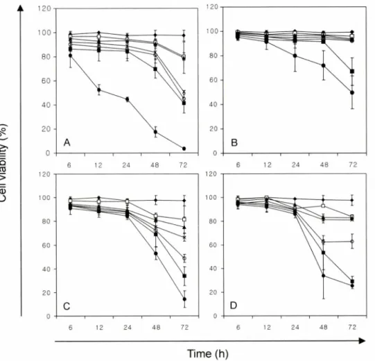
Fig. 2 - Time kinetics of viability of SK-MEL-28 cells treated with various concentrations of cisplatin, heptaplatin, sunpla and mannitol. SK- MEL-28 cells were treated with these compounds at concentrations of 0, 0.001, 0.01, 0.1 1, 10 and 100 |jM and incubated for 6, 12, 24, 48 and 72 h. The results represent cell viability and Bars indicate mean士SD from three independent experiments. It was represented A : cisplatin, B : heptaplatin, C : sunpla D : mannitol and ♦ : 0 |_lM, □ : 0.001 (iM, ▲ : 0.01 |jM, x : 0.1 |iM, O : 1 nM, ■ : 10uM , • : 100 uM.
실험결고 l 및 고찰
Cisplatin, heptaplatin 그리고 선플라에 의한 세포생존를 Cisplatin, heptaplatin 그리고 선폴라 각각의 최종농도를 0, 0.001, 0.01, 0.1, 1, 10 그리고 100 nM이 되게 농도별로 처리하 고 6, 12, 24, 48 및 72시간 동안 시간별로 배양한 후 SK-MEL- 28 세포의 세포생존률을 측정하였다. 그 결과 cisplatiiti: 투여하 지 않은 세포에 비해 cisplatin을 투여한 세포들은 농도와 배양시 간이 증가함에 따라 세포생존률이 감소하는 것으로 나타났다.
0.0이과 0.01 tiM을 투여하였을 때에는 모든 배양시간에서 80%
이상의 세포생존률을 나타냈으며 , 0.1 nM 이상의 농도에서는 72 시간 배양시, 50% 이하로 감소하는 것을 알 수 있었다. 특히 100nM을 투여하였을 때에는 시간이 중가함에 따라 세포의 생존률이 급격히 감소하는 것으로 나타났고, 48시간과 72시간 배양시에는 세포생존률이 각각 17.5土4.33%와 3.7±1.08%로 감 소되어 이들 농도에서는 세포독성이 있는 것으로 평가되어졌다
▲
120(Fig. 2A).
Heptaplatin의 경우, 최종농도가 0.001,0.01, 0.1 그리고 1 |oM 이 되게 처리하여 세포를 배양한 결과, 배양한 모든 시간에서 90% 이상의 세포생존률을 보였다(Fig. 2B). 10 幽 을 투여한 경 우에도 48시간 배양으로 91.5%의 높은 생존률을 보였지만 72시 간 배양시에는 66.9%로 급격히 감소되는 것으로 나타났다. 그리 고 72시간 배양시 , 농도가 중가함에 따라 세포생존률은 감소하 는 것으로 나타났지만 감소의 정도가 cisplatin의 경우보다는 완 화되었으며, 특히 100 nM 투여시 세포생존률이 49.8%로 cisplatin 의 3.7% 보다 매우 높은 생존률을 보였다.
선플라의 경우는 heptaplatin과 부형제로 사용된 mannitol이 1 :2 의 중량비로 혼합되어져 있기 때문에 선플라속에 함유되어 져 있는 heptaplatin의 농도를 0.001, 0.01, 0.1, 1,10 그리고 100|iM로 제조하며 실험하였다. 처리되어진 농도에서 6,12 그 리고 24시간 동안 배양한 결과, 80% 이상의 세포생존률을 나타 내었다(Fig. 2C). 그러나 48시간과 72시간 동안 배양했을 때에는
—,
120348 최수라 • 명평근
농도가 증가함에 따라 세포생존률이 감소하는 경향을 나타내었 다. 이 선플라의 ICso값은 72시간 배양시 10.95|oM 로써,순수한 원료인 heptaplatin(IC50 : 95.35 |jM) 에 비해 더 낮은 농도에서 SK-MEL-28 세포를 죽일 수 있는 효과를 나타내는 것을 알 수 있었다. H eptaplatin 과 선플라의 세포생존률 결과와 ICjo값의 차 이가 생기는 이유를 알아보고자 선플라에만 부형제로 포함되어 져 있는 m annitol 을 SK-MEL-28 세포에 처리하고 그 효과를 확 인해 보았다 (Fig. 2D).
M annit 이에 의한 세포독성을 평가하기 위해 cisplatin, hep
taplatin 그리고 선플라의 경우와 동일한 조건으로 실험한 결과,
0.001, 0.01 그리고 0.1 |oM로 최종농도가 되게 처리한 후 배양한 모든 시간에서 80% 이상의 세포생존률을 보였다. 하지만 48 시 간과 72 시간 배양시 1 ᅣ iM 이상의 농도에서부터는 세포생존를 감소 폭이 중가했으며, 특히 72 시간 배양시 lO O ^M 에서는 25.3%
로 감소된 세포생존률을 나타내었다. 선폴라의 세포생존를 감소 를 나타내는 Fig. 2C 와 m annitol 에 의한 세포생존를 감소를 나 타내는 Fig. 2D 의 결과를 비교했을 때, 그 감소의 유형이 유사 한 것을 확인 할 수 있었으며 선폴라의 더 높은 세포생존를 감 소는 부형제인 m annitol 의 영향인 것으로 사료되어진다. 또한 부 형제인 m annitol 의 IC 50 값은 72 시간 배양시 36.15u M 로써, heptaplatin 에 비해 월등히 낮아진 선플라의 IC ^ 값을 설명해 주
는 것으로 볼 수 있었다.
Flow cytometry에 으ᅵ한 세포주기(cell cycle) 특이성 분석 Cisplatin, heptaplatin 그리고 선플라의 각각의 최종농도가 0,
1, 5,10, 50 그리고 lOOuM이 되게 하여 lx 106 세포에 처리한 후 24시간과 48시간 동안 배양하여,이 약물들이 SK-MEL-28 세 포의 cell cycle에 어떠한 영향을 미치는지 flow cytometer로 분 석하였다.
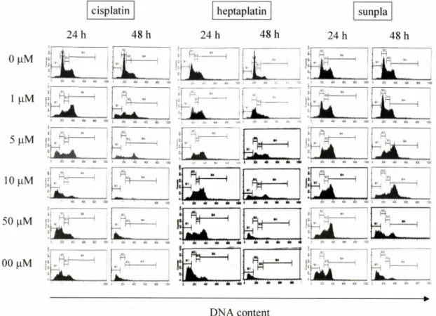
Cisplatin을 처리한 결과,농도와 시간에 의존적으로 죽어가는 세포집단(cell death group)0] 증가함을 알 수 있었다(Fig. 3). 그 리고 이미 많은 연구들을 통해 보고되어진 바와 같이 cisplatin에 의해 cell cycle 중 G2/M기가 정체되는 것을 확인 할 수 있었다.
Fig. 3과 Table II, III에 나타난 값에서 알 수 있듯, 10|oM 처리 후 24시간과 48시간 동안 배양하였을 때, 각각 54.68±2.32%와 71.13±4.30%로 cell death group의 유동율이 가장 높은 것으로 나타났고,50 uM과 100 幽에서 24시간 배양시 23.91± 1.41%와 26.54±1.38%로 48시간 배양시 54.01±4.85와 74.08±7.58%로 오히려 cell death group의 유동율이 감소함을 볼 수 있었다. 또 한 G2/M기의 분석결과,24시간 배양시 lp M 에서 64.53±6.05%
로 정체율이 가장 높았으며, 48시간 배양시에는 5^M 처리시 42.08±1.79%로 정체율이 가장 컸다. 반면,G0/G1 기와 찌 는 24
cisplatin | heptaplatin sunpla
24 h 48 h 24 h 48 h 24 h 48 h
ac «: N C K£ -n 우 ' 췌 싸 !!^ 예ᄀ T*" ~ i.
니 ! V 그 , 1느 1H 1
DNA content
Fig. 3 - Cell cycle analysis of SK-MEL-28 cells by cisplatin, heptaplatin and sunpla. Cells were treated with concentrations of 0, 1 , 5 , 10,50 and 100 나 M of these compounds for 24 h and 48 h. M l to M4 represent cell death group, G0/G1, S and G2/M phase, respectively.
J. Pharm. Soc. Korea
0 1.590 U ±0.38 3.95
±1.59 22.04 b ±4.69 , 八 54.68 iU ±2.32 CA 23.91 50 ±1.41 100 26.54 iuu ±1.38
Table I - IC50 value of compounds about SK-MEL-28 cells for 72 hrs
Compound icso m )
Cisplatin 10.92
Heptaplatin 95.35
Sunplaa) 10.95
Mannitol 36.15
a)The mixture of heptaplatin and mannitol (w : w = l : 2) to make a product.
시간과 48시간 배양시 cisplatin을 처리한 모든 농도에서의 유동 율이 cisplatin을 처리하지 않은 세포의 유동율보다 낮은 유동율 을 보였음으로 정체의 효과는 없는 것으로 확인되었다.
Heptaplatin에 의한 cell cycle 분석 결과, cisplatin의 결과와 유사함을 보였다. G2/M기를 정체하는 효과를 보였으며 , 농도와 시간에 의존적.0■로 cell death group0] 증가하는 것으로 나타났 다(Fig. 3). Heptaplatin을 처리하여 24시간과 48시간 동안 배양 한 세포 모두 처리한 가장 높은 농도인 100|iM에서 각각 4479
±9.78%와 82.75±2.49%로 가장 높은 cell death group 유동율 이 나타났으며,cisplatin보다는 더 높은 농도에서 cisplatin과 유 사한 cell death group 유동율을 얻을 수 있었다. 그리고 G2/M 기는 24시간 배양시 10|iM에서 47.47± 10.42%로, 48시간 배양 시 5 ᅣ iM에서 39.01 ±0.56%로 정체률이 가장 높았다. 또한 G0/
G1 기와 크기를 분석한 결과,이 두 간기에서는 정체되는 부분이 없었으며, heptaplatin을 처리하지 않은 세포보다 처리하여 배양 한 세포의 G0/G1 기 유동율이 heptaplatin의 농도가 증가할수록 감소하는 경향을 보였다. 이 는 24시간 배양시 10|aM 농도까지 는 heptaplatin을 처리하지 않은 세포와 비슷한 유동를을 보이다 가 그 이상의 농도에서는 감소하였으며 , 48시간 배양시에는 5(iM
이상의 농도에서부터 유동율이 감소하였다(Tables II, III).
선플라에 의한 cell cycle을 분석한 결과,이 경우도 역시 SK- MEL-28 세포에 G2/M기의 정체를 일으켰으며, 농도와 시간에 의존적으로 cell death groupi- 중가시키는 것으로 나타났다(Fig.
3). Heptaplatin과 선플라의 결과를 비교해 볼 때,50nM과 100 HM을 처리한 후 24시간 배양시 heptaplatin의 경우 각각 16.36
±6.92%와 44.79±9.78%의 cell death group 유동율을 보인 반 면, 선폴라에서는 11.39±3.39%와 32.14±6.09%로 약간 낮게 나 타났으며 , 48시간 배양시에는 heptaplatin의 경우, 50과 100 |iM 에서 각각 77.9±6.43%와 82.75±2.49%로 높게 나타난 반면, 선 플라는 37.5±8.69%와 58.47±8.90%로 비교적 낮은 cell death group 유동율을 나타내었다. G2/M기는 24시간과 48시간 배양시 모두 10|^M을 처리하였을 때, 49.7±2.43%와 62.53±1.70%로 가장 정체가 심했다(Tables II,III). 이는 세포생존률 결과로부터 얻은 IC50값과는 상반된 결과로,선플라는 혼합되어진 mannitol 에 의한 直과로 인해 heptaplatin만을 처리한 세포에서 보다 더 높은 세포독성을 유발시키는 것으로 생각되어지며,heptaplatin 은 cisplatin과 같이 DNA에 선택성을 나 타 내 며 , 부형제인 mannitol은 heptaplatin0] 세포내의 DNA를 공격하는 것을 방해 하는 것으로 사료되어진다. Cisplatin과 heptaplatin의 경우에서 와 같이 선플라에 의해서도 세포의 G0/G1 기와 드기가 정체되지 않았다. 24시간과 48시간 배양시 모두, 선플라의 농도가 증가할 수록 G0/G1 기의 유동율은 감소하였지만, 50|jM과 100|iM을 처 리하였을 때에는 24시간 배양시 24.36±3.84%께서 30.57±2.25%
로, 48시간 배양시 24,57±7.54에서 30.26±5.83%로 증가함을 나타냈다. 그리고 S기를 분석한 결과, 24시간 배양시에는 50|oM 까지 거의 일정한 유동율을 보였으며, 48시간 배양시에는 농도
Table II - Cell cycle analysis3 of SK-MEL-28 cell cultured for 24 h after treatment of cisplatin, heptaplatin and sunpla
Cisplatin Heptaplatin Sunpla
Cell
death G0/G1 S G2/M Cell
death G0/G1 S G2/M Cell
death G0/G1 G2/M
SK-MEL-28 cells were treated with various concecntrations of cisplatin, heptaplatin and sunpla for 24 h. And cell cycle analysis was performed by flow cytometer. Data represent percentage of mean±S.D from three independent experiments.
aCell cycle analysis was performed by flow cytometer.
bSK-MEL-28 cells were incubated for 24 h after treatment of compounds.
cSK-MEL-28 cells were treated with cisplatin, heptaplatin and sunpla at concentration of 0 , 1 , 5 , 10,50 and 100 (iM.
.5 . 31
29 . 60
15 . 46
.7 . 43
.9 .94
03 .83 s
±3 32. s
±35
j
25.
±3 56
. 16
04 . 99
84 . 94
75 . 3 7
45 . 97
46 . 76 21.
±6 22. u 23.
±s5
22. s
±2 66
. 90
84 . 2 9
17 . 69
24 . 77
36 .84
57 . 25
s s s s s s
11 . 93
08 . 21
31 . 98
47 0 .4 2 .5
. 33
38 . 06 71
. 80
83 . 79
97 . 91
.5 . 09
67 . 46
05 . 98 76
. 14
34 . 34
26 .97
52 . 25
76 . 96
31 . 18 51.
±3 40.
±1 24.
±1 22.
±6 21.
±1 35.
±6 64
. 43 5 . 25
94 . 08
12 .84
. 36
. 9 2
. 79
. 78 w
±0 2.
±0 2 .9
±l 4.
±l 1 6±6 44
±9 24
. 49
53 . 05
18 . 69
98 . 57
99 . 11
18 . 02 31.
±4 64.
±6 42.
±3 13.
±1 15.
±3 20.
±2 62
. 63
83 . 61
47 . 45
31 . 79
02 . 55
18 .19 18.
±3 18.
±0 14.
±1 8.
±0 15.
±1 19.
±1 42
. 72
33 . 1 1
57 . 32
25 . 05
43 . 55
.1 . 90 48.
±1 14.
±3 20.
±5 24.
±2 44.
±8 36
±0
350 최수라 • 명평근
-6 04 64 13 82 44 .5 78 21 05 08 1 6 4. 0. 2. 6 . 0. 11 0.
2 .2 1 1 0.
1±2±1±1±2±±
2.14
±0.63 4.65
±1.45 10.97
±0.67 29.83
±2.62 77.9
±6.43 82.75
±2,49 19.36
±0.44 15
±0.77 7.59
±0.83 2.23
±0.62 3.52
±1.72 4.31
±1.74
SK-MEL-28 cells were treated with various concecntrations of cisplatin, heptaplatin and sunpla for 24h. And cell cycle analysis was performed by flow cytometer. Data represent percentage of mean ±S.D. from three independent experiments.
aCell cycle analysis was performed by flow cytometer.
SK-MEL-28 cells were incubated for 24 h after treatment of compounds.
cSK-MEL-28 cells were treated with cisplatin, heptaplatin and sunpla at concentration of 0 , 1, 5, 10, 50 and 100 (iM.
가 증가할수록 유동율이 감소하는 경향을 보였다 (Tables II, III).
그러므로 Cell cycle 이 G 2/M 기에서 정체된다 함은 Cdc2 와 cyclin A 또는 cyclin B 의 결합에 문제가 생겼다 함을 의미하며, 특히 M 기의 특이적인 유비키틴화 효소인 APC 가 cyclin B 를 유 비키틴화 하는데, 이 cyclin B 가 유비키틴화 되지 않거나, 이 유 비키틴화된 cyclin B 가 프로테아좀에 의해 분해되지 않으면 cell cycle 은 정체되게 된다.20> 이들의 과정은 apoptosis 를 일으켜 암 세포를 죽일 수 있는 하나의 방법이며,특히 현재 항암제로 사용 되는 cisplatin 처럼 그 제 3 세대 화합물인 heptaplatin 과 그 제형 인 선플라가 유사한 결과를 나타냄을 보인 바, 현재 이들이 SK- MEL-28 세포에 대하여 세포사멸과 어떠한 기전으로 apoptosis 가 일 에 지 의 연구가 진행되고 있는 중이다.
결 론
현재 세계적으로 매년 약 5% 의 비율로 성장하고 있는 피부암 에 대한 항암제를 개발하기 위하여 피부암 세포인 인체 흑색종 세포주 (SK-MEL-28) 에 대해 한국 신약 1 호인 sunpla 의 주성분 인 hepataplatin 과 cisplatin, 그리고 heptaplatin 에 m annitol 을' 첨 가해서 제형으로 만든 sunpla 들에 의한 피부암 세포성장억제에 대한 비교 분석과 이들이 어떠한 세포주기를 억제함으로써 항암 효과를 나타내는지를 분석한 결과는 다음과 같다.
1. Cisplatin, heptaplatin 그리고 선폴라 각각의 최종농도를 0, 0.001, 0.01, 0.1, 1, 10 그리고 10 0|oM 이 되게 농도별로 처리하 고 72 시간 배양한 후 SK-MEL-28 세포의 세포생존률을 측정한 결과 cisplatin 의 IC 50 값은 10.92|iM 로써,선폴라 (IC 50= 10.95
|iM)S|- 비슷하며 선플라는 순수한 원료인 h 印 taplatin(IC50 :
95.35 nM ) 에 비해 더 낮은 농도에서 SK-MEL-28 세포를 죽일 수 있는 효과를 나타내는 것을 알 수 있었다. 한편 부형제인 m annitol 의 ICso 값은 36.15nM 로써, heptaplatin 에 비해 월등히 낮아진 선폴라의 I Q 값에 영향을 미치는 것으로 볼 수 있었다.
2. Cisplatin, heptaplatin 그리고 선플라의 각각의 최종농도가 0, 1, 5, 10, 50 그리고 100 nM이 되게 하여 SK-MEL-28 세포 (lx 106 세포)에 처리한 후 24시간과 48시간 동안 배양하여, 이 약물들이 SK-MEL-28 세포의 cell cycle 에 어떠한 영향을 미치 는지 flow cytometer 로 분석한 결과 cisplatin 과 heptaplatin 그 리고 선플라에 의해 cell cycle 중 G2/M기가 정체되는 것을 확 인 할 수 있었다.
이상의 결과로 보아 cisplatin 과 heptaplatin 그리고 선플라가 피부암세포에 유사한 항암성 효과을 나타난다고 사료되어진다.
감사의 말씀
본 연구는 한국과학재단 우수연구센터 (R11-2002-100-03003- 0) 지원과 충남대학교 암연구소의 일부지원을 받아 수행되었기 에 이에 감사드립니다. 또한 heptapaltin 과 sunpla 를 제공해주신 이화여자대학교 약학대학 김대기 박사께 감사드립니다.
문 헌
1) Kaufmann, S. H. : Induction of endonucleolytic DNA cleavage in human acute myelogenous leukemia cells by etoposide, camptoyhecin, and other cytotoxic anticancer drugs : a cautionary note. Cancer Res. 49, 5870 (1989).
2) Barry, M. A., Behnke, C. A. and Eastman, A. : Activation of Table III - Cell cycle analysis of SK-MEL-28 cell cultured for 48 h after treatment of cisplatin, heptaplatin and sunpla
Cisplatin Heptaplatin Sunpla
dea!L 00/01 S G2/M d2 fh 00/01 S G2/M deafh G0/G1 S G2/M
73 . 85
52 . 21 6 . 60
53 . 70
24 .27
11 . 79 .4
. 42
05 . 79
81 . 49
27 . 29
63 . 10
06 . 08
s
±4 说 S S
?
±0
± 31
°
11 . 13
58 . 16
99 . 77
13 . 14
57 . 54
26 . 83 8
2 03
40D8 02 37 24 .5 69 47 90 3.
±.1 3.
±.2 3.
±.1 9 .3
±.3 37
±.8 58.
±.8 3
.4 4 . 3 2 . 45 1 .6 9 . 01 0 .5 6 7 .8 1 .5 7 . 26 4 .6 7 . 5 2 0 . 3 6 1
± 17± 39± 3
± 6
± o
± 96
. 70
19 . 67
57 . 63
21 . 68
42 . 5 2
66 . 27 63.
±7 55.
±1 32.
±0 19.
±0 13.
±1 15.
±2 .
23
. 96
. 46
. 79
.35
.80
. 53
. 21
. 97
.14
. 28
. 84 10.
9.1
5.1
9.2
6.3
8.4
6+12+11
±
1
±±31
± 07
. 47
82 . 49
95 . 48
13 . 30
01 .85
08 . 58 S
O
7.1
5.3
14.
4.4
4.7 2
± 1
± 3
± 7
± 5
± 7
±