Open Access
Adjuvant Treatment after Surgery in Stage IIIA Endometrial Adenocarcinoma
Original Article
Purpose
We evaluated the role of adjuvant therapy in stage IIIA endometrioid adenocarcinoma patients who underwent surgery followed by radiotherapy (RT) alone or chemoradiotherapy (CTRT) according to risk group.
Materials and Methods
A multicenter retrospective study was conducted including patients with surgical stage IIIA endometrial cancer treated by radical surgery and adjuvant RT or CTRT. Disease-free survival (DFS) and overall survival (OS) were analyzed.
Results
Ninety-three patients with stage IIIA disease were identified. Nineteen patients (20.4%) experienced recurrence, mostly distant metastasis (17.2%). Combined CTRT did not affect DFS (74.1% vs. 82.4%, p=0.130) or OS (96.3% vs. 91.9%, p=0.262) in stage IIIA disease compared with RT alone. Patients with age ! 60 years, grade G2/3, and lymphovascular space involvement had a significantly worse DFS and those variables were defined as risk factors. The high-risk group showed a significant reduction in 5-year DFS (! 2 risk factors) (49.0% vs. 88.0%, p < 0.001) compared with the low-risk group (< 2). Multivariate analysis confirmed that more than one risk factor was the only predictor of worse DFS (hazard ratio, 5.45; 95% confidence interval, 2.12 to 13.98; p < 0.001). Of patients with no risk factors, a subset treated with RT alone showed an excellent 5-year DFS and OS (93.8% and 100%, respectively).
Conclusion
We identified a low-risk subset of stage IIIA endometrioid adenocarcinoma patients who might be reasonable candidates for adjuvant RT alone. Further randomized studies are needed to determine which subset might benefit from combined CTRT.
Key words
Endometrial neoplasms, Adjuvant radiotherapy, Adjuvant chemoradiotherapy
+ + + + + + + + + + + + + + + + + + + + + + + + + + + + + + + + + + + + + + + + + + + + + + + + + + + + + + + + + + + + + + + + + + + + + + + + + + + + + + + + + + + + + + + + + + + + + + + + + + + + + + + + + + + + + + + + + + + + + + + + + + + + + + + + + + + + + + + + + + + + + + + + + + + + + + + + + + + + + + + + + + + + + + + + + + + + + + + + + + + + + + + + + + + + + + + + + + + + + + + + + + + + + + + + + + + + + + + + + + + + + + + + + + + + + + + + + + + + + + + + + + + + + + + + + + + + + + + + + + + + + + + + + + + + + + + + + + + + + + + + + + + + + + + + + + + + + + + + + + + + + + + + + + + + + + + + + + + + + + + + + + + + + + + + + + + + + + + + + + + + + + + + + + + + + + + + + + + + + + + + + + + + + + + + + + + + + + + + + + + + + + + + + + + + + + + + + + + + + + + + + + + + + + + + + + + + + + + + + + + + + + + + + + + + + + + + + + + + + + + + + + + + + + + + + + + + + + + + + + + + + + + + + + + + + + + + + + + + + + + +
Correspondence: Won Park, MD, PhD Department of Radiation Oncology, Samsung Medical Center,
Sungkyunkwan University School of Medicine, 81 Irwon-ro, Gangnam-gu, Seoul 06351, Korea Tel: 82-2-3410-2616
Fax: 82-2-3410-2619
E-mail: [email protected]
Co-correspondence: Sei Kyung Chang, MD Department of Radiation Oncology,
CHA Bundang Medical Center, CHA University, 59 Yatap-ro, Bundang-gu, Seongnam 13496, Korea
Tel: 82-31-780-5426 Fax: 82-31-702-8803 E-mail:[email protected]
Received September 18, 2015 Accepted October 7, 2015 Published Online October 29, 2015
Mee Sun Yoon, MD, PhD
1Seung Jae Huh, MD, PhD
2Hak Jae Kim, MD
3Young Seok Kim, MD, PhD
4Yong Bae Kim, MD, PhD
5Joo-Young Kim, MD, PhD
6Jong-Hoon Lee, MD
7Hun Jung Kim, MD
8Jihye Cha, MD
9Jin Hee Kim, MD, PhD
10Juree Kim, MD, PhD
11Won Sup Yoon, MD, PhD
12Jin Hwa Choi, MD
13Mison Chun, MD, PhD
14Youngmin Choi, MD
15Kang Kyoo Lee, MD, PhD
16Myungsoo Kim, MD, PhD
17Jae-Uk Jeong, MD
1Sei Kyung Chang, MD
18Won Park, MD, PhD
2*A list author’s a!liations appears at the end
of the paper.
Introduction
Stage IIIA endometrial cancer represents a wide range of tumor involvement, including the uterine serosa, adnexa, parametria, and peritoneal space, based on the staging sys- tem of the International Federation of Gynecology and Obstetrics (FIGO) in 1988 [1-3]. This variability might have an influence on survival for stage IIIA endometrial cancer.
In addition, the heterogeneity of histology, such as papillary serous or clear cell carcinoma, makes it difficult to compare outcomes between studies. This variability has led to a wide range of 5-year overall survival (OS) rates for stage IIIA endometrial cancer, varying from 39% to 76% [3-6].
The optimal adjuvant management for patients with advanced endometrial cancer has yet to be defined, particu- larly for IIIA patients as incidence is low, representing only 2.6% of patients with endometrial cancer [7]. Radiotherapy (RT) has traditionally been employed in advanced endome- trial carcinoma to improve locoregional control [8,9]. Several studies have evaluated the role of combined modality adju- vant therapy compared with a single modality in advanced endometrial cancer and have suggested that combined chemoradiotherapy (CTRT) can provide additional benefits compared with computed tomography (CT) or RT alone [10-13]. In contrast, a randomized study failed to show an improvement in disease-free survival (DFS) and OS in patients treated with combined CTRT compared with adju- vant RT alone for high-risk endometrial cancer [14]. Those studies included only a small proportion of stage IIIA patients. Thus, the effects of different adjuvant therapies for stage IIIA endometrial cancer remain controversial. In addi- tion, these previous studies involved a small portion of IIIA populations, patient’s stage was determined based on the 1988 FIGO stage, and both endometriod and non-endometri- oid adenocarcinoma were included.
The current study included a group of patients with stage IIIA endometrioid adenocarcinoma with favorable histology only, using the revised 2009 FIGO stage to overcome the aforementioned variabilities. The revised FIGO IIIA, which was published in 2009, excluded positive peritoneal cytology alone and denoted parametrial involvement separately as stage IIIB. Several studies have attempted to analyze risk fac- tors for poor outcomes [1,5,6,9,15,16]; however, few studies have evaluated the role of particular adjuvant therapies according to risk group for revised 2009 FIGO stage IIIA dis- eases. Here, we evaluated survival outcomes according to risk factors and sought to determine the benefit of combined modality treatment after surgery in the high-risk subset of stage IIIA patients.
Materials and Methods
All patients with stage IIIA endometrial cancer treated with surgery, followed by adjuvant RT or combined CTRT, in Korea between January 1990 and December 2011 were evaluated. All patients were staged based on the revised 2009 FIGO criteria for endometrial cancer. Patients eligible for analysis had endometrioid adenocarcinoma only.
Nineteen patients with non-endometrioid type adenocar- cinoma, such as papillary serous, clear cell, adenosquamous, or carcinosarcoma were excluded. In addition, five patients with only positive peritoneal cytology, and those who underwent adjuvant CT alone (n=3) or inadequate RT (n=2) were also excluded. The remaining 93 stage IIIA patients who underwent surgery followed by adjuvant RT alone or combined CTRT were analyzed retrospectively.
The clinical and pathologic data of 93 patients from 18 institutions were collected. After approval by the Korean Radiation Oncology Group (KROG 13-17), the medical and RT records of the patients were reviewed retrospectively.
1. Treatment
All patients underwent hysterectomy and bilateral salp- ingo-oophorectomy with pelvic and/or aortic lymphadenec- tomy, followed by postoperative RT or combined CTRT.
Pelvic lymph node dissection was performed in 77 patients (82.8%) and paraaortic lymph node (PALN) dissection or sampling was performed in 31 patients (33.3%).
All patients underwent external pelvic RT, delivered to the tumor bed and regional lymphatics with 10-MV photons using the four-field box technique. The total dose to the pelvis ranged from 45 Gy to 54 Gy in 1.8 Gy daily fractions, 5 days per week. Twenty-one patients (21.6%) underwent additional vaginal brachytherapy using Fletcher-Suit after- loading applicators. Two to six fractions of 3-5 Gy were delivered to the vaginal surface or 5 mm from the vaginal surface. Two patients (2.2%) were treated with an extended- field RT encompassing a volume of PALN, usually located at the T12-L1 interface. Another two patients received total abdominal RT of 30 Gy in 1.5 Gy fractions followed by a boost to the pelvis with the doses of 19.8 Gy or 24 Gy in 1.8 Gy fractions.
Adjuvant combined CTRT was performed in 36 patients
(38.7%). Thirty patients were treated with concurrent CTRT
(83.3%), four patients with CT followed by RT (11.1%), and
two patients with RT followed by CT (5.6%). Patients receiv-
ing adjuvant CT were treated with cisplatin-based CT (n=19,
52.8%), carboplatin plus paclitaxel (n=8, 22.2%), paclitaxel
alone (n=6, 16.7%), or other drugs (n=3, 8.3%). Adjuvant
CT was delivered with a median of six cycles (range, 1 to 9
cycles), although two patients received only one cycle, and another two patients only received two cycles. Five patients did not complete the full course of CT because of G3 hema- tologic toxicity (n=2), patient refusal (n=1), or no available data on toxicity (n=2).
In adjuvant RT alone, the average treatment period was 42.9 days (range, 36 to 60 days) and 46.9 days (range, 35 to 117 days) for the CTRT group. No significant difference in the RT period was observed between the two treatment groups (p=0.057).
2. Statistical analysis
Locoregional recurrence was defined as a recurrence in the pelvis, vagina, or paraaortic lymphatic region. Failure was defined as biopsy-proven recurrence or progression of dis- ease on serial imaging studies. Time to recurrence and death was calculated from the date of surgery until failure or death from any cause. DFS was defined as alive without disease recurrence at the time of censoring. Survival curves were cal- culated using the Kaplan-Meier method, and comparison of the curves was performed using a log-rank test. The chi- square test, Fisher exact test, and independent samples t test were used for comparison of characteristics between the two groups. Multivariable analysis was performed using the Cox proportional hazards model. Statistical analyses were per- formed using SPSS ver. 21 (IBM Co., Armonk, NY), and p-values less than 0.05 were considered statistically signifi- cant.
Results
1. Patient characteristics and pattern of failure
The tumor and treatment characteristics of all 93 patients, 57 (61.3%) who received postoperative RT alone and 36 (38.7%) who received CTRT, are shown in Table 1. The median age of patients in the CTRT group was older than that in the RT alone group. A larger portion of patients in the CTRT group showed involvement of serosa (27.8% vs. 19.3%, p=0.361) and lymphovascular space (30.6% vs. 15.8%, p=0.214).
A higher proportion of patients in the CTRT group under- went paraaortic lymphadenectomy compared to the RT alone group (52.8% vs. 21.1%, p=0.002). The extent of irradi- ation field of adjuvant RT and vaginal brachytherapy did not differ significantly in the two groups.
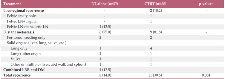
The median follow-up period was 62 months (range, 3 to 188 months). Eight patients (14.0%) in the RT alone group
and 11 patients (30.6%) in the CTRT group relapsed during the follow-up period (p=0.054) (Table 2). The majority of recurrences were distant metastases including abdominal recurrence, 75% (6/8) in the RT alone group and 81.8%
(9/11) in the CTRT group. In the RT alone group, one patient developed combined local and distant metastases with pelvic lymph node and liver metastasis.
2. DFS and OS
The 5-year OS and DFS were 93.7% and 79.0%, respec- tively. Univariate analyses showed statistically significant difference in DFS with regard to age (p < 0.001), tumor grade (p=0.029), and lymphovascular involvement (p=0.041) (Table 3). No significant difference in OS (91.9% vs. 96.3%, p=0.262) or DFS (82.4% vs. 74.1%, p=0.130) was observed between the RT alone group and the combined CTRT group.
Five-year DFS was lower in patients with serosal involve- ment than in those without, but there was no statistical significance (81.5% vs. 67.2%, p=0.180). Involvement of adnexa did not show a significant relationship with DFS. Of the 93 patients, 57 (61.3%) had solitary adnexal or serosal involvement. Patients with serosal involvement had a worse 7-year DFS (66.7%) compared to those with solitary adnexal involvement (90.5%), but this difference did not reach statis- tical significance (p=0.629).
The above mentioned variables (age ! 60 years, tumor grade 2/3, and lymphovascular space involvement) for prog- nosis in DFS were defined as risk factors. Of 77 patients with available data for risk factors, 25 patients (32.5%) had no risk factors, 30 (38.9%) had one, 16 (20.8%) had two, and six (7.8%) had three. A significant correlation was observed between the number of risk factors and DFS. The 5-year DFS was 91.7% in patients with no risk factors, 84.8% in patients with one, 55.0% in patients with two, and 33.3% in patients with three (p=0.001). The 5-year OS according to the number of risk factors was 95.0% in patients with no risk factors, 92.0% in patients with one, 93.8% in patients with two, and 80.0% in patients with three (p=0.542).
The patients were subgrouped according to low-risk (patients with no or one risk factor) and high-risk (patients with two or three risk factors) groups. The proportion of high-risk patients in the combined CTRT group was more than twice that of the RT alone group (41.9% vs. 19.6%, p=0.033). Thirty-seven patients (80.4%) in RT alone were clas- sified as low risk and nine (19.6%) as high risk. In CTRT, 18 patients (58.1%) were classified as low risk, while 13 patients (41.9%) as high risk. A significant reduction in 5-year DFS was observed in the high-risk group (49.0% vs. 88.0%, p < 0.001) compared with the low-risk group (Fig. 1).
Five-year OS was not significantly lower in the high-risk
group (90.7%) versus the low-risk group (93.5%) (p=0.618).
For RT alone, 5-year DFS and OS were lower in the high- risk group than in the low-risk group (66.7% vs. 84.6%, p=0.133 and 77.8% vs. 93.5%, p=0.149, respectively), but the differences did not reach statistical significance (Fig. 2).
In the CTRT group, the low-risk group showed an improved 5-year DFS (94.4% vs. 38.5%, p=0.001) compared with the high-risk group, but there was no significant difference in OS (93.3% vs. 100%, p=0.465).
3. Multivariate analysis
To examine the role of adjuvant multi-modality therapy, the risk groups were analyzed according to treatment modal- ities. No significant difference in 5-year OS (93.5% vs. 93.3%, p=0.785) or DFS (84.6% vs. 94.4%, p=0.852) was observed between the RT alone group and the combined CTRT group in the low-risk group. In the high-risk group, combined CTRT showed a trend for improved 5-year OS (100% vs.
77.8%, p=0.086) compared with RT alone, but the difference did not reach statistical significance. Combined CTRT did Table 1. Clinical and pathologic characteristics in stage IIIA endometrial adenocarcinoma according to adjuvant modality
Characteristic All (n=93) RT alone (n=57) CTRT (n=36) p-value
Age, median (range, yr) 51 (25-83) 50 (25-83) 54 (35-75) 0.281
Age (yr)
< 60 74 (79.6) 46 (80.7) 28 (77.8) 0.733
! 60 19 (20.4) 11 (19.3) 8 (22.2)
Grade
I 41 (44.1) 28 (49.1) 13 (36.1) 0.291
II 30 (32.3) 15 (26.3) 15 (41.7)
III 20 (21.5) 12 (21.1) 8 (22.2)
Unknown 2 (2.2) 2 (3.5) 0 (
Serosal involvement
Negative 70 (75.3) 44 (77.2) 26 (72.2) 0. 361
Positive 21 (22.6) 11 (19.3) 10 (27.8)
Unknown 2 (2.2) 2 (3.5) 0 (
Adnexal involvement
Negative 16 (17.2) 11 (19.3) 5 (13.9) 0.501
Positive 77 (82.8) 46 (80.7) 31 (86.1)
Lymphovascular involvement
Negative 59 (63.4) 38 (66.7) 21 (58.3) 0.214
Positive 20 (21.5) 9 (15.8) 11 (30.6)
Unknown 14 (15.1) 10 (17.5) 4 (15.1)
Tumor size
a), mean (range, cm) 4.6 (0.4-12) 4.4 (1.5-12) 5.0 (0.4-10.2) 0.327 Lymph node dissection
Pelvic LN dissection 77 (82.8) 44 (77.2) 33 (91.7) 0.072
Paraaortic LN dissection 31 (33.3) 12 (21.1) 19 (52.8) 0.002
Type of external beam radiotherapy
Whole pelvis 89 (95.7) 54 (94.7) 35 (97.2) 0.500
Whole pelvis+paraaortic field 2 (2.2) 1 (1.8) 1 (2.8)
Whole abdomen 2 (2.2) 2 (3.5) 0 (
Additional vaginal brachytherapy
No 72 (77.4) 46 (80.7) 26 (72.2) 0.341
Yes 21 (21.6) 11 (19.3) 10 (27.8)
CTRT sequence
Concurrent 30 (83.3)
Sequential 6 (16.7)
Values are presented as number (%) unless otherwise indicated. RT, radiotherapy; CTRT, chemoradiotherapy; LN, lymph
node.
a)Data were available in 70 patients with measurable primary tumor.
not affect 5-year DFS (38.5% vs. 66.7%, p=0.274) compared with RT alone for the high-risk group.
In multivariate analysis including age, grade, lympohovas- cular involvement, number of risk factors, and type of adju-
vant modality, more than one risk factor was the only pre- dictor of worse DFS (hazard ratio [HR], 5.45; 95% confidence interval [CI], 2.12 to 13.98; p < 0.001). In multivariate analysis for OS, only old age was predictive of OS (HR, 5.8; 95% CI, Table 2. Patterns of failure according to adjuvant treatment
Table 3. Factors affecting survival in univariate analysis
Treatment RT alone (n=57) CTRT (n=36) p-value
a)Locoregional recurrence - 2 (18.2) -
Pelvic cavity only - 1 (
Pelvic LN+vagina - 1 (
Pelvic LN+paraaortic LN 1 (12.5) -
Distant metastasis 6 (75.0) 9 (81.8) -
Peritoneal seeding only 2 ( 2 (
Solid organs (liver, lung, vulva, etc.)
Lung only 1 ( 4 (
Lung+other organ 1 ( 1 (
Vulva 1 ( 1 (
Other or multiple (liver, abd wall, and spleen) 1 ( 1 (
Combined LRR and DM 1 (12.5) - -
Total recurrence 8 (14.0) 11 (30.6) 0.054
Values are presented as number (%). RT, radiotherapy; CTRT, chemoradiotherapy; LN, lymph node; LRR, locoregional recurrence; DM, distant metastasis.
a)Chi-square test.
Parameter Disease-free survival Overall survival
5-Year (%) p-value 5-Year (%) p-value
Age (< 60 yr vs. ! 60 yr)
< 60 86.6 < 0.001 96.7 0.028
! 60 48.9 81.4
Grade (G1 vs. G2/3)
G1 90.0 0.029 94.4 0.387
G2/G3 68.9 93.3
LVI
No 83.2 0.041 91.9 0.699
Yes 57.9 94.4
Serosal involvement
No 81.5 0.180 91.8 0.747
Yes 67.2 100
Adnexal involvement
No 73.8 0.790 100 0.991
Yes 79.1 92.5
Size (diameter, cm)
" 5 84.0 0.175 95.5 0.482
> 5 77.3 89.4
Adjuvant treatment
RT alone 82.4 0.130 91.9 0.262
CTRT 74.1 96.3
LVI, lymphovascular involvement; RT, radiotherapy; CTRT, chemoradiotherapy.
1.15 to 29.61; p=0.034). In multivariate analysis, combined CTRT did not show a statistical difference compared to RT alone in DFS or OS (Table 4).
4. Treatment related toxicities
Twelve patients (33.3%) in the CTRT group experienced grade 3-4 acute toxicity. Grade 3-4 acute hematologic toxici- ties were observed in 11 patients in the CTRT group and a grade 3 acute intestinal obstruction was found in one patient.
In the RT alone group, one patient (1.8%) treated with extended-field RT had grade 4 acute hematologic toxicity.
No patients in the CTRT group had grade 3-4 late toxicity and one patient in the RT alone group treated with whole abdominal irradiation had a grade 3 late leg edema.
Discussion
The current study showed the impact of a number of risk factors for stage IIIA endometrioid adenocarcinoma. Patients with more than one risk factor (high-risk group) had a worse prognosis. Indeed, more than one risk factor was the only strong negative prognostic factor in multivariate analysis.
Patients in the high-risk group were 4 to 5 times more likely to develop a distant metastasis compared to those in the low- risk group (40.9% vs. 9.1%, p < 0.001), therefore, the addition
of more intense systemic therapy to those in the high-risk group may improve the outcome. The current study showed that combined CTRT showed a trend with an improved 5-year OS (100% vs. 77.8%, p=0.086) compared with RT alone, although CTRT did not affect 5-year DFS compared with RT alone for the high-risk group. Several studies have suggested that combined CTRT can provide additional ben- efits compared with CT or RT alone [10-13]. The retrospective study in stage III and IV endometrial cancer by Alvarez Sec- ord et al. [13] reported that adjuvant CT alone was associated with poor 3-year OS (33%) and progression-free survival (19%) compared to either RT alone (70% and 59%) or com- bined therapy (79% and 62%). In a multicenter retrospective study conducted with 78 patients with stage III endometrial cancer treated with adjuvant CT and/or RT, the 3-year relapse-free survival rates were 86.5% for the combined CTRT group, 65.8% for the CT alone, and 44.1% for RT alone, suggesting that combined CTRT modality may induce an advantage in relapse-free survival compared to RT or CT alone [17]. Randomized trials were conducted for compari- son of sequential CTRT with RT alone. The first trial was run by the Italian Oncology Group with 153 patients (IIB-IIIC, 65% stage III), and the second by the Nordic Society of Gynaecological Oncology/European Organisation for the Research and Treatment of Cancer, which included patients with mainly high-risk early stage disease (only 1.6% stage III). In the Italian trial, no statistically significant difference in DFS or OS was observed between treatment groups. How- ever, in joint pooled analysis in these two randomized trials,
Di se as e- fre e su rv iv al (% )
100 80 60 40 20
0 0
Time (mo)
24 48 72 96 120
p < 0.001
A
Low-risk group 5-year: 88.0%
High-risk group 5-year: 49.0%
Ov er al l s ur vi va l ( % )
100 80 60 40 20
0 0
Time (mo)
24 48 72 96 120
p =0.618
B
Low-risk group 5-year: 93.5%
High-risk group 5-year: 90.7%
Fig. 1. Kaplan-Meier survival curve of disease-free survival (A) and overall survival (B) according to the low-risk group
with no or one risk factor and the high-risk group with two or three risk factors (risk factor: age ! 60 or ! G2 or lymphovas-
cular space invasion).
Di se as e- fre e su rv iv al (% ) 100
80 60 40 20
0 0
Time (mo)
24 48 72 96 120
p=0.133 Low-risk group 5-year: 84.6%
High-risk group 5-year: 66.7%
Ov er al l s ur vi va l ( % )
100 80 60 40 20
0 0
Time (mo)
24 48 72 96 120
p =0.149
A
Low-risk group 5-year: 93.5%
High-risk group 5-year: 77.8%
Di se as e- fre e su rv iv al (% )
100 80 60 40 20
0 0
Time (mo)
24 48 72 96 120
p=0.001 Low-risk group 5-year: 94.4%
High-risk group 5-year: 38.5%
Ov er al l s ur vi va l ( % )
100 80 60 40 20
0 0
Time (mo)
24 48 72 96 120
p =0.465
B
Low-risk group 5-year: 93.3%
High-risk group 5-year: 100%
Fig. 2. Subgroup analysis according to treatment modality. Disease-free survival and overall survival in the radiotherapy alone group (A) and the chemoradiotherapy group (B). The low-risk group with no or one risk factor and the high-risk group with two or three risk factors (risk factor: age ! 60 years or ! G2 or lymphovascular space invasion).
Table 4. Multivariate analysis
Parameter Disease-free survival Overall survival
HR (95% CI) p-value HR (95% CI) p-value
Age (< 60 yr vs. ! 60 yr) - 0.340 5.83 (1.15-29.61) 0.034
Grade (G1 vs. G2G3) - 0.903 - 0.834
LVI (no vs. yes) - 0.884 - 0.437
No. of risk factors
a)(< 2 vs. ! 2) 5.45 (2.12-13.98) < 0.001 - 0.093
RT alone vs. CTRT - 0.488 - 0.177
HR, hazard ratio; CI, confidence interval; LVI, lymphovascular space invasion; RT, radiotherapy; CTRT, chemoradiotherapy.
a)
Risk factor: age ! 60 years or ! G2 or LVI.
sequential addition of CT to RT improved at least progres- sion-free survival (78% vs. 69%, p=0.009), and trended a ben- efit of overall survival (82% vs. 75%, p=0.07) compared with RT alone [12].
An important question is whether or not it is reasonable to recommend adjuvant RT alone without chemotherapy in low-risk patients. Prior study has suggested that a subgroup of women with stage IIIA who have endometrioid tumor, no lymphovascular space involvement, and positive peritoneal cytologic finding only show an excellent prognosis [15].
Havrilesky et al. [6] found that among 24 patients with peri- toneal cytology only, non-serous histology, and grade 1-2 disease, no recurrence was found in 12 patients receiving adjuvant treatment, while one patient showed recurrence without adjuvant treatment. Our study included patients according to the 2009 FIGO staging system and excluded those with peritoneal cytology only. For 25 patients with no risk factors, 17 patients (68%) were treated with adjuvant RT alone and eight patients (32%) with adjuvant combined CTRT, resulting in excellent 5-year DFS (91.7%) and OS (95.0%), respectively. No difference in DFS and OS was observed between RT alone and the CTRT group. Thus, a subset of stage IIIA patients with no risk factors might be treated with adjuvant RT alone.
Several studies have reported that patients with extrauter- ine spread limited to the adnexa showed favorable 5-year DFS rates, ranging from 71% to 89.6% [3,18,19]. By contrast, serosal involvement had a relatively poor prognosis [15,20].
A retrospective study of 15 patients with solitary serosal invasion had a poor 5-year DFS (41.5%) due to the high inci- dence of distance metastases [20]. A multicenter retrospec- tive comparative analysis in Netherlands published the outcomes for 67 patients with stage IIIA endometrial carci- noma, evaluating differences in outcome between serosa and adnexal involvement [18]. Those with involvement of the serosa alone had a worse 7-year distant metastasis-free sur- vival (58.7%) compared to adnexal involvement alone (72.7%), but this difference was not significant (p=0.399).
Authors concluded that either adnexal or serosal involve- ment showed a comparable disease-specific survival and only presence of lymphovascular invasion was significant in multivariate analysis (HR, 3.6; p=0.038). The current study was composed of a relatively large homogeneous subgroup of surgically staged IIIA patients with only adnexal or serosal involvement. Of the 93 patients in our study, 57 (61.3%) had solitary adnexal or serosal involvement. Consistent with pre- vious studies, patients with serosal involvement had a worse 7-year DFS (66.7%) compared to those with solitary adnexal involvement (90.5%) (p=0.629).
In this multicenter analysis, we failed to demonstrate additional benefit of combined adjuvant CTRT compared with adjuvant RT alone for stage IIIA endometrial cancer.
While these findings might suggest a comparable survival outcome between combined CTRT and RT alone groups, our study is limited by the difference of high-risk features between the groups. Although there was no statistically sig- nificant difference in median age, grade, and lymphovascu- lar involvement between the RT alone group and the combined CTRT group, the CTRT groups did show a trend with an older age, higher grade, and more involvement of lymphovascular space than the RT alone group. In addition, a subset of patients with more than one risk factor, the high- risk group, in the combined CTRT group was more than twice that of the RT alone group (41.9% vs. 19.6%, p=0.033).
These factors might lead to comparable survival outcomes between the RT alone group and the combined CTRT group.
Another limitation of this study is the retrospective design with heterogeneity of the CT agent, sequence of combined CTRT and surgical technique with paraaortic lymphadec- tomy. This variability leads to difficulty in determining the additional benefit of CTRT in stage IIIA endometrioid ade- nocarcinoma.
For gynecologic Oncology group 258 (GOG258), the cur- rent phase III randomized trial for adjuvant modality, the CTRT followed by carboplatin+paclitaxel is being compared to combination of carboplatin and paclitaxel in stage III and IVA patients. Multi-center randomized trials are required to further evaluate the role of adjuvant combined modality, according to risk groups, particularly in stage IIIA endome- trial adenocarcinoma which is an uncommon entity.
Conclusion
In conclusion, the current study showed a favorable out- come in a subset of low-risk patients compared to the high- risk group in stage IIIA endometrioid adenocarcinoma treated with adjuvant RT alone or adjuvant combined CTRT.
We found that patients classified as high risk (! 2 risk factors) showed a significant reduction in 5-year DFS compared with the low-risk group (49.0% vs. 88.0%, p < 0.001) with an increase in the development of distant metastasis (40.9% vs.
9.1%, p < 0.001). We identified a subset of stage IIIA patients without risk factors who might be reasonable candidates for adjuvant RT alone.
Conflicts of Interest
Conflict of interest relevant to this article was not reported.
1. Jobsen JJ, ten Cate LN, Lybeert ML, van der Steen-Banasik EM, Scholten A, van der Palen J, et al. The number of metastatic sites for stage IIIA endometrial carcinoma, endometrioid cell type, is a strong negative prognostic factor. Gynecol Oncol.
2010;117:32-6.
2. Kato T, Watari H, Endo D, Mitamura T, Odagiri T, Konno Y, et al. New revised FIGO 2008 staging system for endometrial cancer produces better discrimination in survival compared with the 1988 staging system. J Surg Oncol. 2012;106:938-41.
3. Connell PP, Rotmensch J, Waggoner S, Mundt AJ. The signif- icance of adnexal involvement in endometrial carcinoma.
Gynecol Oncol. 1999;74:74-9.
4. Denschlag D, Tan L, Patel S, Kerim-Dikeni A, Souhami L, Gilbert L. Stage III endometrial cancer: preoperative pre- dictability, prognostic factors, and treatment outcome. Am J Obstet Gynecol. 2007;196:546.e1-7.
5. Lum MM, Belnap TW, Frandsen J, Brown AP, Sause WT, Sois- son AP, et al. Survival analysis of cancer patients with FIGO stage IIIA endometrial cancer. Am J Clin Oncol. 2015;38:
283-8.
6. Havrilesky LJ, Secord AA, O'Malley DM, Broadwater G, Bae- Jump V, Cohn DE, et al. Multicenter analysis of recurrence and survival in stage IIIA endometrial cancer. Gynecol Oncol.
2009;114:279-83.
7. Lewin SN, Herzog TJ, Barrena Medel NI, Deutsch I, Burke WM, Sun X, et al. Comparative performance of the 2009 inter- national Federation of gynecology and obstetrics' staging sys- tem for uterine corpus cancer. Obstet Gynecol. 2010;116:
1141-9.
8. Choi EC, Kim JH, Kim OB, Byun SJ, Park SG, Kwon SH. Post- operative radiotherapy for endometrial cancer. Radiat Oncol J. 2012;30:108-16.
9. Schorge JO, Molpus KL, Goodman A, Nikrui N, Fuller AF Jr.
The effect of postsurgical therapy on stage III endometrial car- cinoma. Gynecol Oncol. 1996;63:34-9.
10. Kuku S, Williams M, McCormack M. Adjuvant therapy in stage III endometrial cancer: treatment outcomes and survival:
a single-institution retrospective study. Int J Gynecol Cancer.
2013;23:1056-64.
11. Nakayama K, Nagai Y, Ishikawa M, Aoki Y, Miyazaki K. Con- comitant postoperative radiation and chemotherapy following surgery was associated with improved overall survival in patients with FIGO stages III and IV endometrial cancer. Int J Clin Oncol. 2010;15:440-6.
12. Hogberg T, Signorelli M, de Oliveira CF, Fossati R, Lissoni AA, Sorbe B, et al. Sequential adjuvant chemotherapy and radiotherapy in endometrial cancer: results from two ran- domised studies. Eur J Cancer. 2010;46:2422-31.
13. Alvarez Secord A, Havrilesky LJ, Bae-Jump V, Chin J, Calin- gaert B, Bland A, et al. The role of multi-modality adjuvant chemotherapy and radiation in women with advanced stage endometrial cancer. Gynecol Oncol. 2007;107:285-91.
14. Kuoppala T, Maenpaa J, Tomas E, Puistola U, Salmi T, Gren- man S, et al. Surgically staged high-risk endometrial cancer:
randomized study of adjuvant radiotherapy alone vs. sequen- tial chemo-radiotherapy. Gynecol Oncol. 2008;110:190-5.
15. Mariani A, Webb MJ, Keeney GL, Aletti G, Podratz KC.
Assessment of prognostic factors in stage IIIA endometrial cancer. Gynecol Oncol. 2002;86:38-44.
16. AlHilli MM, Mariani A, Bakkum-Gamez JN, Dowdy SC, Weaver AL, Peethambaram PP, et al. Risk-scoring models for individualized prediction of overall survival in low-grade and high-grade endometrial cancer. Gynecol Oncol. 2014;133:
485-93.
References Author Details
1
Department of Radiation Oncology, Chonnam National University Hwasun Hospital, Chonnam National University Medical School, Hwasun,
2Department of Radiation Oncol- ogy, Samsung Medical Center, Sungkyunkwan University School of Medicine, Seoul,
3Department of Radiation Oncol- ogy, Seoul National University Hospital, Seoul,
4Department of Radiation Oncology, Asan Medical Center, Seoul,
5Depart- ment of Radiation Oncology, Yonsei University College of Medicine, Seoul,
6Center for Uterine Cancer, Research Insti- tute and Hospital, National Cancer Center, Goyang,
7Depart- ment of Radiation Oncology, St. Vincent's Hospital, College of Medicine, The Catholic University of Korea, Suwon,
8
Department of Radiation Oncology, Inha University School of Medicine, Incheon,
9Department of Radiation Oncology,
Yonsei University Wonju College of Medicine, Wonju,
10
Department of Radiation Oncology, Dongsan Medical Cen- ter, Keimyung University School of Medicine, Daegu,
11
Department of Radiation Oncology, Cheil General Hospital and Women’s Healthcare Center, Kwandong University Col- lege of Medicine, Seoul,
12Department of Radiation Oncol- ogy, Korea University College of Medicine, Seoul,
13Depar- tment of Radiation Oncology, Chung-Ang University Hospital, Seoul,
14Department of Radiation Oncology, Ajou University School of Medicine, Suwon,
15Department of Radiation Oncology, Dong-A University School of Medicine, Busan,
16Department of Radiation Oncology, University of Wonkwang School of Medicine, Iksan,
17Department of Radiation Oncology, Incheon St. Mary’s Hospital, College of Medicine, The Catholic University of Korea, Incheon,
18