서 언
생체조직의 방어기작중 하나인 염증반응은 감염에 의한 외 인적인 요소 및 조직의 자극과 기능저하에 따른 내인적인 요소 에 의해 유도됨에 따라 매우 다양한 질병의 생리학적 또는 병리 학적 과정에 관여한다(Medzhitov, 2008). 이러한 염증반응은 주로 현대사회에서 심혈관질환, 치매, 암 발생, 비만, 대사성증 후군 같은 만성질환의 주요원인으로 알려졌다(Chung et al., 2009). 대식세포(macrophage)는 생체 내에서 숙주의 항상성 (homeostasis)을 유지하며 많은 종류의 숙주 반응에 관여하며, NO (nitric oxide)와 cytokine (염증매개물질)의 분비를 통해 염 증 반응 시 생체를 방어하는 역할을 한다(Higuchi et al., 1990).
NO는 NOS (nitric oxide synthase)에 의해 생성되며 일반적으
로 정상적인 농도로 존재할 때 신경계와 면역계사이에서 전달 물질로 작용하지만, 과한 NO의 생성은 조직의 손상과 신경 퇴 행성 질병같은 염증성 질환을 유발시키게 된다(Jaffery and Snyder, 1995; Choi et al., 2010; Lowenstein et al., 1993).
PGE2 (prostaglandin E2)는 COX2 (cyclooxygenase 2)에 의해 대사되는 염증 매개인자로써 신체의 redness(발적), swelling (종창), pain(통증)의 원인으로, NO와 마찬가지로 염증을 매개 하는 주요 원인으로 알려져 있다(Ji et al., 2004; Emanuela et al., 2011).
현재까지 식물에서 얻어진 추출물을 이용해 항염증 효능이 있는 천연소재를 탐색하는 여러 연구들이 진행되고 있으며, 이 를 탐색하는 것은 기능성 식품, 화장품, 질병의 예방과 치료에 유용하게 이용될 것으로 보고되고 있다(Gou et al., 2018; Kim et al., 2018; Lee et al., 2018; Park et al., 2018).
아까시 나무(Robinia pseudacacia L.)의 꽃에는 유리당과 무
아까시 나무 고온추출물의 항염증 효과
노종현, 강병만, 정원석*
한약진흥재단
Anti-inflammatory Effect of the Robinia pseudoacacia L.
High Temperature Extract
Jong Hyun Nho, Byoung Man Kang and Won Seok Jung*
National Development Institute of Korean Medicine, Jangheung-gun 59338, Korea
Abstract - This study was conducted to compare anti-inflammatory effect of Robinia pseudoacacia L. using different extraction methods (water extraction, ethanol extraction and high temperature extraction). We investigated anti-inflammatory effect of Robinia pseudoacacia L. extract (RP1, water extract; RP2, ethanol extract; RP3, high temperature extract) on lipopolysaccharide (LPS)-stimulated inflammation using Raw 264.7 cell. Cells were treated with various concentrations (12.5, 25, 50, 100 or 200 ㎍/㎖) of water extract, ethanol extract and high temperature extract. Cytotoxicity was not observed on Raw 264.7 cells, LPS-stimulated production of NO (nitric oxide), PGE
2(prostaglandin E
2) and cytokines (TNF-α, IL-6 and IL-1β) was reduced by RP3 treatment more than RP1 and RP2. In conclusion, these results indicated that inflammation on Raw 264.7 cells was improved by RP3. Treatment of RP3 could be used to natural medicine for improving inflammatory response. However, further experiment is required to observe how the high temperature extraction at 500℃
for 48 h influences on alteration of active ingredient in Robinia pseudoacacia L., and conducts the inflammation signal pathway on Raw 264.7 cells.
Key words -
Extraction, Inflammation, Raw 264.7 cells, Robinia pseudoacacia L.
*교신저자: E-mail [email protected] Tel. +82-61-860-2873
ⓒ 본 학회지의 저작권은 (사)한국자원식물학회지에 있으며, 이의 무단전재나 복제를 금합니다.
Original Research Article
기질의 함량과 항산화 효과를 나타내는 비타민C (ascorbic acid) 가 많이 함유되어 있다고 알려져 있으며(Kwon et al., 1995), 항 산화 활성과 항박테리아 효과에 대한 연구가 진행되어 있다 (Youn et al., 2017). 아까시 나무추출물은 치아 우식을 유발하는 Streptococcus mutans와 치주염을 유발하는 Porphyromonas gingivalis를 억제한다고 알려져 있지만(Patra et al., 2015), 생 리활성 수준에서 어떠한 효능이 나타나는지 알려지지 않았다.
천연소재의 추출방법은 물 증류 또는 수증기 증류와 같은 증 류추출법, 각기 다른 용매를 사용한 용매추출법, 기계를 이용한 압착법이 흔하게 이용되고 있지만(Shin et al., 2012), 추출을 통해 얻어지는 추출물은 추출효율이 낮고 추출 시간이 길며, 유 효성분 파괴와 변화 같은 단점이 나타날 뿐만 아니라(Kwon, 2002; Park et al., 2004), 여러 천연소재의 추출방식에 따라서 분리되는 성분과 수율의 차이가 나타난다고 여러 연구를 통해 밝혀져 있다(Lee et al., 2013; Woo et al., 2010; Park and Hong, 2012).
따라서 본 연구는 그람음성균에 존재하며 내독소를 일으키 는 물질인 LPS (lopopolysaccaride)를 마우스유래 대식세포인 Raw 264.7 세포주에 처리하여 염증반응을 유발시키고, 추출방 법이 각기 다른 아까시 추출물들(물 추출물, 에탄올 추출물, 고 온 추출물)을 처리하여 염증반응을 억제할 수 있는지 조사해보 고자 하였다.
재료 및 방법
실험 재료와 추출물의 제조
본 연구에 사용된 아까시 나무는 충청북도 충주 천등산 일원 에서 10월경 채취 하였으며, 한경대학교 이상각박사의 동정을 받아 5년 이상된 주간을 채취하여 껍질을 제거하였다. 분리된 아까시 나무는 고온추출하기 위해 20 ㎝ 이하로 절단한 뒤에 약 20 ㎏ 정도를 뒤집어진 원형 옹기 안에 넣고 500℃로 약 48시간 동안 가열하여 filter paper (Thermo, Waltham, MA, USA)를 통해 바닥으로 떨어지는 아까시 나무 원액을 약 10 ℓ (약 50%) 얻었다. 아까시 나무 원액은 다시 filter paper를 이용하여 필터 하고 그중에서 1 ℓ를 동결 건조하여 약 232 g의 파우더를 수득하 여 PBS (phosphate buffered salin)에 일정 농도가 되도록 녹여 실험에 사용하였다. 물 추출물은 분리된 아까시 나무를 무게 당 10배의 증류수를 가하여 60℃에서 48시간 동안 교반하면서 유 효성분을 추출한 뒤 감압농축하여 동결건조한 것을 일정농도가 되도록 PBS에 녹여 filter paper로 필터한 뒤 시료로 사용하였
다. 마찬가지로, 에탄올 추출물은 분리된 아까시 나무를 무게 당 10배의 에탄올을 넣고 환류 냉각추출방법을 통해 70℃ 조건 에서 3 시간 3회 반복하여 추출하였다. 추출된 시료는 filter paper를 이용하여 여과하고, 여과액을 동결 건조하여 시료를 PBS에 녹인 뒤 사용하였다.
세포배양
Raw 264.7 세포주는 한국세포주은행(KCLB, Seoul, Korea) 에서 분양받아 사용하였다. Dulbecco’s modified Eagel’s medium, DMEM medium (Thermo, Waltham, MA, USA)에 10% fetal bovine serum, FBS (Thermo, Waltham, MA, USA), 1% penicilin/
streptomycin을 첨가하여 사용하였고. 37℃, 5% CO2 조건에서 배양하고 세포를 유지하였다.
세포생존율 분석
세포생존율은 CellTiter 96® AQueous one solution cell proliferation assay kit (Promega, Fitchburg, WI, USA)를 사 용하였으며, 제조사의 프로토콜에 따라 측정하였다. Raw 264.7 세포주를 96 well plate에 2 × 10⁵ cells/㎖ 농도가 되도록 분주 한 뒤 37℃, 5% 배양기에서 24시간 배양한 후 추출물을 각각 12.5, 25, 50, 100, 그리고 200 ㎍/㎖ 농도로 24시간 동안 처리하 였다. 24시간 뒤 MTS 시약 20 ㎕를 넣고 2시간 동안 배양한 후 microplate reader Infinite® 200 PRO (TECAN, Mannedorf, Switzerland)를 이용하여 490 ㎚에서 흡광도를 측정하였다. 세 포생존율은 정상대조군에 대한 생존율로 표시하였고 이에 따라 보호효과가 있는 농도를 확인하였다.
NO (nitric oxide) 농도 측정
LPS로 활성화된 Raw 264.7 세포에서 각 추출물의 NO 생성 억제효과를 측정하기 위해 시료를 여러농도 전처리한 뒤 LPS (500 ng/㎖)를 24시간동안 처리하였다. 그 후 griess reagent system (Promega, Fitchburg, WI, USA)을 사용하여 제조사의 프로토콜에 따라서 수행하였다. Raw 264.7 세포주를 96 well plate에 2 × 10⁵ cells/㎖ 농도가 되도록 분주한 뒤 37℃, 5% 배 양기에서 24시간 배양한 후 추출물을 각각 12.5, 25, 50, 100, 그리고 200 ㎍/㎖ 농도로 24시간 동안 처리하였다. 그 후 세포 배양액과 griess reagent를 1 : 1 비율로 혼합하여 넣고 10분 동안 반응시킨 다음 microplate reader Infinite® 200 PRO (TECAN, Mannedorf, Switzerland)를 이용하여 540 ㎚에서 흡 광도를 측정하였다.
Prostaglandin E2 (PGE2) 농도 측정
추출물의 PGE2 분비 억제효과를 측정하기 위해 PGE2 ELISA kit (Enzo, Farmingdale, NY, USA)를 이용해 실험을 진행하였 다. Raw 264.7 세포주를 96 well plate에 2 × 10⁵ cells/㎖ 농도 가 되도록 분주한 뒤 37℃, 5% 배양기에서 24시간 배양한 후 추 출물을 각각 25, 50, 100, 그리고 200 ㎍/㎖ 농도로 24시간 동안 처리하였다. 그 후 세포배양 상층액을 세포와 분리하기 위해 원 심분리기로 4℃, 13,000 × g 조건에서 10분간 원심분리 하였 고, 세포가 완전히 제거된 세포배양액을 이용해 제조사에서 제 공한 프로토콜에 따라 PGE2 농도를 측정하였다.
Cytokines (TNF-α, IL-1β, IL-6) 농도 측정
LPS로 자극된 Raw 264.7 세포주에 대해 추출물의 염증성매 개인자 억제 효과를 확인하기 위해 R&D system (R&D system, Minneapolis, MN, USA)사의 mouse TNF-alpha DuoSet ELISA (DY410), mouse IL-6 DuoSet ELISA (DY406), mouse IL-1 beta/IL-1F2 DuoSet ELISA (DY401)를 사용하였다. Raw 264.7 세포주를 96 well plate에 2 × 10⁵ cells/㎖ 농도가 되도록 분주 한 뒤 37℃, 5% 배양기에서 24시간 배양한 후 추출물을 각각 25, 50, 100, 그리고 200 ㎍/㎖ 농도로 24시간 동안 처리하였다. 그 후 세포배양 상층액을 세포와 분리하기 위해 원심분리기로 4℃, 13,000 × g 조건에서 10분간 원심분리 하였고, 세포가 완전히 제거된 세포배양액을 이용해 제조사에서 제공한 프로토콜에 따 라 각각 염증인자에 대한 농도를 측정하였다.
통계처리
통계처리는 평균 ± 표준편차(mean ± SD)로 나타냈다. 유의 성을 검정하기 위해 SPSS (Statistical Package for Social Science Inc., Chicago, IL, USA) 통계 프로그램을 사용하였다.
일원변량분석(one way ANOVA)을 실시하였으며, 유의성이 있 는 경우 p<0.05 수준에서, Duncan's multiple range test (DMRT)를 실시하였다.
결과 및 고찰
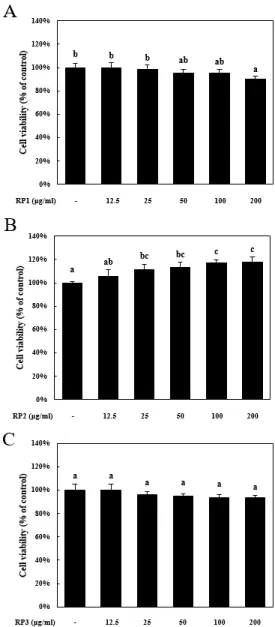
추출물의 Raw 264.7 세포에 대한 세포독성 평가 각 추출의 세포독성을 평가하기 위해 Raw 264.7 세포주를 이 용하여 추출물의 농도별(12.5, 25, 50, 100, 그리고 200 ㎍/㎖) 세포 생존율을 측정하였다. 각 시료별 처리 24간 후 정상대조군 의 생존율에 비례하여 세포독성을 판단하였다(Fig. 1). RP1(아
까시 나무 물 추출물)은 200 ㎍/㎖ 농도에서 정상대조군에 비해 세포생존율이 90.2 ± 2.6%로 유의적으로 감소하였다. RP2(아 까시 나무 에탄올 추출물)은 세포독성이 나타나지 않았으며, RP3(아까시 나무 고온 추출물)은 정상대조군에 비해 세포생존 율이 최고농도인 200 ㎍/㎖ 농도에서 93.5 ± 2.1% 수준으로, 최 저농도(12.5 ㎍/㎖; 세포생존율, 96.4 ± 2.4%)부터 농도 의존적
Fig. 1. Cytotoxic evaluation of extracts on Raw 264.7 cells.
Raw 264.7 cells were treated with extracts (RP1; water extract, RP2; ethanol extract and RP3; high temperature extract) for various concentration. Cell viability was measured by MTS assay. Means values ± SD from triplicate separated experiments are shown. *Means with difference letters are significantly different at p<0.05 by duncan’s multiple range test.
으로 감소되는 경향을 나타내었지만 이는 유의적인 차이가 나 타나지 않았다(Fig. 1). 이는 아까시 추출물(RP1, RP2 그리고 RP3)이 Raw 264.7 세포주에 대해 큰 세포독성을 나타내지 않는 다고 생각되며, 이 결과를 바탕으로 실험 농도를 세포독성이 크 게 나타나지 않는 12.5, 25, 50, 100, 그리고 200 ㎍/㎖로 정하고 여러 실험을 진행하였다.
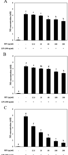
추출물의 NO(nitric oxide)생성 억제효과 분석
아까시 나무 추출물이 LPS 처리에 의해 증가되는 nitric oxide 생성에 미치는 효과를 확인하기 위해 NO assay를 진행하 였다. 추출물은 앞서 세포생존율 실험에서 정한 12.5, 25, 50, 100, 그리고 200 ㎍/㎖ 농도로 전처리한 뒤 LPS를 500 ng/㎖의 농도로 24시간 동안 처리했다. LPS 단독 처리군은 정상대조군 에 비교해 NO 생성량이 각각 실험에서 유의적으로 증가하였다.
RP1 (아까시 나무 물 추출물)은 200 ㎍/㎖ 농도에서 NO 생성이 3.09 ± 0.35 nM로 LPS 단독 처리군에 비해 약 26% 정도 감소하 였고(Fig. 2A), RP2(아까시 나무 에탄올 추출물)은 3.29 ± 0.05 nM로 약 24% 감소하였다(Fig. 2B). RP3 (아까시 나무 고온 추출 물)은 최저농도인 12.5 ㎍/㎖의 농도에서 NO 생성이 3.26 ± 0.20 nM로 최소 농도부터 NO생성을 억제하는 효과를 나타냈 고, 200 ㎍/㎖ 농도에서 NO 생성이 0.62 ± 0.23 nM로 LPS 단독 처리군에 비해 약 87% 정도 감소하였다(25 ㎍/㎖; 2.29 ± 0.10 nM, 50 ㎍/㎖; 1.45 ± 0.02 nM, 100 ㎍/㎖; 0.89 ± 0.05 nM).
결과를 종합하면 추출물 중 고온 추출물이 LPS 처리에 의해 증 가된 NO의 생성을 가장 많이 억제하였으며 효과는 농도의존적 으로 나타났다. 앞서 언급한 것처럼 NO는 과하게 생성될 시 염 증반응을 매개할 뿐만 아니라, 세포독성을 나타내기 때문에 숙 주방어 작용에서 체내 세포에 세포독성을 일으키는 매개체로 작용할 수 있다(Kim and Kim, 2002; Blaise et al., 2005). 이 결과를 바탕으로 아까시 고온 추출물(RP3)이 물 추출물(RP1)과 에탄올 추출물(RP2)보다 NO 억제 활성이 가장 높은 것으로 생 각되지만, 이는 Raw 264.7 세포주 내에 생성된 NO를 확인한 결과로 NO 합성에 관여하는 nitric oxide synthase의 활성 또 는 단백질 발현량을 추가로 확인하여 추출물이 어떤 단계에서 작용하는지 알아보는 추가적인 실험이 필요할 것이라고 생각 된다.
추출물의 PGE2 생성 억제효과 분석
다음으로 아까시 나무 추출물이 LPS 처리에 의해 증가되는 PGE2 생성을 억제할 수 있는지 확인하기 위해 PGE2에 대한
ELISA assay를 수행했다. 앞서 실험들과 마찬가지로 추출물들 을 각각 12,5 25, 50, 100, 그리고 200 ㎍/㎖ 농도로 Raw 264.7 세포주에 전 처리한 뒤 LPS를 500 ng/㎖의 농도로 24시간 동안 처리하였다. PGE2의 농도를 확인했을 때, RP1은 LPS 단독 처리 군의 PGE2 농도가 2714.1 ± 18.8 pg/㎖로 증가하였지만 200 ㎍/
Fig. 2. Effect of extracts on LPS-stimulated NO production in Raw 264.7 cells. After pretreatment with 500 ng/㎖ LPS (lipopolysaccharide) for 30 min, Raw 264.7 cells were treated with various concentration of extract (RP1; water extract, RP2;
ethanol extract and RP3; high temperature extract) for 24 h.
Culture medium was subjected to NO assay. Data represent the mean ± SEM of three independent experiments. *Means with difference letters are significantly different at p<0.05 by duncan’s multiple range test.
㎖ 농도에서 856.5 ± 21.5 pg/㎖로 약 68.4% 정도 감소하였다 (Fig. 3A). RP2는 831.9 ± 49.6 pg/㎖ 농도로 LPS 단독 처리군 대비 70.6% 정도(Fig. 3B), RP3는 농도별 PGE2 억제율이 각각
12.5 ㎍/㎖; 1836.70 ± 18.90 pg/ml, 25 ㎍/㎖; 959.33 ± 56.68, 50 ㎍/㎖; 864.99 ± 56.68, 100 ㎍/㎖; 827.54 ± 73.23로 나타 났으며, 최저농도인 12.5 ㎍/㎖를 처리했을 때 약 32.3%의 억제 효과가 나타나기 시작했고 최고농도인 200 ㎍/㎖농도는 317.7
± 16.0 pg/㎖ 농도로 LPS 단독 처리군 대비 86.3% 억제효과를 나타냈다(Fig. 3C). 결과를 종합하면 RP3(아까시 나무 고온 추 출물)의 PGE2 억제 효과가 가장 좋은 것으로 나타났다. 앞서 언 급했던 추출방법에 따른 유효성분 변화에 관한 연구처럼(Woo et al., 2010), 이 결과도 추출법에 따라 큰차이는 아니지만 효능 의 차이가 나타나는 것으로 확인된다. 하지만 이 결과를 바탕으 로 PGE2의 대사에 관여하는 COX2 (cyclooxygenase 2)의 단백질 발현량과 활성도를 확인하는 추가 실험이 필요하다고 생각된다.
추출물의 TNF–α 생성 억제효과 분석
아까시 나무 추출물들이 LPS 처리에 의해 증가되는 TNF-α 농도를 감소시킬 수 있는지 확인하기 위해 TNF-α에 대한 ELISA assay를 수행했다. 앞서 실험들과 마찬가지로 추출물들을 각각 12,5 25, 50, 100, 그리고 200 ㎍/㎖ 농도로 Raw 264.7 세포주에 전 처리한 뒤 LPS를 500 ng/㎖의 농도로 24시간 동안 처리하였 다. PGE2의 농도를 확인했을 때, RP1은 LPS 단독 처리군의 TNF-α 농도가 3782.3 ± 117.8 pg/㎖로 증가되었을 때 1817.5
± 218.0 pg/㎖의 농도로 감소하여 약 51.9% 정도 감소되었다 (Fig. 4A). RP2의 TNF-α 농도는 1539.0 ± 130.3 pg/㎖로 LPS 단독 처리군에 비해 약 59.3% 정도(Fig. 4B), RP3는 처리농도별 TNF-α의 수치가 12.5 ㎍/㎖; 2861.43 ± 155.96 pg/㎖, 25 ㎍/
㎖; 2129.96 ± 73.89 pg/㎖, 50 ㎍/㎖; 1899.05 ± 96.16 pg/㎖, 100 ㎍/㎖; 1717.05 ± 113.13 pg/㎖로 나타났다. 최저농도인 12.5
㎍/㎖를 처리했을 때 약 24.4%의 억제효과가 나타나기 시작했 으며 최고농도인 200 ㎍/㎖에서 TNF-α의 농도가 1354.5 ± 74.1 pg/㎖로 약 64.1% 정도 감소되었다(Fig. 4C). TNF-α는 감 염, 패혈증 또는 류마티스 관절염과 같은 만성 염증성질환에서 농도가 증가된다고 알려져 있으며, 세포외부에 존재하는 TNFR1 (tumor necrosis factor receptor 1)과 TNFR2 (tumor necrosis factor receptor 2)와 상호작용하여 염증관련 신호전달을 촉진 시킨다(Bradley, 2008; Popa et al., 2007). 이 결과를 바탕으로 LPS로 유도시킨 염증반응에서 RP3(아까시 나무 고온 추출물)이 TNF-α의 농도를 억제시키는 효과가 가장 크게 나타났으며, 이 는 고온 추출시 물 추출과 에탄올 추출에 비해 아까시 나무의 유 효 성분 추출 비율에 영향을 미칠 수 있다고 생각되지만 이는 성 분 분석실험과 같은 추가적인 연구가 필요할 것이라고 생각된다.
Fig. 3. Effect of extracts on LPS-stimulated PGE2 production in Raw 264.7 cells. After pretreatment with 500 ng/㎖ LPS (lipopolysaccharide) for 30 min, Raw 264.7 cells were treated with various concentration of extract (RP1; water extract, RP2;
ethanol extract and RP3; high temperature extract) for 24 h.
Culture medium was subjected to ELISA assay. Data represent the mean ± SEM of three independent experiments. *Means with difference letters are significantly different at p<0.05 by duncan’s multiple range test.
추출물의 IL-6 생성 억제효과 분석
다음으로 아까시 나무 추출물들이 LPS 처리에 의해 증가되 는 IL-6농도를 감소시킬 수 있는지 확인하기 위해 IL-6에 대한 ELISA assay를 수행했다. 앞선 실험들과 마찬가지로 추출물들
을 각각 12,5 25, 50, 100 그리고 200 ㎍/㎖ 농도로 Raw 264.7 세포주에 전 처리한 뒤 LPS를 500 ng/㎖의 농도로 24시간 동안 처리하였다. IL-6의 농도를 확인했을 때, RP1은 LPS 단독 처리 군의 IL-6 농도가 611.3 ± 12.4 pg/㎖로 증가되었을 때, 239.0 Fig. 4. Effect of extracts on LPS-stimulated TNF–α production
on Raw 264.7 cells. After pretreatment with 500 ng/㎖ LPS (lipopolysaccharide) for 30 min, Raw 264.7 cells were treated with various concentration of extract (RP1, RP2 and RP3) for 24 h. Culture medium was subjected to ELISA assay. Data represent the mean ± SEM of three independent experiments.
*Means with difference letters are significantly different at p<0.05 by duncan’s multiple range test.
Fig. 5. Effect of extracst on LPS-stimulated IL-6 production on Raw 264.7 cells. After pretreatment with 500 ng/㎖ LPS (lipopolysaccharide) for 30 min, Raw 264.7 cells were treated with various concentration of extract (RP1, RP2 and RP3) for 24 h. Culture medium was subjected to ELISA assay. Data represent the mean± SEM of three independent experiments.
*Means with difference letters are significantly different at p<0.05 by duncan’s multiple range test.
± 9.8 pg/㎖으로 감소비율이 약 60.9%로 확인되었다(Fig. 5A).
반면 RP2는 213.2 ± 1.5 pg/㎖로 감소비율이 약 65.1%로(Fig.
5B), RP3는 처리 농도별 IL-6의 수치가 각각 12.5 ㎍/㎖; 529.89
± 20.74 pg/㎖, 25 ㎍/㎖; 473.06 ± 10.51 pg/㎖, 50 ㎍/㎖;
339.71 ± 13.47 pg/㎖, 100 ㎍/㎖; 246.15 ± 10.12 pg/㎖로 나타 났다. 최저 농도인 12.5 ㎍/㎖에서 IL-6의 억제율이 LPS처리군 과 비교하였을 때 약 13%로 최고농도인 200 ㎍/㎖는 186.4 ± 14.1 pg/㎖로 감소비율이 69.5%로 나타났다(Fig. 5C). 결과를 종합하면 앞선 결과들과 마찬가지로 감소비율의 큰 차이는 나 타나지 않았지만 고온추출에 의해 얻어진 RP3가 가장 억제효과 가 큰 것으로 확인 되었을 뿐만 아니라 농도의존적으로 IL-6의 수치를 감소시켰다.
추출물의 IL-1β 생성 억제효과 분석
마지막으로 추출물들이 LPS 처리에 의해 분비되는 IL-1β농 도를 감소시킬 수 있는지 확인하기 위해 IL-1β에 대한 ELISA assay를 세포배양액을 이용해 실험을 수행하였다. 앞선 실험들 과 마찬가지로 추출물들을 각각 12,5 25, 50, 100, 그리고 200
㎍/㎖ 농도로 Raw 264.7 세포주에 전 처리한 뒤 LPS를 500 ng/㎖
의 농도로 24시간 동안 처리하였다. RP1은 LPS 단독 처리군의 IL-1β의 농도가 352.6 ± 16.9 pg/㎖로 증가되었을 때, 86.4 ± 15.7 pg/㎖이며 감소비율이 약 75.4%로 나타났다(Fig. 6A). RP2 는 IL-1β 농도가 29.1 ± 26.6 pg/㎖이며 감소비율이 약 63.3%
로(Fig. 6B), RP3는 처리 농도별 IL-1β 수치가 각각 12.5 ㎍/㎖;
283.54 ± 15.84 pg/㎖, 25 ㎍/㎖; 214.41 ± 14.73 pg/㎖, 50 ㎍/㎖;
143.16 ± 26.63 pg/㎖, 100 ㎍/㎖; 105.16 ± 7.07 pg/㎖로 나타 났다. 최저 농도인 12.5 ㎍/㎖에서 IL-1β의 억제율이 LPS처리 군과 비교하였을 때 약 19%로, 최고농도인 200 ㎍/㎖ 61.7 ± 7.8 pg/㎖로, 감소비율이 약 82.4%로 확인되었다.
IL-1β는 염증조절복합체(inflammasome)에 의해 조절되는 염증매개인자로써 NLRP3 (NACHT, LRR and PYD domains- containing protein 3), ASC (PYCARD, Apoptosis-associated speck-like protein containing a CARD), caspase-1과 같은 단 백질들에 의해 조절되고 류마티스 관절염이나 통풍성 관절염같 은 면역질환에서 증가된다고 알려져 있다(Borthwick, 2016;
Ren and Torres, 2009). 모든 결과를 종합하였을 때 RP3 (아까 시 나무 고온 추출물)은 LPS에 의해 증가된 NO의 생성, 염증 매 개인자(IL-6, IL-1β, TNF-α 그리고 PGE2)를 억제하는 것으로 확인되었으며, 이를 통해 염증반응을 개선시킬 수 있는 천연 추 출물로써 이용될 수 있다고 생각한다. 하지만 Raw 264.7 세포
주는 염증조절복합체를 구성하는 ASC 단백질이 발현되지 않고 다른 경로를 통해 IL-1β를 분비하기 때문에(Pelegrin et al., 2008; Ahn et al., 2017), 설치류에서 직접 macrophage를 일차
Fig. 6. Effect of extracts on LPS-stimulated IL-1β production in Raw 264.7 cells. After pretreatment with 500 ng/㎖ LPS (lipopolysaccharide) for 30 min, Raw 264.7 cells were treated with various concentration of extract (RP1, RP2 and RP3) for 24 h. Culture medium was subjected to ELISA assay. Data represent the mean± SEM of three independent experiments.
*Means with difference letters are significantly different at p<0.05 by duncan’s multiple range test.
배양(primary culture)하여 관련 신호전달 경로를 확인하는 추 가 실험이 필요할 것이라고 생각된다.
적 요
본 연구는 아까시나무의 물 추출물, 에탄올 추출물 및 고온 추출물을 이용하여 마우스 대식세포주인 Raw 264.7 세포에 대 해 염증억제 효과가 있는지 알아보고자 수행하였다. RP1(아까 시 나무 물 추출물), RP2(아까시 나무 에탄올 추출물) 및 RP3(아 까시 나무 고온 추출물)은 세포생존율 분석에서 200 ㎍/㎖의 농 도까지 Raw 264.7 세포에 세포독성을 나타나지 않았다. NO 생 성 억제효과를 분석하였을 때 LPS 처리군 과 비교하여 RP3는 약 87% 정도의 억제효과를 나타내 RP1과 RP2에 비해 NO 억제 활성이 가장 높았다. 뿐만 아니라 RP3는 RP1과 RP2와 비교하여 염증매개인자의 억제율이 각각 PGE2 (86.3%), TNF-α (64.1%), IL-6 (65.1%) 및 IL-1β (63.3%)로 가장 높았다. 이는 RP3의 처 리가 LPS에 의해 증가된 염증매개인자의 분비를 억제함을 통해 항염증 효과가 있을 것으로 생각되며, 염증관련 신호전달경로 에 직접적으로 작용할 가능성이 있는 것으로 판단된다. 하지만 Raw 264.7 세포주는 염증조절복합체를 구성하는 ASC 단백질 이 발현되지 않아 다른 신호전달 경로를 통해 염증매개인자를 분비하기 때문에, 설치류의 대식세포를 직접 일차배양(primary culture)하여 이에 관련된 신호전달경로를 확인하는 추가 실험 이 필요하다고 사료된다.
References