ISSN 1225-6552, eISSN 2287-7630 https://doi.org/10.7853/kjvs.2017.40.1.1
< Original Article >
Veterinary Service
Available online at http://kjves.org
*Corresponding author: Bo-Young Jeon, Tel. +82-33-760-5108, Fax. +82-33-760-2561, E-mail. [email protected]
Random amplified polymorphic DNA analysis of bacterial pathogens using universal rice primers
Sezim Monoldorova1, Jinsol Kim1, Joon Hee Kim2, Bo-Young Jeon1*
1Department of Biomedical Laboratory Science, College of Health Science, Yonsei University, Wonju 26493, Korea
2School of Food Science and Technology, College of Biotechnology & Natural Resource, Chung-Ang University, Seoul 06974, Korea
(Received 10 January 2017; revised 15 March 2017; accepted 15 March 2017)
Abstract
Molecular typing of pathogenic microorganisms is important for epidemiological investigation of in- fectious disease outbreaks. In this study, we applied Universal Rice Primers (URP) that were originated from repetitive sequences in rice chromosomal DNA to random amplified polymorphic DNA (RAPD) analysis of pathogenic bacteria such as Escherichia coli, Listeria monocytogenes, and Salmonella sp. Of the twelve URP primers examined to date, seven primers (URP-2, -3, -4, -5, -6, -8, and -9) generated reproducible and polymorphic PCR products ranging from 1 to 13 bands. One of them, URP-6 was very effective in differentiating seven E. coli serotypes, seven L. monocytogenes clinical isolates, and eight Salmonella subspecies (ssp.) serovars. The results thus indicate that RAPD analysis using URP primers might be useful in typing bacterial pathogens including E. coli, L. monocytogenes, and Salmonella strains.
Key words : RAPD analysis, URP primers, E. coli, Listeria monocytogenes, Salmonella
INTRODUCTION
Identification of bacterial pathogens at the strain or subspecies level is important for epidemiological studies (Chang, 2013; Ranjbar et al, 2014). In Escherichia coli, Listeria monocytogenes, and Salmonella subspecies (ssp.), distinction by serological and biochemical methods is sometimes difficult (Liu et al, 2016; Chen et al, 2011).
Therefore, rapid and easy-to-perform typing methods within strains or serovars are of great value for epidemiological investigations during outbreaks, and for planning specif- ic and effective control measures.
In recent years, molecular typing of pathogenic bac- teria has been widely used in epidemiological studies.
Common typing methods include restriction fragment length polymorphism (RFLP), multilocus enzyme elec- trophoresis, pulse-field gel electrophoresis, ribotyping,
and variable number tandem repeat typing (Chen et al, 2011; Ranjbar et al, 2014; Liu et al, 2016). In addition, random amplified polymorphic DNA (RAPD) analysis has been widely explored for molecular typing (Poursha- fie et al, 2008; Grover et al, 2016). RAPD analysis has also been called arbitrarily primed PCR (AP-PCR) and is based on genomic DNA amplification using a single primer with an arbitrary nucleotide sequence (Waldron et al, 2002). Multiple PCR products resulting from RAPD analysis are separated by size using conventional agarose gel electrophoresis. The DNA banding patterns of different isolates can then be compared. RAPD analy- sis has been widely used to differentiate between bacte- rial strains, because it has a good discriminatory ability, is faster, relatively simple, and more economical than other genomic typing methods (Franklin et al, 1999;
Reinoso and Bettera, 2016). Because RAPD analysis rapidly generates molecular fingerprints, it is suitable for
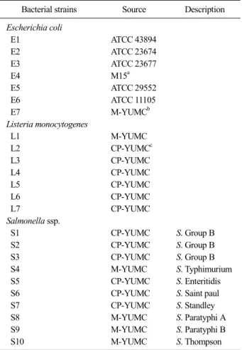
Table 1. Bacterial strains used in this study
Bacterial strains Source Description Escherichia coli
E1 ATCC 43894
E2 ATCC 23674
E3 ATCC 23677
E4 M15a
E5 ATCC 29552
E6 ATCC 11105
E7 M-YUMCb
Listeria monocytogenes
L1 M-YUMC
L2 CP-YUMCc
L3 CP-YUMC
L4 CP-YUMC
L5 CP-YUMC
L6 CP-YUMC
L7 CP-YUMC
Salmonella ssp.
S1 CP-YUMC S. Group B
S2 CP-YUMC S. Group B
S3 CP-YUMC S. Group B
S4 M-YUMC S. Typhimurium
S5 CP-YUMC S. Enteritidis
S6 CP-YUMC S. Saint paul
S7 CP-YUMC S. Standley
S8 M-YUMC S. Paratyphi A
S9 M-YUMC S. Paratyphi B
S10 M-YUMC S. Thompson
aPurchased from Qiagen (Germantown, MD, USA).
bOriginated from Department of Microbiology, College of Medicine, Yonsei University.
cOriginated from Department of Clinical Pathology, College of Medicine, Yonsei University.
large-scale screening of bacterial isolates for epidemio- logical monitoring, provided that suitable primers are available for each pathogen.
Kang et al. (2002) reported that Universal Rice Primers (URP), originally derived from repetitive sequences in the rice genome, gave amplification profiles with high reproducibility in plants. These primers provided higher reproducibility than arbitrary 10-mer primers those have been commonly used in current RAPD analysis (Lim et al, 2007; Lim et al, 2009) because URP primers can be used under highly stringent PCR conditions, specifically wit an annealing temperature higher than 55°C. There are some reports of RAPD analysis using URP primers in eukaryotes, including mushroom, Allium spp., potato, and fish (Lim et al, 2007; Kim et al, 2007; Lim et al, 2009). We assumed that RAPD analysis using UPR pri- mers would be useful for the molecular typing of bacte- rial pathogens.
In the present study, we performed RAPD analysis using UPR primers to develop an effective molecular typing method on bacterial pathogens, such as E. coli, L. monocytogenes, and Salmonella spp.
MATERIALS AND METHODS
Bacterial strains
The bacterial strains investigated in this study were listed in Table 1 and included seven strains of E. coli, seven strains of L. monocytogenes, and nine strains of Salmonella ssp. The organisms were obtained from the American Type Culture Collection (ATCC) (Rockville, MD, USA) and the Departments of Clinical Pathology and Microbiology at Yonsei University College of Medicine. E. coli M15 strain was purchased from Qiagen (Germantown, MD, USA).
Preparation of purified bacterial genomic DNA Genomic DNA was prepared from bacteria using ce- tyl trimethyl ammonium bromide (CTAB) as described (Murray and Thompson, 1980). Briefly, the bacterial strains were cultured in Luria Bertani (LB) broth over-
night and harvested by centrifugation at 12,000×g for 5 min. The pellet was resuspended in 567 L TE buffer (10 mM Tris-HCl, 1 mM EDTA, [pH8.0]). Thirty L 20% sodium dodecyl sulfate and 3 L proteinase K (20 mg/ml in distilled water) were added to the suspension and incubated at 37°C for 1 hour. The reaction mixture was incubated with 100 L 5M NaCl and 80 L CTAB-NaCl solution (10% CTAB in 0.7M NaCl) at 65°C for 10 min. The solution was extracted with phe- nol-chloroform-isoamyl alcohol (25:24:1) solution, and DNA was precipitated with isopropanol by incubation at
−20°C for 2 hours. Finally, the DNA was harvested by centrifugation at 12,000 ×g for 20 min, washed with 70% ethanol, and resuspended in 30 L of TE buffer.
RNA was removed by incubating the solution with
Fig. 1.RAPD patterns for screening URP primers with E. coli E1 gemomic DNA. Lane M: 1 kb ladder; lanes 1∼12: URP primers (1∼
12).
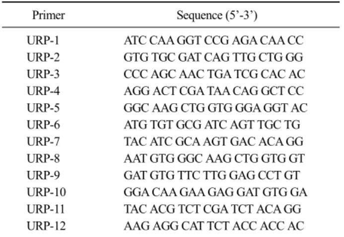
Table 2. Oligonucleotide sequences of Universal Rice Primers used in this study
Primer Sequence (5’-3’)
URP-1 ATC CAA GGT CCG AGA CAA CC URP-2 GTG TGC GAT CAG TTG CTG GG URP-3 CCC AGC AAC TGA TCG CAC AC URP-4 AGG ACT CGA TAA CAG GCT CC URP-5 GGC AAG CTG GTG GGA GGT AC URP-6 ATG TGT GCG ATC AGT TGC TG URP-7 TAC ATC GCA AGT GAC ACA GG URP-8 AAT GTG GGC AAG CTG GTG GT URP-9 GAT GTG TTC TTG GAG CCT GT URP-10 GGA CAA GAA GAG GAT GTG GA URP-11 TAC ACG TCT CGA TCT ACA GG URP-12 AAG AGG CAT TCT ACC ACC AC
RNAse I (1 g/mL) at 37°C for 2 hours. The DNA concentration was measured with a spectrophotometer (Pharmacia Biotech., Piscataway, NY, USA) at 260 nm.
The DNA was kept at −20°C until use.
Primers and PCR amplification
The PCR amplification for RAPD analysis was done with URP primers (Table 2, SRILS UniPrimerTM Kit, Seoulin Bioscience, Seoul, Korea). The PCR reaction was performed with a 50 L PCR mixture containing 10 mM Tris-HCl, 50 mM KCl, 1.5 mM MgCl2, 0.01%
gelatin, 200 M of each dNTP, 200 ng primer, 2.5 unit Taq DNA polymerase (Perkin-Elmer Biosystems, San Francisco, CA, USA), and 50 ng genomic DNA as a template. PCR amplification was carried out in a Perkin-Elmer 9600 thermocycler (Norwalk, CT, USA) using the following reaction profile (one cycle of 4 min at 94°C, 35 cycles of 1 min at 94°C, 1 min at 55°C, 2 min at 72°C; one final extension for 7 min at 72°C).
The PCR reaction was repeated twice. The PCR prod- ucts were analyzed by electrophoresis in 1.8% agarose gel by staining with ethidium bromide.
GelCompar analysis of DNA fingerprints
The DNA fingerprint banding patterns were scanned and analyzed with GelCompar software (AAB Software, Fullerton, CA, USA). Degrees of homology were de- termined by Dice comparisons, and clustering correlation
coefficients were calculated by the unweighted pair group method with arithmetic averages.
RESULTS
Selection of informative URP primers
To identify primers that generate informative PCR profiles, twelve URP 20-mer primers were examined with E. coli E1 (ATCC 43894) genomic DNA. Of the twelve primers, seven (URP-2, -3, -4, -5, -6, -8, and -9) produced informative profiles (Fig. 1), and among them three primers (URP-4, -5, -8) generated one or two PCR products. Five primers (URP-1, -7, -10, -11, -12) did not produce PCR products. Among the seven primers that gave a profile, URP-6 was selected as a represent in the following experiments to differentiate E. coli, L.
monocytogenes, and Salmonella strains because URP-6 showed the regular polymorphic bands to L. mono- cytogenes and Salmonella strains (data not shown).
RADP analysis of E. coli strains using URP-6 RAPD fingerprint patterns of seven E. coli strains were analyzed using URP-6 (Fig. 2). E. coli genomic DNA amplification using URP-6 resulted in 13 distinct DNA bands ranging from approximately 170 bp to 2.6
Fig. 2. (A) RAPD fingerprint profiles of E. coli strains with URP-6.
Lane M: lambda DNA/EcoRI+HindIII; lanes 1∼7: E. coli E1∼7. (B) GelCompar software analysis of E. coli strains with URP-6. Clustering was done with the unweighted pair group with arithmetic averages (UPGMA) algorithm using fine correlation on gel tracks.
Fig. 3.(A) RAPD fingerprint profiles of L. monocytogenes with URP-6. Lanes M: 1kb DNA ladder; lane 1∼7: L. monocytogenes L1∼7. (B) GelCompar software analysis of L. monocytogenes with URP-6. Clustering was done with UPGMA algorithm using fine cor- relation on gel tracks.
kb. Interestingly, in the RAPD pattern of E. coli E1 strain, there was a distinctive band (750 bp) in lane 2, 3, 4, and 7. RAPD profile analysis by the Pearson product-moment correlation method and the unweigh- ted-pair-group method with arithmetic averages cluster- ing clearly distinguished six strains of E. coli (Fig. 2B).
One E. coli strain (E7) gave the same pattern as E. coli E4 strain (M15 strain).
RADP analysis of L. monocytogenes using URP-6 Genomic DNA amplification of seven strains of L.
monocytogenes using URP-6 resulted in RAPD patterns consisting of 13 DNA bands ranging from 220 bp to 2.1 kb (Fig. 3). Analysis of the RAPD profiles clearly distinguished two clusters with a similarity coefficient of 54% (Fig. 3B). A similarity coefficient was between 67%
and 85% for these clusters. Therefore, the results showed that URP-6 could clearly distinguish six strains of L.
monocytogenes.
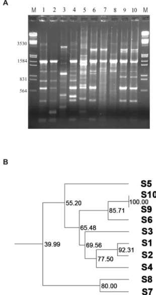
RAPD analysis of Salmonella ssp. using URP-6 Genomic DNA amplification of ten Salmonella ssp.
strains using URP-6 resulted in RAPD patterns consist- ing of 19 DNA bands ranging from approximately 0.2
Fig. 4. (A) RAPD fingerprint profiles of Salmonella strains with URP-6. Lanes M: lambda DNA/EcoRI+HindIII; lanes 1∼3:
Salmonella S5∼7; lane 4: S3; lane 5: S10; lanes 6∼7: S8∼9; lanes 8
∼9: S1∼2; lane 10: S4. (B) GelCompar software analysis of Salmonella strains with URP-6. Clustering was done with UPGMA al- gorithm using fine correlation on gel tracks.
to 3.0 kb (Fig. 4). PCR products from the Salmonella ssp. strains using URP-6 showed that all except Salmonella S3 strain shared a 1,570 bp DNA band. Strains belong- ing to Salmonella Group B, Salmonella S2 (S. Paratyphi B) and S4 (S. Thompson) gave a cluster with a nearly congruent pattern, thus indicating a close relationship. In analysing the RAPD profiles, a coefficient of similarity between 40 and 92% was found for seven of the eight strains (Fig. 4B).
DISCUSSION
The high sensitivity and specificity of PCR-based mo- lecular techniques have made a revolutionary impact on the diagnosis and epidemiology of infectious diseases.
RAPD fingerprinting technique is a particularly powerful typing method utilizing arbitrary oligonucleotides (arbitrary PCR) to prime DNA synthesis at a low annealing tem- perature, divulging genomic diversity. Although arbitrary PCR is a powerful and general tool, it has low reprodu- cibility (Kim et al, 2010; Ranjbar et al, 2014; Soler-García et al, 2014).
We found that RAPD analysis using UPR primers could be applied to bacterial pathogens, though these primers were derived from repetitive sequences in rice genomes (Kang et al, 2002; Lim et al, 2007). RAPD analysis using URP primers on bacterial pathogens such as E. coli, Salmonella ssp., and Listeria monocytogenes, was specific and reproducible (data not shown). The re- production of the RAPD analysis using URP primers may be due to the more stringent PCR conditions (55°C annealing temperature) (Jana et al, 2005; Lim et al.
2007).
As a representative of URP primers, RAPD analysis using URP-6 was powerful to recognize differences be- tween bacterial pathogens, such as E. coli, L. mono- cytogenes, and Salmonella ssp. at the strain or sub- species level. This result is consistent with other reports that RAPD analysis using URP-6 showed a polymorphic DNA band pattern from the genus Aquilegia (Kang et al, 2002; Kim et al, 2007). RAPD analysis with URP-6 might be used to discriminate other pathogenic bacteria.
CONCLUSION
In conclusion, RAPD analysis using URP-6 is a reli- able, reproducible, and rapid strategy to distinguish the pathogenic bacteria, including E. coli, L. monocytogenes, and Salmonella ssp.
COMPETING INTERESTS
The authors declare that they have no competing interests.
ACKNOWLEDGMENTS
We thank Dr. Yoon-Seop Jeong for kind providing the bacterial strains, and Prof. Hyeyoung Lee and Prof.
Sang-Nae Cho for helpful discussion and critical reading of the manuscript. This work was supported by Basic Science Research Program through the National Research Foundation of Korea (NRF) funded by the Ministry of Education, Science and Technology (No. NRF-2014R1- A1A2055172).
REFERENCES
Chang K. 2013. Molecular Epidemiology of Cryptococcus neofor- mans/Cryptococcus gattii Complex Isolates from Pigeon Droppings in Korea. J Exp Biomed Sci 19: 213-223.
Chen S1, Li J, Saleh-Lakha S, Allen V, Odumeru J. 2011.
Multiple-locus variable number of tandem repeat analy- sis (MLVA) of Listeria monocytogenes directly in food samples. Int J Food Microbiol 148: 8-14.
Franklin RB, Taylor DR, Mills AL. 1999. Characterization of mi- crobial communities using randomly amplified poly- morphic DNA (RAPD). J Microbiol Methods 35: 225- 235.
Grover A, Sharma PC. 2016. Development and use of molecular markers: past and present. Crit Rev Biotechnol 36:
290-302.
Jana TK, Singh NK, Koundal KR, Sharma TR. 2005. Genetic dif- ferentiation of charcoal rot pathogen, Macrophomina phaseolina, into specific groups using URP-PCR. Can J Microbiol 51: 159-164.
Kang HW, Park DS, Go SJ, Eun MY. 2002. Fingerprinting of di- verse genomes using PCR with universal rice primers
generated from repetitive sequence of Korean weedy rice. Mol Cells 13: 281-287.
Kang HW, Park DS, Park YJ, Lee BM, Cho SM, Kim KT, Seo GS, Go SJ. 2002. PCR Based Detection of Phellinus lin- teus using Specific Primers generated from Universal Rice Primer (URP) Derived PCR Polymorphic Band.
Mycobiology 30: 202-207.
Kim DM, Kwon JY, Lim KB, Kim HH. 2007. Genetic Relationship of 12 Taxa of Genus Aquilegia Using URP- RAPD. Flower Res J 15: 58-63.
Kim SH, Cho SW, Hwang SY, Jeon SY, Kim YK. 2010.
Molecular Identification Patterns of Clinical Isolates from Korean Patients Infected with Dermatophytes. J Exp Biomed Sci 16: 187-192.
Lim SH, Kim JG, Kang HW. 2009. Novel SCAR primers for spe- cific and sensitive detection of Agrobacterium vitis strains. Microbiol Res. 164: 451-460.
Liu Y, Shi X, Li Y, Chen Q, Jiang M, Li W, Qiu Y, Lin Y, Jiang Y, Kan B, Sun Q, Hu Q. 2016. The evaluation and ap- plication of multilocus variable number tandem repeat analysis (MLVA) for the molecular epidemiological study of Salmonella enterica subsp. enterica serovar En- teritidis infection. Ann Clin Microbiol Antimicrob 15: 4.
Pourshafie MR, Saifi M, Mousavi SF, Sedaghat M, Nikbakht GH, Rubino S. 2008. Clonal diversity of Salmonella enterica serotype Typhi isolated from patients with typhoid fever in Tehran. Scand J Infect Dis 40: 18-23.
Ranjbar R, Karami A, Farshad S, Giammanco GM, Mammina C.
2014. Typing methods used in the molecular epidemiol- ogy of microbial pathogens: a how-to guide. New Mi- crobiol 37: 1-15.
Reinoso EB, Bettera SG. 2016. Random amplified polymorphic DNA PCR in the teaching of molecular epidemiology.
Biochem Mol Biol Educ 44: 391-396.
Soler-García AA, De Jesús AJ, Taylor K, Brown EW. 2014.
Differentiation of Salmonella strains from the SARA, SARB and SARC reference collections by using three genes PCR-RFLP and the 2100 Agilent Bioanalyzer.
Front Microbiol 5: 417.
Waldron J, Peace CP, Searle IR, Furtado A, Wade N, Findlay I, Graham MW, Carroll BJ. 2002. Randomly Amplified DNA Fingerprinting: A Culmination of DNA Marker Technologies Based on Arbitrarily-Primed PCR Am- plification. J Biomed Biotechnol 2: 141-150.