• 검색 결과가 없습니다.

Nailbed malignant melanoma in three dogs

N/A
N/A
Protected

Academic year: 2021

Share "Nailbed malignant melanoma in three dogs"

Copied!
4
0
0

로드 중.... (전체 텍스트 보기)

전체 글

(1)

pISSN 2466-1384 eISSN 2466-1392 Korean J Vet Res (2019) 59(4):219~222 https://doi.org/10.14405/kjvr.2019.59.4.219

219 CASE REPORT

Nailbed malignant melanoma in three dogs

Hyoung-Nam Jo

1,†

, Myeong-Won Suh

1,†

, Joo-Ok Lee

2

, Jae-Hoon Kim

1,

*

1

College of Veterinary Medicine and Veterinary Medical Research Institute, Jeju National University, Jeju 63243, Korea

2

Ekklim Anima Hospital, Daegu 41165, Korea

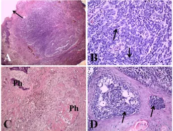
Abstract: Three 8-12-year-old male cocker spaniels presented with an appendicular mass accompanied by pain, inflammation, lameness, and loss of nail in the digits of the forelimb or hindlimb. A histological examination revealed dermal masses of the digit composed of numerous neoplastic cells with marked pleomorphism and high mitosis. The neoplastic cells showed a strong invasive tendency into the epidermis and adjacent bony tissues, such as distal phalanx. Immunohistochemistry of the neoplastic cells in three masses revealed positive reactions for Melan-A. These three dogs were diagnosed as nailbed malignant melanoma in the digit based on the history, clinical signs, and histopathologic features.

Keywords: digit, distal phalanx, dog, Melan-A, nailbed malignant melanoma

Melanoma is a malignant tumor arising from melanocytes, which produce melanin and are derived from neuroectodermal melanoblasts in the epidermis or adnexa structures, but primarily from the external root sheath of the hair follicle [1]. Malignant melanoma is common in dogs but uncommon in other domestic animals [1]. Canine melanocytic tumors occur most frequently in the oral cavity and the haired skin, or around the lips [1,2]. In dogs, this tumor accounts for 3% of all neoplasm and up to 7% of all malignant tumors [2].

Nailbed (human term ‘subungual’) malignant melanoma is derived from the melanocytes of the nailbed epithelium [1]. Nailbed malignant melanoma is common only in dogs and accounts for approximately 8% of malignant mela- noma cases [1]. The digits affected by subungual melanoma often have deformed nails, growth of a mass from the nailbed, or paronychia [1,2].

Several cases of malignant melanoma have been reported in the oral cavity, limbs, and digits of dogs in Korea [3-5]. Only two cases of malignant mela- noma on the digit of the forelimb and lower limb in dogs have been described in Korea [3,5], but an immunohistochemical diagnosis was not applied in those cases. This report describes the histopathological and immunohisto- chemical features in three cases of nailbed malignant melanoma in the digits of dogs in Korea.

The first dog (case one) presented with an abnormal digital swelling mass and pain, and the second dog (case two) had a rapid grown digital mass near nail and lameness (Table 1). After being bitten by another dog one month ear- lier, an abnormal mass appeared in that digit of the second dog. The third dog (case 3, Fig. 1) presented to the animal hospital with a four-month history of swelling, inflammation, and loss of nail in the 4th digit of the right forelimb.

No previous trauma to the digit could be recalled in case one and three.

Abnormal growth of the mass without any significant changes to the distal phalange at the affected digits was observed in these dogs using a radio- graphic examination according to field clinicians (Fig. 2A). No recurrence of neoplasm and metastasis was observed eight months after surgery in case 3 (Fig. 2B). Unfortunately, follow-up monitoring could not be performed in the other two dogs.

The masses, with/without distal and middle phalanges in the digits of dogs, were excised completely under local anesthesia. After surgery, the masses were fixed in 10% buffered formalin and submitted to the pathology labora-

*Corresponding author Jae-Hoon Kim

College of Veterinary Medicine and Veterinary Medical Research Institute, Jeju National Uni- versity, Jeju 63243, Korea

Tel: +82-64-754-3387 Fax: +82-64-702-9920 E-mail: kimjhoon@jejunu.ac.kr

The first two authors contributed equally to this work.

ORCID Jae-Hoon Kim

https://orcid.org/0000-0002-4410-9126 Conflict of Interest

The authors declare no conflicts of interest.

Received: July 12, 2019

Revised: October 1, 2019

Accepted: October 17, 2019

(2)

220 Hyoung-Nam Jo, Myeong-Won Suh, Joo-Ok Lee, Jae-Hoon Kim

tory at the College of Veterinary Medicine in Jeju National University. After decalcification in formic acid-formalin solution for two or three days, the submitted digit masses were trimmed, embedded in paraffin, sectioned at 3 µm, and stained with hematoxylin and eosin for the optical micros- copy examination. To determine the origin of the tumor cells, the primary antibody for Melan-A (Dako, Denmark) was used in additional paraffin-embedded sections for immuno- histochemistry (IHC).

Grossly, the requested digits showed severe irregular swell- ing with a pale red color. A solitary pale red nodular mass was found beneath the area of the nail in the cut-surface of the affected digits in three dogs. The size of the masses ranged from 10 × 5-10 mm in two dogs (cases one and two) and 15 × 15 mm in the case three dog.

A histological examination of the digit masses revealed diffuse hyperplastic and hyperkeratotic (ortho and parakera- tosis) stratified squamous epithelium with/without ulceration and underneath inflammation on the surface (Fig. 3A). The dermal masses were composed of numerous neoplastic cells (major epithelioid and minor spindle cells types) with marked pleomorphism. These neoplastic cells had enlarged or irregular sized nuclei and prominent nucleoli, as well as moderate amounts of pale cytoplasm with/without dark brown or black melanin pigmentation. Occasionally, spindle- type neoplastic cells formed interwoven or whorled pattern distribution in some areas of the mass. A large number of mitotic figures (1-2/high power field) was observed through-

out the mass (Fig. 3B). The neoplastic cells had strong inva- sive tendency into the adjacent distal phalanx; therefore, many neoplastic cells were found in the bone marrow (Fig.

Table 1. Clinical data of canine nailbed malignant melanomas

No. of dog Breed Age (yr) Sex Body weight (kg) Location

1 Cocker spaniel 8 Castrated M 13 2nd digit of right forelimb

2 Cocker spaniel 12 Castrated M Unknown 5th digit of right hindlimb

7 Cocker spaniel 12 M 13 4th digit of right forelimb

M, male.

Fig. 1. Severe irregular swelling mass with a pale red color, ulceration, and loss of nail (arrow) in the 4th digit of the right forelimb (A) Case 3. Surgical biopsy including the 3rd and 2nd phalanx was performed (B).

Fig. 2. Radiographs showed abnormal cutaneous mass (arrow) in the 4th digit of the right forelimb two months before the sur- gical excision (A) Case 3. No recurrence of the neoplasm was observed in the amputated digit eight months after surgery (B).

Fig. 3. Histopathologic findings of nail bed malignant melano-

mas in dogs. (A) Note the severe epidermal ulceration (arrow)

and dermal neoplastic mass in case one (H&E stain, ×40). (B)

Neoplastic cells had high mitotic figures (arrows) in case three

(H&E stain, ×400). (C, D) Neoplastic melanocytes showed strong

invasion to adjacent distal Ph in case one and covered epidermis

(arrows) in case three (H&E stain, ×200, ×400). H&E, hema-

toxylin and eosin; Ph, phalanx.

(3)

Nailbed malignant melanoma in dogs 221

3C). In addition, cases two and three exhibited junctional activity, as well as the presence of small nests of neoplastic melanocytes within the epidermis (Fig. 3D). The distal pha- langeal bone showed severe atrophy and surrounding fibro- sis because of the invasion of neoplastic cells. According to IHC, the neoplastic epithelioid and spindle cells of the der- mal mass and epidermis in three dogs showed strong posi- tive reactions for Melan-A (Fig. 4). Based on the clinical and gross findings, histopathology, and IHC, these cases were diagnosed as nailbed malignant melanoma in the digits of dogs.

Generally, melanocytic tumors of non-haired skin in dogs, including the oral cavity and lip, have been considered to have a poorer prognosis than those of the haired skin. Com- pared to cutaneous melanocytic tumors in dogs, digital malignant melanomas have a poorer prognosis and higher mortality than those found elsewhere on the skin [6,7]. The majority of melanomas are in the oral and mucocutaneous junction of the lips, and approximately 50% of digital mela- nomas originating from the nail bed were reported to be malignant [1,8]. The reported metastatic rate of malignant melanomas from the canine digits was higher than that in the other cutaneous sites with a reported metastatic rate of 38–

58% and a one-year survival rate of 42–70% [9]. Nailbed melanoma can invade the third phalanx and spread to the regional lymph nodes, particularly the popliteal lymph node and lung [1,10]. In these cases, the strong invasion of neo- plastic cells in the bone marrow in the third phalanx was observed in all dogs. On the other hand, no distant metasta- sis was detected using a radiographic examination.

According to the literature, squamous cell carcinoma (SCC) and melanoma are the first and second most common malignant neoplasms in the canine digits, respectively [9]. In that paper, the metastatic rate of canine digital melanoma was

higher, and the prognosis was poorer than that for dogs with digital SCC. The age of dogs with digital melanoma ranged from 4–14 years at the time of surgical amputation, with an average age of 9.9 years. Females (n = 31) were affected more commonly than males (n = 18) [9]. On the other hand, the three dogs with nailbed malignant melanoma in this study were all male (two castrated and one intact) with an average age of 10.7 years.

Several etiological factors, including trauma, chemical exposure, hormones, and genetic susceptibility, can be asso- ciated with the occurrence of canine melanoma [11]. The influence of these factors may result in reactive hyperplasia of the covered epithelium, with a disruption of the regular keratinocyte-melanocyte interactions and the proliferation of initiated cells. Unlike human cases, ionizing solar radiation exposure might not be related closely to the initiation of canine melanoma [12]. Based on a previous report, canine digital tumors occurred more often on the forelimbs (n = 38;

59%) than on the hindlimbs (n = 26; 41%), and more than 60% of the SCC, malignant melanoma, and other malignant tumors were found on the forelimbs [13]. Compared to the hindlimbs of dogs, plausible mechanisms for explaining this site predilection include the increased weight bearing on the forelimbs as well as the increased potential for exposure to carcinogens during digging behavior [13]. The cocker span- iel, Scottish terrier, and giant schnauzer as well as dogs with a heavily pigmented oral cavity are canine breeds with an increased risk of developing melanoma in the digits [1,9,11].

Two cases (case one and two) of nail bed malignant mela- noma in this study were found in the right forelimb of cocker spaniels weighing 9 kg and 13 kg, respectively. On the other hand, the other case existed in the right hindlimb of a cocker spaniel with an unknown weight. This dog had experienced a biting wound, a type of trauma, with another dog at the 5th digit of the right hindlimb before the neoplastic mass was observed. Therefore, a previous traumatic wound might be closely related to the occurrence of nailbed melanoma in this dog.

Melanocytic tumors are generally composed of one of the following cell types: epithelioid, spindle, mixed, dendritic, and round cells [7,12]. All these cell types may occur in neo- plastic masses, either alone or in combination. Melanomas may be difficult to diagnose because of the marked pleomor- phism of neoplastic melanocytic cells. Therefore, IHC stain- ing will be helpful in defining the precise origin of the tumor cells. The most common commercial antibodies used are the S-100 protein, melanoma-associated antigen (Melan-A/

MART-1 or PNL2), vimentin, neuron specific enolase, tyros- inase-related proteins 1 and 2 (TRP-1 and TRP-2) [1,11,12].

According to the literature, Melan-A, PNL2, TRP-1, and TRP-2 are highly sensitive and 100% specific IHC markers for the diagnosis of canine oral melanomas [12,14]. In this study, dermal masses of canine digits were composed of two types of neoplastic cells: major epithelioid and minor spindle cells. In addition, both neoplastic cells expressed strong pos- Fig. 4. Immunohistochemical results for Melan-A in digital

masses. Numerous neoplastic melanocytes in the dermis (A) Case 1, (B) Case 2 and epidermis (C) Case 3 showed strong positive reactions for Melan-A (immunohistochemistry stain,

×200 [A and B] and ×400 [C]).

(4)

222 Hyoung-Nam Jo, Myeong-Won Suh, Joo-Ok Lee, Jae-Hoon Kim

itive reactions for Melan-A antibody.

Nailbed malignant melanoma should be differentiated from melanocytoma, SCC, and other carcinomas of an undeter- mined origin. Cutaneous melanocytoma is benign tumor aris- ing from melanocytes, and most of them are easy to diagnosis due to the presence of melanin pigments and low mitosis [1]. There is no single diagnostic method and no uni- versally accepted criteria capable of distinguishing benign from malignant melanocytic tumors [6,12]. The histopatho- logic grade may help define the aggressiveness of neoplastic cells and determine the prognosis. The mitotic index (MI;

number of mitotic figures per 10 consecutive non-overlap- ping high-power fields) is the most commonly evaluated his- tologic factor for the patient prognosis [6]. In addition, other parameters for a morphologic evaluation are an evaluation of the cell size and shape, nuclear size and shape, chromatin pattern, prominence of nucleoli, and lack of pigment [10]. A previous study of digital melanocytic tumors of dogs reported that the histological criteria for a malignancy were based on a high MI and nuclear and nucleolar pleomorphism [7]. Generally, in dogs, the presence of more than three mito- ses per 10 high power fields is indicative of a malignancy [6]. The characteristic histopathological features, including marked cellular pleomorphism, anisokaryosis, lack of brown pigments, invasion, and high MI, indicate that the three cases of canine digit masses in this study were malignant mela- noma rather than melanocytoma. In addition, the differential diagnosis for digital SCC and other carcinomas was also excluded in these cases according to the results for IHC.

For canine nailbed tumors, amputation of the affected toe is usually required due to the extensive bone involvement.

Dogs treated with surgery (digit amputation or lumpectomy) alone show longer survival times. In addition, a variety of chemotherapeutic agents have been used to delay the spread of digital tumors, including dimethyl triazeno imidazole car- boxamide, melphalan, doxorubicin, and cyclophosphamide [13]. To overcome the poor response rates of chemotherapy, a murine xenogeneic DNA vaccine in conjunction with local- regional control, such as amputation, was evaluated for the canine digit melanoma [15].

All canine nailbed melanomas should be considered malig- nant tumors. Although nailbed malignant melanoma usually grows slowly, many cases will have already invaded and destroyed the adjacent phalanx at the time of initial examina- tion. Therefore, a histopathology examination and initial imaging analysis, such as magnetic resonance imaging and high-resolution ultrasonography with color Doppler study for nailbed mass should be performed to achieve a rapid and accurate diagnosis and select the appropriate treatment.

References

1. Goldschmidt MH, Goldschmidt KH. Epithelial and melanocytic

tumors of the skin. In: Meuten DJ (ed.). Tumors in Domestic Animals. 5th ed. pp. 123-134, Wiley Blackwell, Ames, 2017.

2. Taulescu M, Sevastre B, Marcus I, Moussa R, Bolfa P, Gal A, Cosmina CU, Cătoi C. Evaluation of malignancy criterias in dogs melanocytic neoplasms. Bulletin UASVM. Vet Med 2009;66:121-128.

3. Han KB, Cho IH, Kim HS, Kim HY. Metastatic malignant melanoma in digit of the dog. Korean J Vet Res 2001;41:

227-231.

4. Park SA, Yi NY, Jeong MB, Kim WT, Kim SE, Park YW, You MH, Choi MC, Yoon JH, Kim DY, Seo KM. A retrospective study of oral tumors in dogs: 44 cases (2002- 2007). J Vet Clin 2009;26:36-40.

5. Woo GH, Kim HY, Park JW, Bak EJ, Kim JY, Choi CU.

Histopathologic and electron microscopic findings of canine malignant melanoma from the lower limb and digit. Korean J Vet Serv 2007;30:533-538.

6. Smedley RC, Spangler WL, Esplin DG, Kitchell BE, Bergman PJ, Ho HY, Bergin IL, Kiupel M. Prognostic markers for canine melanocytic neoplasms: a comparative review of the literature and goals for future investigation. Vet Pathol 2011;48:54-72.

7. Spangler WL, Kass PH. The histologic and epidemiologic bases for prognostic considerations in canine melanocytic neoplasia. Vet Pathol 2006;43:136-149.

8. Aronsohn MG, Carpenter JL. Distal extremity melanocytic nevi and malignant melanomas in dogs. J Am Anim Hosp Assoc 1990;26:605-612.

9. Wobeser BK, Kidney BA, Powers BE, Withrow SJ, Mayer MN, Spinato MT, Allen AL. Diagnoses and clinical outcomes associated with surgically amputated canine digits submitted to multiple veterinary diagnostic laboratories. Vet Pathol 2007;44:355-361.

10. Schultheiss PC. Histologic features and clinical outcomes of melanomas of lip, haired skin, and nail bed locations of dogs. J Vet Diagn Invest 2006;18:422-425.

11. Ramos-Vara JA, Beissenherz ME, Miller MA, Johnson GC, Pace LW, Fard A, Kottler SJ. Retrospective study of 338 canine oral melanomas with clinical, histologic, and immuno- histochemical review of 129 cases. Vet Pathol 2000;37:597- 608.

12. Smith SH, Goldschmidt MH, McManus PM. A comparative review of melanocytic neoplasms. Vet Pathol 2002;39:651- 678.

13. Henry CJ, Brewer WG Jr, Whitley EM, Tyler JW, Ogilvie GK, Norris A, Fox LE, Morrison WB, Hammer A, Vail DM, Berg J; Veterinary Cooperative Oncology Group (VCOG).

Canine digital tumors: a veterinary cooperative oncology group retrospective study of 64 dogs. J Vet Intern Med 2005;

19:720-724.

14. Smedley RC, Lamoureux J, Sledge DG, Kiupel M. Immuno- histochemical diagnosis of canine oral amelanotic melanocytic neoplasms. Vet Pathol 2011;48:32-40.

15. Manley CA, Leibman NF, Wolchok JD, Rivière IC, Bartido

S, Craft DM, Bergman PJ. Xenogeneic murine tyrosinase

DNA vaccine for malignant melanoma of the digit of dogs. J

Vet Intern Med 2011;25:94-99.

수치

Fig. 3. Histopathologic findings of nail bed malignant melano- melano-mas in dogs. (A) Note the severe epidermal ulceration (arrow) and dermal neoplastic mass in case one (H&E stain, ×40)

참조

관련 문서

이번 STEAM R&E STEAM R&E에 참여한 경험을 통해 어떻게 연구를 진행해 나가야 하는지 알 수 있었고, STEAM R&E STEAM R&E에 참여하며 얻은

In this study, hydrogen peroxide(H 2 O 2 ) was used for producing low molecular weight sodium alginates(LMWSAs) under e-beam irradiation or controlled

Relationship between lymph node metastasis and lymphatic invasion, diagnosed by immunohistochemical staining and H&E staining in gastric

Increased immunolocalization was observed in the stratum lacunosum-moleculare (black arrows in B and C) and in stratum pyramidale (white arrows in G and

In order to conduct a more detailed and dense analysis of the overall operation of the Creation & Production Center, the case of the creation

Three small identical spheres A, B, and C, which can slide on a horizontal, frictionless surface, are attached to three 200-mm-long strings, which are tied to a ring G. Which

 & È .: on/ ùW†÷ø0  BBBBBBBBBBBBBBBB E'.  ùW†÷ø0

 Effects of salt on hydrate-containing phase equilibria were experimentally determined for H-Lw equilibria and predicted for three-phase ( H-I-V C2H6 , H-L W -V C2H6 , H-L W