Morphological and Functional Correlates in Goldmann-Favre Syndrome: A Case Series
4
0
0
전체 글
(2) Korean J Ophthalmol Vol.26, No.2, 2012. was a gross delay in implicit time in both eyes with 30 Hz f licker. Fundus f luorescein angiography (FFA) revealed window defects corresponding to areas of atrophy of the retinal pigment epithelium (RPE). The patient underwent laser photocoagulation to the lattice in his left eye. The patient returned for a follow-up visit 18 months later. His visual acuity was stable. Cataract formation was noted in his right eye. He had developed a bicycle wheel pattern of foveal schisis in his left eye. Imaging with spectral domain optical coherence tomography (SD-OCT; Copernicus, Optopol Technologies, Zawierci, Poland) of the right eye revealed lamellar macular holes with macular schisis, microcystic spaces, and vitreo-macular traction. SD-OCT imaging of the left eye revealed cystoid macular edema with inner layer schisis. Foveal thickness was 77 microns in the right eye and 672 microns in the left eye. Microperimetry (Nidek Technologies, Padova, Italy) showed a reduced mean retinal sensitivity of 0.0 db in the right eye and 5.5 db in the left eye, with a central dense scotoma in both eyes (Fig. 1). Pedigree construction and venous blood sampling was done for cytogenetic analysis. The chromosomes were stained by Giemsa-trypsin banding and scanned using IKAROS software (MetaSystems, Altltussheim, Germany). The pedigree showed an autosomal recessive inheritance pattern with one additional affected male in the family. Twenty-five plates, which were screened for the proband, revealed normal karyotypes. However, genetic analysis could not be performed.. A. Case 2 A 41-year-old male presented with decreased vision, haloes, and visual distortion that he had been experiencing for the past 13 years. His brother (case 3) was known to have the same problems. The patient’s best corrected visual acuity, with +0.50 DS / -4.50 DC ×100, was 6 / 36 in the right eye, and, with +0.50 DS / -4.00 DC ×80, was 6 / 15 in the left eye. Anterior segment examination was normal. Indirect ophthalmoscopy revealed vitreous floaters, macular schisis, and diffuse RPE alterations in both eyes. Also revealed was a peripheral hole in the inferotemporal quadrant in the left eye. The patient’s color vision was analyzed with the Farnsworth D-15 test [6]. The total error score for the Farnsworth test was 36 in the right eye and 19 in the left eye. The test results revealed tritanomaly in the right eye and diffuse color defect in the left eye. SD-OCT imaging of both eyes showed incomplete posterior vitreous detachment, elevated foveal contours, and foveal schisis. It also showed alteration of the photoreceptor layer. Foveal thickness was 292 microns in the right eye and 490 microns in the left eye. Microperimetry showed a reduced mean retinal sensitivity of 7.7 db in the right eye and 8.2 db in the left eye, with a central dense scotoma in both eyes (Fig. 2). Case 3 A 36-year-old male presented with decreased vision, haloes, and visual distortion that he had been experienc-. B. Fig. 1. (A) Fundus of the right eye shows lamellar macular holes with microcystic spaces and clumping of retinal pigment epithelium. Microperimetry shows grossly reduced retinal sensitivity. At the time of examination, time domain (Stratus) optical coherence tomography (OCT) showed confluent macular cystoid changes and foveal retinoschisis. Spectral domain OCT (SD-OCT) on the patient’s followup visit revealed lamellar macular holes with macular schisis, microcystic spaces, and vitreomacular traction. (B) The left eye fundus shows a bicycle wheel pattern of foveal schisis. Microperimetry shows dense central scotomas. Images from time domain OCT and SDOCT show cystic maculopathy with foveal schisis. SD-OCT images were taken at the time of the follow-up visit revealed cystoid macular oedema with inner layer schisis. 144.
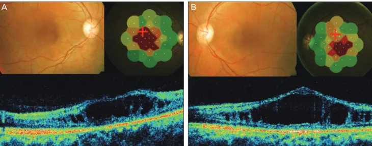
(3) M Bhandari, et al. SD-OCT and Microperimetry in Goldmann-Favre Syndrome. A. B. Fig. 2. (A) The right eye color fundus shows a bicycle wheel pattern in the fovea that is suggestive of foveal schisis with diffuse retinal pigment epithelium alterations. Microperimetry shows dense central scotoma with reduced sensitivity. Spectral domain optical coherence tomography (SD-OCT) shows foveal schisis with elevated foveal contour. (B) The left eye fundus shows a bicycle wheel pattern in the fovea that is suggestive of foveal schisis. Microperimetry shows reduced foveal sensitivity. SD-OCT imaging illustrates foveal schisis with elevated foveal contour.. A. B. Fig. 3. (A) The right eye color fundus shows macular schisis and diffuse retinal pigment epithelium (RPE) alterations. At the time of examination, microperimetry revealed central scotoma. Spectral domain optical coherence tomography (SD-OCT) showed epiretinal membrane. The foveal contour was altered with cystic spaces suggestive of retinal schisis. (B) The left eye color fundus shows macular schisis with diffuse RPE alterations. At the time of examination, SD-OCT revealed altered foveal contours with a tenting up of the fovea with intraretinal cystic spaces suggestive of retinal schisis. Microperimetry showed dense central scotoma with grossly reduced retinal sensitivity.. ing for the past 4 years. His brother (case 2) was known to have the same complaints. The patient’s best corrected visual acuity, with +4.50 DS / -0.50 DC × 90, was 6 / 12 in the right eye, and, with +5.00 DS / -1.00 DC × 90, was 6 / 24 in the left eye. Anterior segment examination was normal. Indirect ophthalmoscopy revealed vitreous floaters, macular schisis, diffuse RPE alterations, and peripheral schisis in both eyes. The patient’s total error score for the Farnsworth D-15. color vision test was 28 in the right eye and 31 in the left eye. The test results revealed tritanomaly in the right eye and diffuse color defect in the left eye. SD-OCT imaging of both eyes showed epiretinal membrane. The foveal contour was altered with cystic spaces that were suggestive of retinal schisis. Alteration of the photoreceptor layer was noted. Foveal thickness was 136 microns in the right eye and 171 microns in the left eye. Microperimetry showed a reduced mean retinal sensitivity of 1.0 db in the right eye 145.
(4) Korean J Ophthalmol Vol.26, No.2, 2012. and 0.2 db in the left eye, with a central dense scotoma in both eyes (Fig. 3).. Discussion Goldmann-Favre syndrome is characterized by foveal and/or peripheral retinoschisis. Differential diagnosis includes conditions such as retinitis pigmentosa (RP), Stickler’s syndrome, and familiar exudative vitreo-retinopathy. RP is differentiated from Goldmann-Favre syndrome on the basis of vascular attenuation and electroretinogram. Cystoid macular oedema associated with RP is a potentially treatable condition, and can be distinguished from retinoschisis by OCT and FFA. Ocular management of Goldmann-Favre syndrome includes scatter photocoagulation of peripheral avascular lesions, as well as vitrectomy for clearance of vitreous hemorrhage and relief of retinal traction if present. Genetic counseling is also offered to patients and potential carriers. Because the syndrome is associated with mutations in Arg311Gln NR2E3 in the 15q23 chromosome, it is a potential target for gene therapy [7]. Case 1 followed the temporal sequence of events over a period of 18 months in a patient diagnosed with Goldmann-Favre syndrome. The patients in cases 2 and 3 were clinically diagnosed with Goldmann-Favre syndrome. We are unaware of previous reports about Goldmann-Favre syndrome that use SD-OCT and microperimetry for analysis. The findings vary with the age of the patient and the severity of the disease. Findings from SD-OCT imaging included lamellar macular holes, macular schisis, microcystic spaces, alterations of photoreceptor layers, epiretinal membrane with vitreomacular traction, enhanced foveal thickness, and elevated foveal contours. Microperimetry revealed reduced foveal sensitivity with dense scotomas.. 146. In this series of cases, retinoschisis and macular cystoid changes noted with SD-OCT matched the scotomas shown with microperimetry. Advanced diagnostic techniques such as SD-OCT and microperimetry detect new characteristics, as well as confirm correlations among known features, to elevate our knowledge of the disease.. Conflict of Interest No potential conflict of interest relevant to this article was reported.. References 1. Fishman GA, Jampol LM, Goldberg MF. Diagnostic features of the Favre-Goldmann syndrome. Br J Ophthalmol 1976;60:345-53. 2. Ikaheimo K, Tuppurainen K, Mantyjarvi M. Clinical features of Goldmann-Favre syndrome. Acta Ophthalmol Scand 1999;77:459-61. 3. Khairallah M, Ladjimi A, Ben Yahia S, et al. Elevated macular retinoschisis associated with Goldmann-Favre syndrome successfully treated with grid laser photocoagulation. Retina 2002;22:234-7. 4. Theodossiadis PG, Koutsandrea C, Kollia AC, Theodossiadis GP. Optical coherence tomography in the study of the Goldmann-Favre syndrome. Am J Ophthalmol 2000;129:542-4. 5. Chavala SH, Sari A, Lewis H, et al. An Arg311Gln NR2E3 mutation in a family with classic Goldmann-Favre syndrome. Br J Ophthalmol 2005;89:1065-6. 6. Vingrys AJ, King-Smith PE. A quantitative scoring technique for panel tests of color vision. Invest Ophthalmol Vis Sci 1988;29:50-63. 7. Haider NB, Jacobson SG, Cideciyan AV, et al. Mutation of a nuclear receptor gene, NR2E3, causes enhanced S cone syndrome, a disorder of retinal cell fate. Nat Genet 2000;24:127-31..
(5)
수치

관련 문서
Photonic crystals containing rugate structure result in a mirror with high reflectivity in a specific narrow spectral region and are prepared by applying
Mortality and clinical signs in SD rats about single oral administration after 3 weeks with callus extract ..... Hematological values in SD rats administered orally
Serum uric acid levels and risk for vascular disease in patients with metabolic syndrome... Prevalence if the metabolic syndrome in a Turkish
이승석, 김주하, 엄태중, 최은서, “Optical coherence tomography application by using optical phase shift based on fiber optic sensor", Photonics
Simultaneous Measurements of the Wake Flow of a Circular Cylinder with a Flexible Film and Its Motions using
Although van der Waals equation is still less accurate at high
The following key words were used: artificial vision, blindness, cortical prosthesis, electrical stimulation electronic implants, macular degener- ation, optic nerve,
A series of joint workshops in the nuclear thermal-hydraulic field were agreed with the intention of facilitating the cooperation activities between NPIC and KAERI, and
