• 검색 결과가 없습니다.

is a potent and safe adjuvant that enhances the antigen-presenting capability of dendritic cells

N/A
N/A
Protected

Academic year: 2022

Share "is a potent and safe adjuvant that enhances the antigen-presenting capability of dendritic cells"

Copied!
6
0
0

로드 중.... (전체 텍스트 보기)

전체 글

(1)

Author contributions: Y.J.L., H.G.J. performed acquisition of data, analysis and/or interpretation of data of in vitro experiments and drafting main parts of the manuscript; Y.H., H.G.J. performed and acquisition/analysis of data of in vivo experiments. Y.J.L., H.G.J. revising the manuscript; H.G.J. per- formed conception and design of study.

This is an Open Access article distributed under the terms of the Creative Commons Attribution Non-Commercial License, which permits unrestricted non-commercial use, distribution, and reproduction in any medium, provided the original work is properly cited.

Copyright © Korean J Physiol Pharmacol, pISSN 1226-4512, eISSN 2093-3827

INTRODUCTION

Vaccines are used to obtain an acquired immunity against cer- tain pathogens [1]. A vaccine contains antigen (Ag) and immuno- stimulating agent. Vaccine Ags include disease-causing weakened or killed microorganisms, its toxins, or one of its surface proteins [1]. However, some vaccine Ags do not have enough intrinsic ca- pability to stimulate immune system, and thus the vaccines need to contain the immunostimulating agents, called adjuvants [2].

Vaccine adjuvant is added to enhance the efficacy of vaccine at a variety of steps. It extends the presence of Ag at injection sites and helps antigen-presenting cells (APCs) to uptake Ag, and also stimulates immune responses by the increase of cytokine produc-

tion and immune-related molecules on the surface of APCs [3].

Vaccine adjuvants have been divided into inorganic substances including alum, cytokines, and bacterial products according to their source types [4].

Bordetella pertussis (B. pertussis) and Mycobacterium bovis (M. bovis) have been used as bacteria-derived immunostimulat- ing agents, partly vaccine adjuvants [5,6]. M. bovis is the causative agent of tuberculosis in cattle and Bacillus Calmette-Guerin (BCG) is prepared from an attenuated strain of M. bovis. BCG has been used as a vaccine adjuvant, but its side effects include fibrosis and granuloma, and thus it is not used currently [6-8]. B.

pertussis is the causative agent of pertussis or whooping cough, and its virulence factors include pertussis toxin, filamentous hae-

Original Article

Bordetella bronchiseptica is a potent and safe adjuvant that enhances the antigen-presenting capability of dendritic cells

You-Jeong Lee 1 , Yong Han 1 , and Hong-Gu Joo 1,2, *

1

College of Veterinary Medicine,

2

Veterinary Medical Research Institute, Jeju National University, Jeju 63243, Korea

ARTICLE INFO Received July 16, 2019 Revised November 4, 2019 Accepted November 5, 2019

*Correspondence Hong-Gu Joo

E-mail: jooh@jejunu.ac.kr Key Words

Bordetella bronchiseptica Dendritic cells

Immunostimulation Safety

Vaccines

ABSTRACT We previously demonstrated that Bordetella bronchiseptica (B. bron- chiseptica) antigen (Ag) enhances the Mycoplasma hyopneumoniae Ag-specific immune response. The focus of this study was whether acellular bacterin of B. bron- chiseptica could be used as an adjuvant to increase antigen-presenting capability of dendritic cells (DCs) by increasing the level of activation. The metabolic activity of DCs was increased by B. bronchiseptica, similar to lipopolysaccharide (LPS). Flow cytometry analysis revealed that B. bronchiseptica increases the expression of ma- jor histocompatibility complex class-2, cluster of differentiation (CD)40, CD54, and CD86 which are closely related to DC-mediated immune responses. B. bronchiseptica enhanced the production of cytokines related to adaptive immune responses. Fur- thermore, the survival rate of B. bronchiseptica-injected groups was 100% at 15 and 20 mg/kg doses, whereas that of LPS-injected groups was only 20%, 0% at 15 and 20 mg/kg doses respectively, and so B. bronchiseptica is likely to be safer than LPS.

Taken together, these results indicate that B. bronchiseptica can be used as an adju-

vant to enhance the antigen-presenting capability of DCs. B. bronchiseptica is a can-

didate for producing vaccines, especially in case of DC-mediating efficacy and safety

demands. This study provides researchers and clinicians with valuable information

regarding the usage of B. bronchiseptica as a safe bacteria-derived immunostimulat-

ing agent for developing efficient vaccines.

(2)

magglutinin, pertactin, and tracheal cytotoxin. Pertussis toxin is a major toxin of B. pertussis and has an adjuvant effect such as antibody response [5,9], but can generate the sensitization leading to anaphylaxis [10].

Bordetella bronchiseptica (B. bronchiseptica) is a similar patho- gen to B. pertussis and causes respiratory disease [11]. Whereas B. pertussis is relatively limited to human, B. bronchiseptica af- fects several animals including dogs, cats, and pigs with different strains [12]. Infection of B. bronchiseptica alone does not seri- ously damage to respiratory system of hosts, but the combined infection with other pathogens can cause significant economic loss [13,14]. B. bronchiseptica modulates the phenotypes of mac- rophages, leading to the inhibition of CD4

+

T cell proliferation [15].

In a previous study, we demonstrated that B. bronchiseptica Ag can enhance the production of Mycoplasma (M.) hyopneumoniae Ag-specific immunoglobulin G [16]. In this study, we investigated whether formalin-fixed whole-cell and acellular vaccine of B.

bronchiseptica can improve the Ag-presenting capability of den- dritic cells (DCs) and may be a candidate of vaccine adjuvant with immunostimulatory activity.

METHODS

Animals and materials

C57BL/6 and Balb/c mice were obtained from OrientBio (Seongnam, Korea) and maintained at our animal facility. 8 to 12- week old mice were used for all experiments. Animal experiments in this study were performed in accordance with the Institutional Guideline for Animal Use and Care of Jeju National University (2018-0011, 2018-0045). Lipopolysaccharide (LPS; Sigma-Aldrich, St Louis, MO, USA) was used to as a positive control of immuno- stimulating agent for DCs. The whole-cell bacterin of B. bronchi- septica was prepared by formalin-inactivation and acellular vac- cine with sonication (B. bronchiseptica) [16]. The protein amount of B. bronchiseptica was measured by Bradford assay.

Preparation of bone marrow-derived DCs

Bone marrow cells (BMs) were harvested from femur and tibia of C57BL/6 mice, and were used to culture DCs. BMs obtained after lysing red blood cells were counted and cultured into 6-well plates. To culture DCs, the cells were cultured with RPMI 1640 medium containing 5% fetal bovine serum, 2 mM L-glutamine, 100 U/ml penicillin-streptomycin, and 10 ng/ml mouse recom- binant granulocyte macrophage-colony stimulating factor (GM- CSF; Peprotech, Rocky Hill, NJ, USA). The culture medium was replaced with new medium containing GM-CSF every two days.

In order to minimize the interference of lymphocytes and granu- locytes in DCs, the floating cells were removed on the 2nd, 4th

day and DC precursor cells attached to the bottom were cultured.

After 6 day culture, the floating cells were harvested and used as DCs.

Measurement of DC metabolic activity

To measure the metabolic activity of DCs, the cells were seeded in 96-well plates and treated with LPS or B. bronchiseptica for 3 days. Then, 5 µl/well Cell counting kit-8 (CCK-8; Dojindo Molec- ular Tech., Kumamoto, Japan) solution was added and incubated at 37°C for 4 h. The optical density was measured at 450 nm with a microplate reader (Multiskan FC; Thermo Fisher Scientific, Waltham, MA, USA).

Measurement of cytokines

DCs were cultured in 96-well plates to analyze the production of cytokines such as tumor necrosis factor (TNF)-alpha, inter- leukin (IL)-10, and IL-12. The cells were treated with LPS or B.

bronchiseptica for 3 days. The supernatants of treated cells were collected and used for Enzyme-linked immunosorbent assay (ELISA). The amounts of TNF-alpha, IL-10, and IL-12 were deter- mined by CytoSet kits (Thermo Fisher Scientific) according to the instructions. The optical density was measured at 405 nm.

Flow cytometry analysis

Flow cytometry analysis was performed using surface marker- specific monoclonal antibodies (Abs) to measure the expression of activation and maturation markers of DCs. DCs were cultured in 6-well plates and treated with LPS or B. bronchiseptica for 3 days in the presence of GM-CSF. The treated cells were stained with biotin-labeled anti-MHC class II (I-A

b

), anti-CD40, anti-CD54, or anti-CD86 Ab, and sequentially with streptavidin-fluorescein isothiocyanate (FITC) (all from BD Bioscience, Franklin Lakes, NJ, USA). To measure the antigen uptake capability of DCs, the cells were incubated with FITC-dextran at 250 µg/ml at 37°C for 1 h. All stained cells were analyzed by CytoFLEX and CytExpert software (Beckman Coulter, Brea, CA, USA).

Mixed lymphocyte reaction (MLR)

To evaluate the adjuvanticity of B. bronchiseptica, the cultured DCs were treated with LPS, B. bronchiseptica for 2 days, and then co-cultured with allogeneic spleen cells. Allogeneic spleen cells were harvested from Balb/c mice. The treated DCs were incubat- ed with 50 µg/ml mitomycin C for 30 min before the co-culture setup. The DCs and allogeneic spleen cells were cultured in 96- well plates at a concentration of 1 × 10

5

cells/ml and 2 × 10

6

cells/

ml, respectively. As a control, only allogeneic spleen cells were

cultured. After 5 days, 10 µl/well of CCK-8 solution was added

and incubated at 37°C for 4 h. The optical density was measured

(3)

at 450 nm with a microplate reader.

Injection of LPS or B. bronchiseptica into mice

7-week old Balb/c mice were divided into 6 groups. B. bronchi- septica or LPS was injected at 10, 15 and 20 mg/kg doses respec- tively, by a single intraperitoneal injection. The mice were moni- tored for one week to check mortality rate and weight change.

Statistical analysis

The data of CCK-8 assay and ELISA are presented as mean ± standard deviation and were analyzed with one-way analysis of variance, Tukey’s multiple comparison test (InStat; GraphPad Software, San Diego, CA, USA). A p-value of < 0.05 was consid- ered as significant. **, *** indicate p < 0.01, 0.001 respectively, compared to control.

RESULTS

Effect of B. bronchiseptica on the metabolic activity of DCs

In order to determine whether B. bronchiseptica affects the metabolic activity of DCs, we performed CCK-8 assays. DCs were treated with B. bronchiseptica or LPS for 3 days at a range of con- centration, 0 to 300 ng/ml, and the metabolic activity of DCs was measured. B. bronchiseptica at 33.3 and 100 ng/ml significantly increased the metabolic activity of DCs compared to control (0 ng/ml) (Fig. 1). The highest increase of metabolic activity was ap- peared at 100 ng/ml, an optimal concentration used for DC acti- vation in this study.

B. bronchiseptica increases the expression of immune response-related surface markers of DCs

To determine whether B. bronchiseptica induces the activa- tion and maturation of DCs, we measured the expression level of surface markers on DCs. The cells were treated with 0.1 µg/ml of B. bronchiseptica or LPS for 2 days and stained with surface marker-specific Abs. B. bronchiseptica strongly increased the sur- face markers expression on DCs, which increase the MHC class 2 molecule by 67% compared to control, and also other 3 molecules more than 150% (Fig. 2). This data showed that LPS also had ef- fects on the activation of DCs, but B. bronchiseptica increased the expression level of the 4 surface markers higher than LPS.

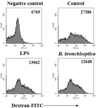
B. bronchiseptica decreases the antigen uptake capability of DCs

Antigen uptake assay was performed to confirm whether B.

bronchiseptica induces the maturation of DCs. The cells were incubated with FITC-dextran at 37°C for 1 h. Both LPS and B.

bronchiseptica showed lower levels of the uptake of FITC-dextran compared to that of control (Fig. 3). The uptake of FITC-dextran in DCs at 4°C was used as negative control.

B. bronchiseptica enhances the cytokine production of DCs

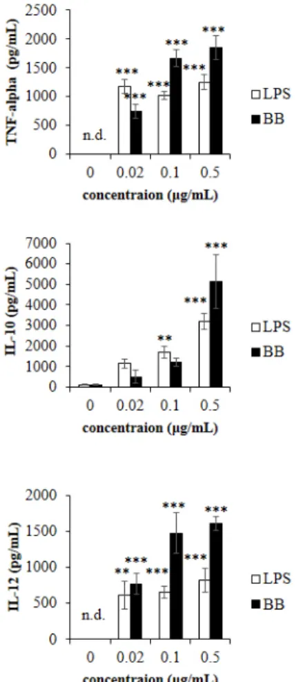
To determine the effect of B. bronchiseptica as an adjuvant for vaccines, we measured the production of TNF-alpha, IL-10, and IL-12 in DCs treated with B. bronchiseptica or LPS. ELISA results demonstrated that both B. bronchiseptica and LPS increased the production of TNF-alpha, IL-10, and IL-12 (Fig. 4). In the case of TNF-alpha, B. bronchiseptica produced less TNF-alpha than LPS at 0.02 µg/ml, but produced more at 0.1 and 0.5 µg/ml. Interest- ingly, IL-10 production was significantly elevated in B. bronchi- septica-treated DCs compared to LPS-treated DCs at 0.5 µg/ml.

B. bronchiseptica enhanced the IL-12 production of DCs higher than LPS in all concentrations.

Effect of B. bronchiseptica on the antigen-presenting capability of DCs

To evaluate whether B. bronchiseptica can increase the antigen- presenting capability of DCs, we co-cultured the treated DCs and allogeneic spleen cells. DCs were pre-treated with B. bronchisep- tica or LPS at 0.1 µg/ml for 2 days, and then co-cultured at a ratio of 1:20 with allogeneic spleen cells harvested from Balb/c mice.

Mitomycin C was used to limit the metabolic activity of DCs it- self and inhibit further maturation during the co-cultures. Both B. bronchiseptica- or LPS-treated DCs significantly increased the metabolic activity and proliferation of allogeneic spleen cells compared to control DCs (Fig. 5). This result demonstrated that B.

Fig. 1. The effect of Bordetella bronchiseptica (B. bronchiseptica)

on the metabolic activity of dendritic cells (DCs). DCs were cultured

in 96-well plates at a concentration of 1.5 × 10

5

cells/ml with 0 to 300

ng/ml B. bronchiseptica or lipopolysaccharide (LPS) for 3 days. The

treated DCs were incubated with 5 µl/well CCK-8 solution at 37°C for 4

h. The optical density (O.D.) was measured at 450 nm by using a micro-

plate reader. **Indicates p < 0.01.

(4)

bronchiseptica can increase the antigen-presenting capability of DCs.

B. bronchiseptica is safe in vivo

To investigate if B. bronchiseptica is safe in vivo, Balb/c mice were injected with 10, 15, and 20 mg/kg of B. bronchiseptica or LPS. After 7 days, the survival rate and weight change were examined. All mice were survived in both B. bronchiseptica- or LPS-injected groups at 10 mg/kg dose. B. bronchiseptica-injected groups showed 100% survival rate at 15 and 20 mg/kg dose, whereas LPS-injected groups showed only 20%, 0% survival rate at 15 and 20 mg/kg dose respectively (Fig. 6). Interestingly, the body weight change of B. bronchiseptica-injected mice was less than 3% decrease. This result indicates that B. bronchiseptica is safer in vivo than LPS.

DISCUSSION

Adjuvants increase the effects of vaccines by enhancing antigen-specific immune responses, such as maximal T cell- dependent immune responses [3]. This effect of vaccine adjuvant can be achieved through several critical events including the ac- tivation of antigen-presenting cells such as DCs [17]. After uptak- ing antigens, DCs migrate to lymph nodes and present antigens to lymphocytes for initiating adaptive immune responses [18]. In previous studies, we demonstrated that B. bronchiseptica antigens

Fig. 2. Effect of Bordetella bronchiseptica (B. bronchiseptica) on the expression of immune response-related surface markers of dendritic cells (DCs). DCs were cultured in 6-well plates at a concentration of 2.5 × 10

5

cells/ml and treated with 0.1 µg/ml B. bronchiseptica or lipopolysaccha- ride (LPS) for 2 days. After the treatment, DCs were stained as described in “Methods”. Numbers of histograms represent the geometric mean fluores- cence intensity.

Fig. 3. Bordetella bronchiseptica (B. bronchiseptica) increases the level of dendritic cell (DC) maturation. DCs were cultured in 6-well plates at a concentration of 5.5 × 10

5

cells/ml and treated with 0.1 µg/

ml of B. bronchiseptica or lipopolysaccharide (LPS) for 2 days. After

treatment, the DCs were incubated with fluorescein isothiocyanate

(FITC)-dextran as described in “Methods”. Numbers of histograms repre-

sent the geometric mean fluorescence intensity. Negative control DCs

was treated at 4°C, and others were treated at 37°C for 1 h.

(5)

increase the production of M. hyopneumoniae-specific IgG [16].

In this study, we investigated whether B. bronchiseptica can be used as an adjuvant to increase the antigen-presenting capability of DCs in several aspects.

This study demonstrated that B. bronchiseptica up-regulated the functions of DCs including metabolic activity. CCK-8 assay showed that B. bronchiseptica increased the metabolic activity of DCs at a level similar to that of LPS. Flow cytometry analysis showed that B. bronchiseptica stimulated the expression of MHC class II, an essential antigen-presenting molecule, on the surface of DCs and increased the expression of DC activation markers such as CD40, CD54, and CD86. In addition, the antigen uptake of B. bronchiseptica-treated DCs was consistently low. The ma- tured DCs have much lower antigen uptake capability than im- mature DCs. Our data collectively indicates that B. bronchisep- tica can increase the degree of activation and maturation of DC, similar to LPS.

TNF-alpha, IL-10 and IL-12 production were measured to determine how much B. bronchiseptica can activate DCs. ELISA showed that B. bronchiseptica strongly increased the production of TNF-alpha, IL-10, IL-12. Our data indicate that B. bronchisep- tica is effective as an adjuvant to activate DCs. Interestingly IL-10 production was significantly elevated in the DCs treated with B.

bronchiseptica compared to LPS at 0.5 µg/ml. IL-10 is a represen- tative anti-inflammatory cytokine and its production is closely connected to the production of inflammatory cytokine including TNF-alpha [19,20]. This result suggests that B. bronchiseptica may have a unique regulatory role in the production of inflam- matory and anti-inflammatory cytokines.

The immune enhancing effect of B. bronchiseptica is similar to or higher than that of LPS. B. bronchiseptica does have less cyto- toxicity than LPS on DCs. B. bronchiseptica is a Gram-negative bacterium containing LPS and is likely to act as an immune stimulating factor. It is also suggested that the LPS of B. bronchi- septica may have a different structure or content compared to

Fig. 4. The effect of Bordetella bronchiseptica (B. bronchiseptica) on the production of immune-related cytokines. Dendritic cells (DCs) were cultured in 96-well plates at a concentration of 5 × 10

5

cells/ml and treated with 0 to 0.5 µg/ml B. bronchiseptica (BB) or lipopolysac- charide (LPS) for 3 days. The supernatant of cells treated with BB or LPS was collected and used for ELISA. The optical density was measured at 405 nm by using microplate reader. TNF, tumor necrosis factor; IL, inter- leukin. **,***Indicate p < 0.01, p < 0.001, respectively.

Fig. 5. Effect of Bordetella bronchiseptica (B. bronchiseptica) on the antigen-presenting capability of dendritic cells (DCs). DCs were treated with 0.1 µg/ml of LPS or B. bronchiseptica (BB) for 2 days. The treated DCs were incubated with 50 µg/ml mitomycin C for 30 min be- fore the co-culture setup. Allogeneic spleen cells were harvested from Balb/c mice. DCs and allogeneic spleen cells were co-cultured in 96- well plates at a 1 × 10

5

cells/ml and 2 × 10

6

cells/ml respectively. After 5 days, CCK-8 assay was performed as described in ‘Methods’. O.D., optical density. **Indicates p < 0.01.

Fig. 6. Bordetella bronchiseptica (B. bronchiseptica) is safe in vivo.

7-week old Balb/c mice were intraperitoneally injected with B. bronchi-

septica (BB) or lipopolysaccharide (LPS) at 10, 15, and 20 mg/kg dose

respectively. The survival rate of the mice groups was determined at 7

day.

(6)

that of E. coli. In the future, we will study which components of B. bronchiseptica including LPS have the immunostimulatory ef- fect.

In addition, we measured the effect of B. bronchiseptica on the antigen-presenting capability of DCs. MLRs measured by CCK-8 assay showed that B. bronchiseptica- and LPS-treated DCs in- creased the metabolic activity and proliferation of allogeneic lym- phocytes. Our data indicate that B. bronchiseptica can enhance the antigen-presenting capability of DCs.

To confirm if B. bronchiseptica is safe in vivo, mice were inject- ed with the same dose of B. bronchiseptica or LPS. Interestingly, B. bronchiseptica-injected mice did not die at all doses (10, 15, 20 mg/kg), whereas LPS-injected mice seriously did at 15 and 20 mg/

kg (survival rate only 20%, 0% respectively). This result suggests that B. bronchiseptica is much safer in vivo than LPS.

Taken together, our data demonstrate that B. bronchiseptica enhances the antigen-presenting capability of DCs and has im- munostimulatory activity as an adjuvant. In addition, the safety of B. bronchiseptica is better than LPS which is already used as a kind of adjuvant (Supplementary Fig. 1). This study shows the availability of B. bronchiseptica as a vaccine adjuvant based on its activity on DCs.

ACKNOWLEDGEMENTS

This work was supported by Korea Institute of Planning and Evaluation for Technology in Food, Agriculture, Forest and Fish- eries (IPET) through Agri-Bio industry Technology Development Program, funded by Ministry of Agriculture, Food and Rural Af- fairs (MAFRA) (Grant number 111065-3, 109014-03-1-CG000).

CONFLICTS OF INTEREST

The authors declare no conflicts of interest.

SUPPLEMENTARY MATERIALS

Supplementary data including one figure can be found with this article online at http://pdf.medrang.co.kr/paper/pdf/Kjpp/

Kjpp2020-24-01-06-s001.pdf.

REFERENCES

1. Karch CP, Burkhard P. Vaccine technologies: from whole organ- isms to rationally designed protein assemblies. Biochem Pharmacol.

2016;120:1-14.

2. Marciani DJ. Vaccine adjuvants: role and mechanisms of action in vaccine immunogenicity. Drug Discov Today. 2003;8:934-943.

3. Del Giudice G, Rappuoli R, Didierlaurent AM. Correlates of adju- vanticity: a review on adjuvants in licensed vaccines. Semin Immu- nol. 2018;39:14-21.

4. Coffman RL, Sher A, Seder RA. Vaccine adjuvants: putting innate immunity to work. Immunity. 2010;33:492-503.

5. Dunne A, Mielke LA, Allen AC, Sutton CE, Higgs R, Cunningham CC, Higgins SC, Mills KH. A novel TLR2 agonist from Bordetella pertussis is a potent adjuvant that promotes protective immunity with an acellular pertussis vaccine. Mucosal Immunol. 2015;8:607- 617.

6. Wangoo A, Brown IN, Marshall BG, Cook HT, Young DB, Shaw RJ.

Bacille Calmette-Guérin (BCG)-associated inflammation and fibro- sis: modulation by recombinant BCG expressing interferon-gamma (IFN-gamma). Clin Exp Immunol. 2000;119:92-98.

7. Filardi MJ, Codish SD, Civerchia L, Howard RK, McKneally MF.

Toxicity of intrapleural Bacillus Calmette-Guérin treatment in ani- mals. Cancer Res. 1979;39:3673-3676.

8. Gordon S, Keshav S, Stein M. BCG-induced granuloma formation in murine tissues. Immunobiology. 1994;191:369-377.

9. Fedele G, Celestino I, Spensieri F, Frasca L, Nasso M, Watanabe M, Remoli ME, Coccia EM, Altieri F, Ausiello CM. Lipooligosaccharide from Bordetella pertussis induces mature human monocyte-derived dendritic cells and drives a Th2 biased response. Microbes Infect.

2007;9:855-863.

10. Samore MH, Siber GR. Pertussis toxin enhanced IgG1 and IgE responses to primary tetanus immunization are mediated by inter- leukin-4 and persist during secondary responses to tetanus alone.

Vaccine. 1996;14:290-297.

11. Zhao Z, Wang C, Xue Y, Tang X, Wu B, Cheng X, He Q, Chen H.

The occurrence of Bordetella bronchiseptica in pigs with clinical respiratory disease. Vet J. 2011 188:337-340.

12. Goodnow RA. Biology of Bordetella bronchiseptica. Microbiol Rev.

1980;44:722-738.

13. Chanter N, Magyar T, Rutter JM. Interactions between Bordetella bronchiseptica and toxigenic Pasteurella multocida in atrophic rhi- nitis of pigs. Res Vet Sci. 1989;47:48-53.

14. Horiguchi Y. Swine atrophic rhinitis caused by pasteurella multo- cida toxin and bordetella dermonecrotic toxin. Curr Top Microbiol Immunol. 2012;361:113-129.

15. Siciliano NA, Skinner JA, Yuk MH. Bordetella bronchiseptica mod- ulates macrophage phenotype leading to the inhibition of CD4+ T cell proliferation and the initiation of a Th17 immune response. J Immunol. 2006177:7131-7138.

16. Yim SH, Hahn TW, Joo HG. Bordetella bronchiseptica antigen enhances the production of Mycoplasma hyopneumoniae antigen- specific immunoglobulin G in mice. J Vet Sci. 2017;18:327-332.

17. Gallucci S, Lolkema M, Matzinger P. Natural adjuvants: endogenous activators of dendritic cells. Nat Med. 1999;5:1249-1255.

18. Théry C, Amigorena S. The cell biology of antigen presentation in dendritic cells. Curr Opin Immunol. 2001;13:45-51.

19. de Waal Malefyt R, Abrams J, Bennett B, Figdor CG, de Vries JE.

Interleukin 10(IL-10) inhibits cytokine synthesis by human mono- cytes: an autoregulatory role of IL-10 produced by monocytes. J Exp Med. 1991;174:1209-1220.

20. Fiorentino DF, Zlotnik A, Mosmann TR, Howard M, O'Garra A.

IL-10 inhibits cytokine production by activated macrophages. J Im-

munol. 1991;147:3815-3822.

수치

Fig. 1. The effect of Bordetella bronchiseptica (B. bronchiseptica)  on the metabolic activity of dendritic cells (DCs)
Fig. 3. Bordetella bronchiseptica (B. bronchiseptica) increases the  level of dendritic cell (DC) maturation
Fig. 4. The effect of Bordetella bronchiseptica (B. bronchiseptica)  on the production of immune-related cytokines

참조

관련 문서

Although L-valyl-ara-C appeared to be beneficial to enhance the cellular uptake of ara-C in Caco-2 cells, prodrug itself exhibited far less

Frequency of conidial germination (A) and appressorium formation (B) of Colletotrichum orbiculare and callose formation of plant cells (C) at the

The untreated surfaces and AA plasma treated with 3D-PCL surfaces were also examined for their in vitro pre-osteoblast (MC3T3-E1) cells proliferation and

Null hypotheses of this study was there is no difference in root canal length determination between conventional and heat-treated Ni-Ti files by 2

The clinical outcome and complication for treating proximal femoral shaft fracture were compared and analyzed through the group treated with closed

Results: The human breast cancer cell subline MCF-7/MX5 cells selected in the presence of 5 µg/ml mitoxantrone (MX) were more resistant to MX (15.7... Western blot and

Control- and ASTRIN- depleted HeLa cells were untreated or treated with 5 Gy. IR and were then fixed at

Recombinant RapA fused with the C-terminus of RapC completely recovered the phenotypes of rapC null cells, indicating that the functions of RapA were modified to become