INTRODUCTION
Chronic critical limb ischemia (CLI) occurs when arterial blood flow to the part or entire foot, is markedly reduced, in most cases as a result of progressive obstructive atherosclerosis. According to Trans-Atlantic Inter-Society Consensus (TASC) (1), there will be approximately 500 to
Endovascular Revascularization for Patients with Critical Limb Ischemia: Impact on Wound Healing and Long
Term Clinical Results in 189 Limbs
Jae-Ik Bae, MD
1, Je Hwan Won, MD
1, Seung Hwan Han, MD
2, Sang Hyun Lim, MD
3, You Sun Hong, MD
3, Jae-Young Kim, MD
4, Ji Dae Kim, MD
1, Jun-Su Kim, MD
5Departments of 1Radiology, 2Orthopedic Surgery and 3Cardiovascular Surgery, Ajou University School of Medicine, Suwon 443-721, Korea; 4DNF Hospital, Seoul 131-875, Korea; 5Department of Family Medicine, Busan Paik Hospital, Inje University College of Medicine, Busan 614-735, Korea
Objective: To evaluate the impact on wound healing and long-term clinical outcomes of endovascular revascularization in
patients with critical limb ischemia (CLI).
Materials and Methods: This is a retrospective study on 189 limbs with CLI treated with endovascular revascularization
between 2008 and 2010 and followed for a mean 21 months. Angiographic outcome was graded to technical success (TS), partial failure (PF) and complete technical failure. The impact on wound healing of revascularization was assessed with univariate analysis and multivariate logistic regression models. Analysis of long-term event-free limb survival, and limb salvage rate (LSR) was performed by Kaplan-Meier method.
Results: TS was achieved in 89% of treated limbs, whereas PF and CF were achieved in 9% and 2% of the limbs, respectively.
Major complications occurred in 6% of treated limbs. The 30-day mortality was 2%. Wound healing was successful in 85%
and failed in 15%. Impact of angiographic outcome on wound healing was statistically significant. The event-free limb survival was 79.3% and 69.5% at 1- and 3-years, respectively. The LSR was 94.8% and 92.0% at 1- and 3-years, respectively.
Conclusion: Endovascular revascularization improve wound healing rate and provide good long-term LSRs in CLI.
Index terms: Critical limb ischemia; Infrapopliteal angioplasty; Diabetic foot
Received September 23, 2012; accepted after revision January 2, 2013.
Corresponding author: Je Hwan Won, MD, Department of Radiology, Ajou University School of Medicine, 164 Worldcup-ro, Yeongtong-gu, Suwon 443-721, Korea.
• Tel: (8231) 219-5863 • Fax: (8231) 219-5862
• E-mail: [email protected]
This is an Open Access article distributed under the terms of the Creative Commons Attribution Non-Commercial License (http://creativecommons.org/licenses/by-nc/3.0) which permits unrestricted non-commercial use, distribution, and reproduction in any medium, provided the original work is properly cited.
Korean J Radiol 2013;14(3):430-438
1000 new cases of CLI every year in a European or North American population of 1 million. Prevalence of CLI seems to increase with the increasing prevalence of diabetes mellitus (2). CLI is difficult to summarize because treatment very much depends on the countries and the institutions to which the patient is referred. However, studies suggest that about 25% of the patients with CLI undergo primary foot amputation even in developed countries (1), and amputation seems to be the common first-line therapy in developing countries, where there is lack of specialized podiatry program (3).
Advances in techniques and devices have led to more widespread use of endovascular revascularization procedures for the restoration of blood flow in CLI, and evidence regarding safety and clinical effectiveness support its application (4-9). Clinical effectiveness of endovascular revascularization has been frequently judged by vessel
pISSN 1229-6929 · eISSN 2005-8330
patency and limb salvage (6-8), but there is paucity of reports on outcomes of the wound. The goal of this study was to evaluate the impact on wound healing as well as long-term clinical outcomes of endovascular revascularization in a large single-center population of patients with CLI.
MATERIALS AND METHODS
Patients
The local Institutional Review Board of the hospital approved this retrospective study and waived the requirements for informed consent and research subject authorization. All patients with CLI (Rutherford category 4-6 [10]) who underwent endovascular revascularization between May 2008 and June 2010 were identified.
During this period, 205 limbs of 167 patients were treated for chronic CLI. The decision to use endovascular revascularization as first line therapy was based upon the clinical examination, anatomic evaluation and judgment in a multidisciplinary meeting of vascular surgeons, orthopedic surgeons and interventional radiologists. Of them, 7 patients who were lost to follow-up and 9 functionally unsalvageable limbs were excluded. Endovascular revascularization of a functionally unsalvageable limb was done to facilitate healing of below the knee amputation (BKA) wound on a decision by attending orthopedic surgeons. Individual limbs were counted separately so that patients undergoing staged, bilateral procedures were recorded and evaluated as two entries. Thus, 189 limbs of 152 patients (age range 41-91, mean 67 year-old) were included in the study.
Demographic and clinical factors are summarized in Table 1.
The indication for endovascular revascularization was rest pain (category 4) in 45 (24%) limbs and non-healing wound (category 5, 6) in 144 (76%) limbs. Chronic renal failure (CRF) was defined as a pre-procedure serum creatinine > 1.7 mg/dL. Histories of coronary arterial obstructive disease, hypertension, and stroke were collected from full medical record review.
Lesions
All lesions were retrospectively analyzed based on the pre-procedure angiography and computed tomography (CT). The distribution of the arterial lesions in a limb was recorded. Femoropopliteal lesion was classified according to the TASC II system (1). Infrapopliteal lesions were classified based on the number of patent tibial (anterior
and posterior) or peroneal artery, because most cases with long segmental occlusion or total occlusion are classified to class D on TASC system. Peroneal artery continuing to the dorsalis pedis artery (n = 3) as an anatomical variation was classed to the anterior tibial artery. Distribution and characteristics of lesion are summarized in the Table 2.
Procedure
All patients were given 300 mg of clopidogrel (Handok Pharmaceutical, Seoul, Korea) and 324 mg of aspirin (Boryung Pharmaceutical, Seoul, Korea) as a loading dose a day before the procedure. Patients with preoperative renal insufficiency were given oral N-acetylcystine prior to procedure. Sedation and analgesia was achieved with an intravenous continuous infusion of remifentanil and local
Table 1. Patient CharacteristicsParameter No. of Patients (n = 152)
Men 121 (80%)
Women 31 (20%)
Diabetes 138 (91%)
Hypertension 80 (53%)
Chronic renal failure 54 (36%)
Coronary artery disease 41 (27%)
Stroke history 21 (14%)
Indication No. of Limbs (n = 189)
Resting pain (Rutherford 4) 45 (24%)
Tissue damage (Rutherford 5 and 6) 144 (76%)
Table 2. Lesion Characteristics
Variables Number (%)
Lesion distribution (189 limbs)
IL + FP 2 (1)
IL + IP 2 (1)
IL + FP + IP 27 (14)
FP 6 (3)
FP + IP 115 (61)
IP 37 (20)
FP lesions (150 limbs)
TASC II A 100 (67)
TASC II B 10 (7)
TASC II C 31 (21)
TASC II D 9 (6)
IP lesions (181 limbs)
Two patent tibial arteries 36 (20)
One patent tibial artery 62 (34)
Peroneal runoff only 57 (31)
No patent crural artery 34 (19)
Note.— IL = iliac, FP = femoropopliteal, IP = infrapopliteal, TASC
= Trans-Atlantic Inter-Society Consensus
anesthesia of the groin. An intravenous heparin bolus (80 IU/kg) was administrated during the procedure. If arterial spasm occurred, 0.1-0.2 mg of nitroglycerin was infused as an intra-arterial in bolus. After the procedure, patients were given a 75 mg clopidogrel daily dose for at least 30 days, along with 81 mg aspirin indefinitely.
All lesions of one limb were treated through 6-7 Fr sheaths in the same session. For an iliac artery lesion (n = 31), a contralateral retrograde common femoral artery (CFA) access was made exclusively to spare the ipsilateral CFA for another possible access. Femoropopliteal artery lesions (n
= 144) were treated through an ipsilateral antegrade CFA access in 110 (78%) or a contralateral CFA access in 34 cases (22%). We approached infrapopliteral artery lesions (n
= 182) through an ipsilateral antegrade CFA access in 179 (98%) except for 3 cases (2%) with obesity and a high CFA bifurcation. No brachial or popliteal punctures were made.
A distal retrograde access of the tibial artery was added to cross the tibial occlusive lesions in 10 cases.
Iliac and femoropopliteal arterial lesions were crossed preferentially with 0.035 inch hydrophilic guide wires (Radifocus, Terumo, Tokyo, Japan). Infrapopliteal arterial lesions were crossed preferentially with hydrophilic 0.018 inch (V18, Boston Scientific, Natick, MA, USA) or 0.016 inch wires (GT, Terumo, Tokyo, Japan). Subintimal crossing was used for complete occlusions that could not be crossed intraluminally.
Percutaneous angioplasty was performed with balloon catheters. Diameters of balloon catheters were decided to match the non-diseased segment adjacent to the lesion on CT or angiography. Non-compliant 6 to 8 mm diameter (Rider, Leventon, Barcelona, Spain) for iliac artery, 4 to 7 mm diameter (Savvy or Powerflex; Cordis, Miami Lakes, FL, USA) for femoropopliteal artery, and 2 to 4 mm (Savvy, Cordis;
Miami Lakes, FL, USA) for infrapopliteal arterial lesions were preferred. Long angioplasty balloons were routinely used for the infrapopliteal arteries to cover all multiple or long segmental lesions. Balloon inflation pressures ranged from 4 to 14 atmospheres and were held for at least 60 seconds. Stents were placed for flow-limiting dissections or suboptimal angioplasty results (residual stenosis > 30%) in the iliac or femoropopliteal arteries. Stents were not placed in the infrapopliteal arteries considering high risk of thrombosis in low-flow vessels. Hemostasis was routinely performed with manual compression 3-6 hours after the procedure. A closure device was not used.
Complications
Complications were defined as minor and major, according to the definitions of the Society of Interventional Radiology (11). Puncture site hematomas were not considered a significant complication unless requiring intervention or blood transfusion. However, any event that prolonged hospital stay during the procedure or required specific medical or surgical treatment immediately after the procedure was recorded as a major complication.
Follow-Up
All patients were routinely prescribed oral aspirin (81 mg) and oral clopidogrel (75 mg) once daily for 3 months if no contraindications to either medication were present. The patients were examined every 1 or 2 weeks after discharge until wound healing, and instructed to visit the physician with irregular intervals thereafter. Checking the status of the wound, pain at rest, and the general condition of the patient were determined from the medical records. For 42 patients whose most recent follow-up data was incomplete, additional information was obtained from telephone calls to the patients and the patient’s family members. Indications for reintervention during the follow-up included recurrence of symptoms, i.e., resting pain, or new wound of the foot.
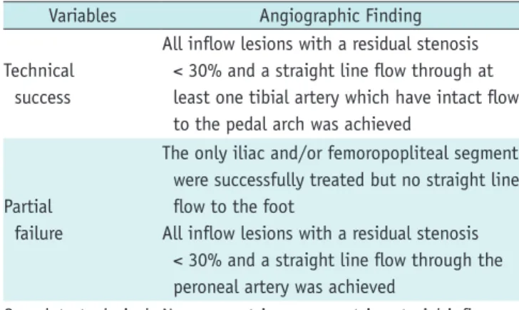
Definitions and End Points
Angiographic outcomes were graded to technical success (TS), partial failure (PF) and complete technical failure (TF), defined in Table 3. A typical case of TS is shown in Figure 1.
For the limbs with non-healing wound, we investigate fate of wound. Healing success was defined when complete healing of initial wound was obtained or when no further amputation was required after planned minimal amputation.
Table 3. Grading of Angiographic Outcomes
Variables Angiographic Finding
Technical success
All inflow lesions with a residual stenosis < 30% and a straight line flow through at least one tibial artery which have intact flow to the pedal arch was achieved
Partial failure
The only iliac and/or femoropopliteal segment were successfully treated but no straight line flow to the foot
All inflow lesions with a residual stenosis < 30% and a straight line flow through the peroneal artery was achieved
Complete technical failure
No apparent improvement in arterial inflow compared with pre-procedure angiography
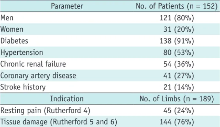
A
E
B
F
C
G
D
H
Fig. 1. Technical success was obtained in 60-year-old male with Rutherford category 5 critical limb ischemia.
A, B. Pre-procedural arteriography of infrapopliteal arteries. Origin of anterior tibial artery (ATA) (arrow) is seen. Posterior tibial artery is not seen. Peroneal artery (arrowheads) is patent. C. Non-healing wounds were gangrenes of 2nd and 4th toes of left foot. D. ATA was recanalized subintimally using 0.016 inch hydrophilic guide wire (arrows) with support of balloon catheter (arrowheads). E. After subintimally crossed wire reentered in distal dorsalis pedis artery (DPA). ATA was dilated with 2 mm and 2.5 mm balloon catheter (arrow). F, G. Recanalized ATA (arrows) and DPA (arrowhead) is evident. H. 2nd and 4th toe gangrenes healed completely 1 month after combined local wound care.
If the wounds persist even after local wound care at the last follow-up, if the patients died before wound could heal, or if progressed to further amputation, outcome was considered to be a healing failure.
During the follow-up, clinical recurrence, new ulceration, new major amputation or re-amputation were taken into account as events as well as the time and cause of death.
Our measure of long-term clinical outcome was functional limb salvage. We considered functional limb salvage successful when the plantar stand was maintained, even when achieved by tarsal-metatarsal amputation (12). Any above-the ankle amputation was considered a failure of limb salvage.
Our definition of event-free limb survival was preservation of functional limb without event. Limb salvage was defined as salvage of a functional limb regardless of subsequent reintervention (6-8).
Statistics
Relationship between angiographic outcome and wound healing was examined by univariate analysis (Fisher’s exact test) and multivariate analysis (multiple logistic regressions). Analysis of the long term effectiveness in limb salvage over time was performed using Kaplan-Meier model and comparisons were made with the log-rank test.
Censored cases are those that did not experience the event of interest, i.e., amputation for a limb salvage analysis or death for a survival analysis, during the study period. The analyses were computed using Statistical Package for the Social Sciences (SPSS) version 18.0 (IBM SPSS, Chicago, IL, USA). Null hypotheses of no difference were rejected if
p-values were less than 0.05.RESULTS
Angiographic Results
The main lesions in iliac arteries were stenoses in 8 and occlusions in 23 limbs (mean length 7.1 ± 3.2 cm).
Iliac lesions were treated successfully in all (100%) with stent placement in 18 limbs (58%). The main lesions in femoropopliteal arteries were stenoses in 100 and occlusions in 50 limbs (mean length 10.6 ± 6.2 cm). The femoropopliteal arterial stenoses were crossed intraluminally and successfully dilated in all. The total occlusions of the femoropopliteal artery were recanalized in 47 limbs (94%);
intraluminally in 15 (32%) and subintimally in 32 (68%), with stent placement in 38 limbs (76%). Of 458 diseased
infrapopliteal arteries in 189 limbs, 330 infrapopliteal arteries were treated endovascularly (average 1.75 arteries per procedure). The main lesions of these arteries were stenoses in 125 and occlusions (mean length 15 ± 6 cm) in 215. The infrapopliteal arterial stenoses were intraluminally crossed and dilated successfully in all (100%). Crossing and dilatation of the infrapopliteal arterial occlusions were successful in 129 arteries (60%); intraluminally in 32 (28%) and subintimally in 97 (72%). Angiographic success was obtained in 169 limbs (89%). PF and complete failure occurred in 16 limbs (9%) and 4 limbs (2%) respectively.
No limbs with partial or complete revascularization failure were suitable for distal bypass surgery.
Complications
Thirteen (7%) minor complications and twelve (6%) major complications were occurred. The 30-day mortality occurred in 4 patients (2%). Acute cardiac attacks including myocardial infarction, although it is unknown whether these were associated with the procedures, occurred during the hospital stay in 6 patients who all expired within 3 months (Table 4).
Follow-Up
Of 144 limbs with non-healing wound, 132 limbs were followed up excluding 12 limbs of 10 patients who expired due to aggravation of comorbidities within 3 months.
Healing succeeded in 112 limbs (85%). Initial wounds healed without amputation in 48 limbs (36%). Planned minimal amputation was performed in 75 limbs; toe amputation in 52 limbs and trans-metatarsal (TM) amputation in 23 limbs.
The planned amputation wound healed in 64 (49%) and failed in 20 limbs (15%). Wounds persisted at the moment of investigation (n = 5) or at their death (n = 4). Further amputations after planned amputation were required in 11 limbs; TM amputation (n = 1), Lisfranc amputation (n
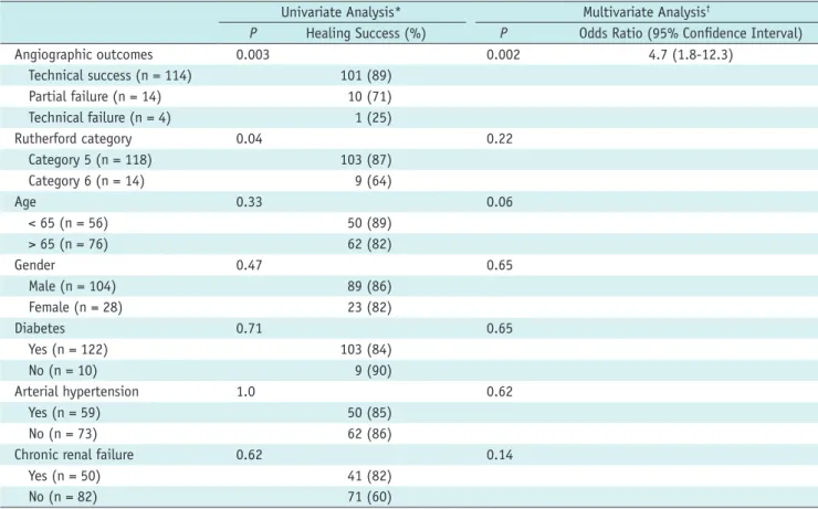
= 2), BKA (n = 8). Relationship between wound healing and angiographic outcome was summarized in Table 5. The healing success rate for PF group was statistically lower than that of TS group but statistically higher than that of CF group. The differences were statistically significant by univariate analysis (p = 0.003) and multivariate analysis adjusted for age, gender, diabetes, arterial hypertension, CRF, and Rutherford category (p = 0.002).
The mean follow-up period was 21 months (range 0-45
months). During the follow-up period, there were BKA in 12
limbs (6%). Recurrences of symptoms occurred in a total
of 53 limbs (28%), of which 50 limbs were managed with repeated endovascular intervention and 3 underwent BKA.
No surgical revascularization was performed in any patient.
Of 50 limbs underwent repeated intervention, 7 limbs (14%) progressed to BKA and 12 patients expired during the follow-up.
The cumulative event-free limb survival rate was 79.3%
and 69.5% at 1- and 3-years respectively (Fig. 2). The
cumulative limb salvage rate (LSR) was 94.8% and 92.0%
at 1- and 3-years retrospectively. The LSR for PF group was statistically lower than that for TS group, but statistically
Table 4. Complications within 1 Month after Procedure
Complications Number (%)
Minor (n = 13)
Coil embolization for arterial perforation 6 Puncture site hematoma/pseudoaneurysm 4
Distal embolization 3
Major (n = 11)
Cardiac function deterioration including
myocardial infarction 6
Upper gastrointestinal hemorrhages required
interventions 3
Puncture site bleeding required embolization 1
Aspiration pneumonia 1
Gastrointestinal perforation with sepsis 1
Table 5. Factors Affecting Healing of Wounds (n = 132)
Univariate Analysis* Multivariate Analysis†
P Healing Success (%) P Odds Ratio (95% Confidence Interval)
Angiographic outcomes 0.003 0.002 4.7 (1.8-12.3)
Technical success (n = 114) 101 (89)
Partial failure (n = 14) 10 (71)
Technical failure (n = 4) 1 (25)
Rutherford category 0.04 0.22
Category 5 (n = 118) 103 (87)
Category 6 (n = 14) 9 (64)
Age 0.33 0.06
< 65 (n = 56) 50 (89)
> 65 (n = 76) 62 (82)
Gender 0.47 0.65
Male (n = 104) 89 (86)
Female (n = 28) 23 (82)
Diabetes 0.71 0.65
Yes (n = 122) 103 (84)
No (n = 10) 9 (90)
Arterial hypertension 1.0 0.62
Yes (n = 59) 50 (85)
No (n = 73) 62 (86)
Chronic renal failure 0.62 0.14
Yes (n = 50) 41 (82)
No (n = 82) 71 (60)
Note.— *Analysis by Fisher’s exact test, †Analysis by multiple logistic regression
Fig. 2. Kaplan-Meier curve shows event-free survival of limbs after endovascular revascularization for critical limb ischemia.
Probabilities of event free limb survival at 1 year and 3 years are 79.3% and 69.5%.
1.0 0.8 0.6 0.4 0.2 0.0
Cumulative event-free limb survival
0 250 500 750 1000 1250 Time (days)
higher than that for TF group (p < 0.001) (Fig. 3).
DISCUSSION
As the prevalence of diabetes increases and endovascular techniques advance, treatment of patients with CLI, most of whom have a non-healing diabetic foot wound with severe peripheral arterial disease (PAD) is a matter of concern. PAD is an important prognostic factor of the diabetic foot. PAD is a part of about 56% of diabetic foot ulcers (DFU), major amputation is more common in DFU limbs with PAD, and the revascularization group of DFU had lower major limb amputation rate (13-15).
This study is a large single-center experience of the effectiveness of endovascular treatment CLI. The mean follow-up of 21 months is comparable to other recent single-center reports (6-9).
Diabetic foot (91%) was the most common cause of CLI.
Rates of comorbidities in the patient population and lesion distribution, characterized by the majority (96%) of the limbs having poor infrapopliteal arteries, are consistent with other studies (3, 7, 16-18).
Rate of subintimal crossing of the occluded
femoropopliteal (68%) or infrapopliteal artery (72%) was high compared with other reports (7, 17, 19-21). The higher subintimal crossing rate is thought to be attributed to operator’s preference of hydrophilic guide wire rather than chronic total occlusion wire, limitation of devices
due to restriction of reimbursement by the national health insurance, and no application of distal artery access until the late phase of the study period.
For the CLI patient with a wound, we wanted to focus on the impact of revascularization on wound healing itself, because avoidance of major amputation alone does not mean the wound is healed. A limb with a superficial toe ulcer can aggravate and turn out to TM amputation, even it is not major amputation, if the pedal arteries are jeopardized by the procedure and a limb with a deep plantar wound can be healed with wound care even after revascularization of the tibial artery fail. In our study, wounds did not heal in 11% even when revascularization of at least one tibial artery was successful, and wound healing in 71% or 25% after revascularization failed partially or completely. This result suggests that just revascularization of one tibial artery is not sufficient to heal a wound if it does not initially improve direct arterial flow to the wound, and stresses the importance of combined local management of wound. Differences of wound healing rate from other studies, in which Giles et al. (9) reported a 57% healing rate and Conrad et al. (6) reported a 57% healing rate, also reflect complexity of pathogenesis, patient’s population and strategies of wound care in CLI.
The healing success rate of CF, PF and TS group gradually increased and the differences between the groups were statistically significant in our results. This means even partially successful revascularization through the peroneal
Fig. 3. Kaplan-Meier curves showing limb salvage rate (LSR) after endovascular revascularization (A) overall LSRs at 1 year and 3 years are 94.8% and 92% respectively. (B) Differences of LSR were statistically significant between technical success and technical failure group (p < 0.001), and between partial failure and technical failure groups (p < 0.001). TS = technical success, PF= partial failure, TF = technical failure 1.0
0.8 0.6 0.4 0.2 0.0
Cumulative limb salvage rate
0 250 500 750 1000 1250 Time (days)
1.0 0.8 0.6 0.4 0.2 0.0
Cumulative limb salvage rate
0 250 500 750 1000 1250 TSPF
TFTS-censored PF-censored TF-censored
Time (days)
A B
artery or treatment of inflow only can significantly improve clinical outcomes. This is partially supported by the study of Faglia et al. (5). They had seven (7%) major amputations in 104 limbs of only peroneal artery revascularization. These data may suggest that quantitative scoring of angiographic outcome rather than dichotomous grading from inflow artery to the wound related artery would be needed to predict short-term and long-term result.
We could not calculate patency rate because many patients received the procedure before establishment of institutional surveillance protocol of revascularized vessel.
The event rate of free limb survival appears similar to other studies. Söder et al. (22) showed that 63% (45 of 72) of the limbs did not require further invasive treatment.
Peregrin et al. (7) reported that the primary LSR, defined as functional limb salvage after the first revascularization with no subsequent intervention, of 76.1% after 1 year of follow-up in a large retrospective study. Our results of LSRs support the prior studies, which have found that despite an inferior primary patency, an endovascular revascularization leads to good LSRs that range from 80% to 95% at 1 to 3 years (4, 6-8, 17, 23).
Rate for major complication (6%) and 30-day mortality (2%) suggest that endovascular revascularization can be performed with lower peri-procedural morbidity and mortality even in high-risk patients (6, 7, 16). Higher upper gastrointestinal bleeding rate (1.6%) than other reported result (0.2%) (15) was thought to be caused by a high prevalence of peptic ulcer in the general population of the area where the study was performed (24).
The limitations of this study were that data were the retrospective design, changes in endovascular techniques and use of the devices during the study period, and the single center nature of the study, possibly biasing indication criteria or strategy. In addition, angiographic outcome and its relationship with wound healing were not analyzed based on the concept of angiosome and wound related artery (25, 26). Also, transcutaneous oxygen measurement as an objective indicator of revascularization was not possible due to lack of facility.
In conclusion, endovascular revascularization is effective to facilitate healing of wounds in CLI and can be performed without significant complications. Although recurrence rate is high, good LSRs are possible. Considering the high prevalence of comorbidities, endovascular revascularization should be chosen as first-line therapy in patients with CLI.
REFERENCES
1. Norgren L, Hiatt WR, Dormandy JA, Nehler MR, Harris KA, Fowkes FG, et al. Inter-Society Consensus for the Management of Peripheral Arterial Disease (TASC II). Eur J Vasc Endovasc Surg 2007;33 Suppl 1:S1-S75
2. Wild S, Roglic G, Green A, Sicree R, King H. Global prevalence of diabetes: estimates for the year 2000 and projections for 2030. Diabetes Care 2004;27:1047-1053
3. Boulton AJ. The diabetic foot: a global view. Diabetes Metab Res Rev 2000;16 Suppl 1:S2-S5
4. Faglia E, Dalla Paola L, Clerici G, Clerissi J, Graziani L, Fusaro M, et al. Peripheral angioplasty as the first-choice revascularization procedure in diabetic patients with critical limb ischemia: prospective study of 993 consecutive patients hospitalized and followed between 1999 and 2003. Eur J Vasc Endovasc Surg 2005;29:620-627
5. Faglia E, Clerici G, Clerissi J, Mantero M, Caminiti M, Quarantiello A, et al. When is a technically successful peripheral angioplasty effective in preventing above-the- ankle amputation in diabetic patients with critical limb ischaemia? Diabet Med 2007;24:823-829
6. Conrad MF, Crawford RS, Hackney LA, Paruchuri V, Abularrage CJ, Patel VI, et al. Endovascular management of patients with critical limb ischemia. J Vasc Surg 2011;53:1020-1025 7. Peregrin JH, Koznar B, Kovác J, Lastovicková J, Novotný J,
Vedlich D, et al. PTA of infrapopliteal arteries: long-term clinical follow-up and analysis of factors influencing clinical outcome. Cardiovasc Intervent Radiol 2010;33:720-725 8. Odink H, van den Berg A, Winkens B. Technical and clinical
long-term results of infrapopliteal percutaneous transluminal angioplasty for critical limb ischemia. J Vasc Interv Radiol 2012;23:461-467, 467.e1
9. Giles KA, Pomposelli FB, Spence TL, Hamdan AD, Blattman SB, Panossian H, et al. Infrapopliteal angioplasty for critical limb ischemia: relation of TransAtlantic InterSociety Consensus class to outcome in 176 limbs. J Vasc Surg 2008;48:128-136 10. Rutherford RB, Baker JD, Ernst C, Johnston KW, Porter JM,
Ahn S, et al. Recommended standards for reports dealing with lower extremity ischemia: revised version. J Vasc Surg 1997;26:517-538
11. Sacks D, Marinelli DL, Martin LG, Spies JB; Society of Interventional Radiology Technology Assessment Committee.
Reporting standards for clinical evaluation of new peripheral arterial revascularization devices. J Vasc Interv Radiol 2003;14(9 Pt 2):S395-S404
12. Garbalosa JC, Cavanagh PR, Wu G, Ulbrecht JS, Becker MB, Alexander IJ, et al. Foot function in diabetic patients after partial amputation. Foot Ankle Int 1996;17:43-48
13. Jeffcoate WJ, Harding KG. Diabetic foot ulcers. Lancet 2003;361:1545-1551
14. Morbach S, Furchert H, Gröblinghoff U, Hoffmeier H, Kersten K, Klauke GT, et al. Long-term prognosis of diabetic foot patients and their limbs: amputation and death over the course of a
decade. Diabetes Care 2012;35:2021-2027
15. Campbell WB, Ponette D, Sugiono M. Long-term results following operation for diabetic foot problems: arterial disease confers a poor prognosis. Eur J Vasc Endovasc Surg 2000;19:174-177
16. Adam DJ, Beard JD, Cleveland T, Bell J, Bradbury AW, Forbes JF, et al. Bypass versus angioplasty in severe ischaemia of the leg (BASIL): multicentre, randomised controlled trial. Lancet 2005;366:1925-1934
17. Romiti M, Albers M, Brochado-Neto FC, Durazzo AE, Pereira CA, De Luccia N. Meta-analysis of infrapopliteal angioplasty for chronic critical limb ischemia. J Vasc Surg 2008;47:975- 981
18. Graziani L, Silvestro A, Bertone V, Manara E, Andreini R, Sigala A, et al. Vascular involvement in diabetic subjects with ischemic foot ulcer: a new morphologic categorization of disease severity. Eur J Vasc Endovasc Surg 2007;33:453-460 19. Alexandrescu V, Hubermont G, Philips Y, Guillaumie B,
Ngongang Ch, Coessens V, et al. Combined primary subintimal and endoluminal angioplasty for ischaemic inferior-limb ulcers in diabetic patients: 5-year practice in a multidisciplinary
‘diabetic-foot’ service. Eur J Vasc Endovasc Surg 2009;37:448- 456
20. Conrad MF, Kang J, Cambria RP, Brewster DC, Watkins MT, Kwolek CJ, et al. Infrapopliteal balloon angioplasty for the treatment of chronic occlusive disease. J Vasc Surg 2009;50:799-805.e4
21. Faglia E, Mantero M, Caminiti M, Caravaggi C, De Giglio R, Pritelli C, et al. Extensive use of peripheral angioplasty, particularly infrapopliteal, in the treatment of ischaemic diabetic foot ulcers: clinical results of a multicentric study of 221 consecutive diabetic subjects. J Intern Med 2002;252:225-232
22. Söder HK, Manninen HI, Jaakkola P, Matsi PJ, Räsänen HT, Kaukanen E, et al. Prospective trial of infrapopliteal artery balloon angioplasty for critical limb ischemia: angiographic and clinical results. J Vasc Interv Radiol 2000;11:1021-1031 23. Kudo T, Chandra FA, Ahn SS. The effectiveness of percutaneous
transluminal angioplasty for the treatment of critical limb ischemia: a 10-year experience. J Vasc Surg 2005;41:423-435;
discussion 435
24. Eun CS, Han DS, Park JY, Jeon YC, Hahm JS, Kim KS, et al.
Changing pattern of antimicrobial resistance of Helicobacter pylori in Korean patients with peptic ulcer diseases. J Gastroenterol 2003;38:436-441
25. Utsunomiya M, Nakamura M, Nakanishi M, Takagi T, Hara H, Onishi K, et al. Impact of wound blush as an angiographic end point of endovascular therapy for patients with critical limb ischemia. J Vasc Surg 2012;55:113-121
26. Neville RF, Attinger CE, Bulan EJ, Ducic I, Thomassen M, Sidawy AN. Revascularization of a specific angiosome for limb salvage: does the target artery matter? Ann Vasc Surg 2009;23:367-373