• 검색 결과가 없습니다.

3D Printer Application for Endoscope-Assisted Spine Surgery Instrument Development: From Prototype Instruments to Patient-Specific 3D Models

N/A
N/A
Protected

Academic year: 2022

Share "3D Printer Application for Endoscope-Assisted Spine Surgery Instrument Development: From Prototype Instruments to Patient-Specific 3D Models"

Copied!
6
0
0

로드 중.... (전체 텍스트 보기)

전체 글

(1)

INTRODUCTION

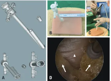
Recently, 3D printers have been applied in various medical fields.

Creating individualized implants for patients has become the most popular application in the surgical field, and the produc- tion of phantom models is also becoming more common.

1,2

Mak-

ing surgical instruments could be a good field of application for 3D printers, although attempts to do so have proven limited.

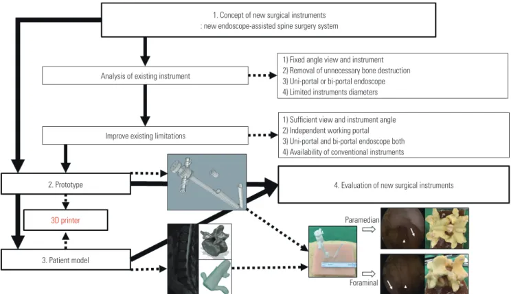
When surgeons have new ideas on surgical instruments, they usually make prototypes using metal, and test their feasibility in cadavers. These processes can be tedious, costly, and time- consuming.

3

In the past, endoscopic spine surgery has primarily been ap- plied for lumbar discectomy. However, with further develop- ment of endoscopic equipment and techniques, the indications thereof have been expanded to spinal stenosis.

4

Since the view and instrument are fixed, conventional endoscopic spine sur- gery has limitations in decompressing foraminal stenosis suf- ficiently, without excessive facet joint resection.
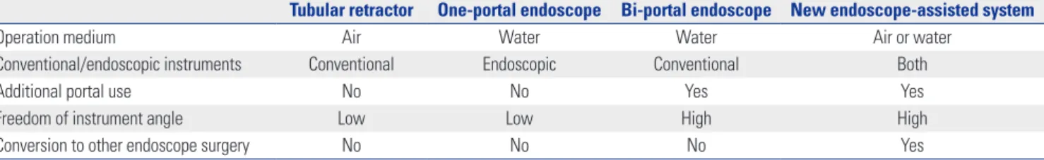
5

To overcome the limitations of endoscopic spine surgery, bi-portal endoscop- ic spine surgery was recently developed. Due to an independent- ly working portal and the widespread availability of convention- al instruments, bi-portal endoscopic spine surgery has become

3D Printer Application for Endoscope-Assisted Spine Surgery Instrument Development: From Prototype Instruments to Patient-Specific 3D Models

Hee-Seok Yang

1

and Jeong-Yoon Park

2

1

Department of Neurosurgery, Seoul Barunsesang Hospital, Seoul;

2

Department of Neurosurgery, Spine and Spinal Cord Institute, Gangnam Severance Hospital, Yonsei University College of Medicine, Seoul, Korea.

Developing new surgical instruments is challenging. While making surgical instruments could be a good field of application for 3D printers, attempts to do so have proven limited. We designed a new endoscope-assisted spine surgery system, and using a 3D printer, attempted to create a complex surgical instrument and to evaluate the feasibility thereof. Developing the new surgical in- struments using a 3D printer consisted of two parts: one part was the creation of a prototype instrument, and the other was the pro- duction of a patient model. We designed a new endoscope-assisted spine surgery system with a cannula for the endoscope and working instruments and extra cannula that could be easily added. Using custom-made patient-specific 3D models, we conducted discectomies for paramedian and foraminal discs with both the newly designed spine surgery system and conventional tubular surgery. The new spine surgery system had an extra portal that can be well bonded in by a magnetic connector and greatly expand- ed the range of access for instruments without unnecessary bone destruction. In foraminal discectomy, the newly designed spine surgery system showed less facet resection, compared to conventional surgery. We were able to develop and demonstrate the use- fulness of a new endoscope-assisted spine surgery system relying on 3D printing technology. Using the extra portal, the usability of endoscope-assisted surgery could be greatly increased. We suggest that 3D printing technology can be very useful for the real- ization and evaluation of complex surgical instrument systems.

Key Words: Endoscope-assisted, 3D printing, paramedian, foraminal, tubular, discectomy

pISSN: 0513-5796 · eISSN: 1976-2437

Received: August 14, 2019 Revised: October 21, 2019 Accepted: November 21, 2019

Corresponding author: Jeong-Yoon Park, MD, PhD, Department of Neurosurgery, Spine and Spinal Cord Institute, Gangnam Severance Hospital, Yonsei University College of Medicine, 211 Eonjuro, Gangnam-gu, Seoul 06273, Korea.

Tel: 82-2-2019-3390, Fax: 82-2-3461-9229, E-mail: [email protected]

•The authors have no potential conflicts of interest to disclose.

© Copyright: Yonsei University College of Medicine 2020

This is an Open Access article distributed under the terms of the Creative Com- mons Attribution Non-Commercial License (https://creativecommons.org/licenses/

by-nc/4.0) which permits unrestricted non-commercial use, distribution, and repro- duction in any medium, provided the original work is properly cited.

Yonsei Med J 2020 Jan;61(1):94-99

https://doi.org/10.3349/ymj.2020.61.1.94

(2)

a breakthrough technique in endoscopic spine surgery.

6

Although bi-portal endoscopic spine surgery has numerous advantages, conventional endoscopic surgery is still useful. Surgeons, how- ever, cannot apply both endoscopic surgery techniques at the same time, due to the completely different designs of the surgi- cal instruments used in each technique. In this study, we aimed to design a new endoscope-assisted spine surgery system that could permit the performance of both conventional and bi-por- tal endoscopic spine surgery using a single surgical instrument.

We also sought to demonstrate the usefulness of 3D printing for the development of complex surgical instruments.

MATERIALS AND METHODS

Workflow of surgical instrument development

For the development of the new surgical instrument, four steps were needed (Fig. 1). The first step was the conceptualization of the new surgical instrument, and the second step involved making a prototype based on new concept and ideas. The third step was to make a patient-specific spine model, and the fourth step was the evaluation of the new surgical instruments. 3D print- ing technique was primarily applied in two parts: making the prototype instrument and the patient-specific model to test the new instrument.

Endoscope-assisted spine surgery instrument concept and design

Two cannulas were joined together: one for an endoscope, and another for surgical instruments. The combined cannula had snowman-shaped opening, with major and minor axis diam- eters of 10.34 mm and 9.16 mm, respectively. The instrument cannula could be blocked with a rubber cap for conversion to bi-portal endoscopic surgery (Fig. 2A), and two cannulas were jointed to reduce diameter and to adjust the endoscopic view (Fig. 2B and C). The new system separated the instrument can- nula from the endoscope, which allowed the use of thicker in- struments (Fig. 2C and D). The instrument cannula was designed to have a 10-mm diameter; therefore, it was possible to use both an endoscope and classical instruments (Fig. 2A). After a 10-mm skin incision, the new endoscope-assisted spine surgery sys- tem was inserted (Fig. 2B).

If an extra portal was needed, we made a 5-mm incision in- dependently and inserted a new working portal using the de- sired angle (Fig. 3A and B). To position the extra working portal in the optimal place, a magnetic connector was designed. The angle between the primary cannula and extra portal was de- signed to be 18 degrees. The extra portal could move together with the endoscope, and the shape of the extra portal could be selected as circular or semicircular (Fig. 3A). After a 5-mm skin incision, the extra working portal was inserted and linked to the magnetic connector of the new endoscope-assisted spine sur- gery system (Fig. 3B, C, and D). We used standard 4-mm di-

1. Concept of new surgical instruments : new endoscope-assisted spine surgery system

Paramedian

Foraminal

4. Evaluation of new surgical instruments Analysis of existing instrument

1) Fixed angle view and instrument 2) Removal of unnecessary bone destruction 3) Uni-portal or bi-portal endoscope 4) Limited instruments diameters

1) Sufficient view and instrument angle 2) Independent working portal

3) Uni-portal and bi-portal endoscope both 4) Availability of conventional instruments Improve existing limitations

2. Prototype

3. Patient model 3D printer

Fig. 1. Workflow diagram of the development and evaluation of new surgical instruments. The solid arrow indicates the conventional application, and the

dotted arrow indicates the application of endoscope-assisted spine surgery.

(3)

ameter and 0-degree endoscopes for the bi-portal endoscope system.

Customized patient-specific 3D spine model

We made a customized patient-specific spine model. The pro- cess for making the spine model consisted of five steps: 1) 3D modeling of spine components using MR and CT scans, 2) mak- ing 3D-printed spine components via 3D printer, 3) making sili- cone molds using 3D-printed spine components, 4) making spine components in consideration of physical properties, and 5) assembly of the patient-specific model.

MR images and CT scans of patients were obtained using a picture archiving and communication system (PACS; IN- FINITT, Seoul, Korea). Imaging data were imported to 3D Slic- er software v 10.0 (https://www.slicer.org), free, open source software for medical imaging computing and 3D reconstruc- tion, according to the Digital Imaging and Communications in Medicine format (Fig. 4A). After the 3D digital images were calculated and reconstructed, every component was separated and exported to a 3D printer (Creatable D3, A-Team Ventures, Seoul, Korea) (Fig. 4B). After the 3D-printed spine components were obtained, silicon molds were made (Fig. 4C). Then, we de- composed the spine structures and assigned these components into two groups. The first group comprised structures that need- ed to have their shape and rigidity preserved, such as bone, and the other group comprised structures that had similar elas- ticity to human tissue, such as discs, nerves, and ligaments. We used various silicones and polymers, based on manufacturer

specifications and reported papers (Fig. 4D).

7

Using each sili- cone mold, vertebral bodies were cast with urethane foam (FOAM-iT5 and FOAM-iT10, Smooth-On, Macungie, PA, USA), intervertebral discs, paraspinal muscles with polyurethane rubber (VytaFlex 10, Smooth-On), neural structures, and liga- ment flavum with silicone (Dragon skin 10, Smooth-On) (Fig.

A

B

D

C

Fig. 2. New endoscope-assisted spine surgery system. (A) Two tubes were joined together: one for an endoscope and the other for an instru- ment working cannula. The instrument working cannula could be blocked with a rubber cap to maintain proper saline pressure. (B) 3D-printed pro- totypes of the new endoscope-assisted spine surgery system. A 10-mm incision was needed for patient model application. (C) Photograph of a surgical test using the new endoscope-assisted spine surgery system.

An endoscopic drill was inserted at the working cannula. (D) Operation field with the new system. Similar operational views with conventional endoscopic spine surgery. The arrow head is the ligamentum flavum at the inter-laminar space, and the arrows indicate the lamina of the superi- or and inferior vertebral body.

A

B

D

C

Fig. 3. New endoscope-assisted spine surgery system with an additional working portal. (A) After blocking of the instrument working cannula, only the endoscope cannula was used. Various shapes (semicircular or circu- lar) of an additional working portal could be used. (B) 3D-printed proto- type of the new endoscope-assisted spine surgery system with an addi- tional working portal. A 10-mm incision and 5-mm additional incision were needed for patient model application. (C) Photograph of a surgical test using the new endoscope-assisted spine surgery system with a semicircular additional working portal. Conventional punch and instru- ments could be used. (D) A conventional drill was inserted at the working cannula. The operation field was well-visualized with the new system with the additional working portal. Instrument movement was freer, com- pared to conventional endoscopic spine surgery, such as bi-portal endo- scopic spine surgery. The arrow head indicates the ligamentum flavum at the inter-laminar space, and the arrow shows the lamina of the superior vertebral body.

Fig. 4. Patient-based model created with the 3D printing technique. (A) 3D modeling of spine components using patient MRI data. (B) Printing spine components using a 3D printer. (C) Making silicone molds for patient- based 3D-printed spine components. (D) Making spinal components con- sidering physical properties (vertebra, discs, nerves, ligaments) using pa- tient-based spine component molds. (E) Making a patient model after assembling the body frame, spine components, and skin with soft tissue.

A B C

D E

(4)

4D). Supportive structures were cast with polyurethane rub- ber (PMC-770, Smooth-On), body frames were cast with ure- thane foam (FOAM-iT5), and skin with muscle components was cast with polyurethane rubber (VytaFlex 10) (Fig. 4E). Af- ter making all spine components, every part was assembled (Fig.

4E). We made two pairs of patient-specific spine models for lumbar 4–5 paramedian disc herniation and lumbar 4–5 fo- raminal disc herniation on the left.

Comparison according to surgical instrumentation and approach

The surgical results were compared according to surgical in- strumentation and methods. Surgical methods were paramedi- an discectomy and foraminal discectomy (Wiltse posterolateral spinal approach).

8

Surgical results of the new endoscope-as- sisted spine surgery system were compared with a tubular re- tractor (16-mm diameter, Medtronic Sofamor Danek, Memphis, TN, USA) using a standard surgical technique (Figs. 5 and 6).

Until sufficient nerve decompression was obtained, a tubular retractor and the new system were used to perform discectomy using two different approaches (paramedian, foraminal dis- cectomy), and the degrees of bone resection were compared after surgery.

RESULTS

Using a conventional tubular retractor and the new system, dis- cectomies were performed through paramedian and forami- nal approaches using two pairs of customized patient-specific 3D spinal models (Supplementary Videos 1–4, only online). The paramedian approach using the tubular retractor was per- formed using a conventional method (Fig. 5, Supplementary Video 1, only online).

9

The paramedian approach using the new endoscope-assisted spine surgery system at the lumbar 4–5 and the left paramedian disc herniation was also performed (Fig. 6, Supplementary Video 3, only online). After making a 10-mm incision in the skin layer near the midline, we inserted the new system (Figs. 2C and 6), and a 4-mm endoscope was positioned into the endoscope cannula. After saline inflow was implemented, discectomy was performed using conventional full endoscope instruments (Fig. 2C and D). This should have provided us with enough vision for sufficient decompression, and the view and resolution were very similar to conventional endoscopic spine surgery (Fig. 2D). An additional 5-mm inci- sion was made (Fig. 3B), and the extra portal with magnetic con- nector was positioned properly (Fig. 3B and C). Circular and semi-circular shapes of extra portals were available, and discec- tomy was possible using various conventional surgical instru- ments (Fig. 3D, Supplementary Video 3, only online). Since the surgical instruments had gone down the working portal wall, the instrument could be placed safely.

The foraminal approach using the tubular retractor was per- formed according to a conventional method (Fig. 5, Supple- mentary Video 2, only online). Foraminal discectomy was per- formed with the new endoscope-assisted spine surgery system using the lumbar 4–5, left foraminal disc herniation model. Af- ter partial resection of the superior articular process, exiting nerve roots and foraminal discs were identified, and disc ma- terials were removed with pituitary forceps via the extra portal (Fig. 6, Supplementary Video 4, only online). The authors com- pared the bone destruction of these spine models after discec- tomy (Figs. 5 and 6). Comparison of the conventional tubular re- tractor and the new endoscope-assisted spine system showed that sufficient neural decompression and discectomy were achieved by both surgical approaches. However, damage to the superior articular process was significantly reduced with the foraminal approach using the new surgical system (Figs. 5 and 6, foraminal approach).

DISCUSSION

Herein, we described our process of developing a complex sur- gical system using a 3D printer, and confirmed the usefulness of the 3D printer for the development of surgical instruments.

Endoscopic spine has rapidly evolved since it was initially de- scribed by Ruetten, et al.

10

With various reports of decompres- Paramedian

Foraminal

Fig. 5. Discectomy with a tubular retractor. A paramedian approach is shown above, and a foraminal approach is shown below. The arrows in- dicate the nerve root, and arrow heads show the discs.

Paramedian

Foraminal

Fig. 6. Discectomy with the new endoscope-assisted spine surgery sys-

tem. A paramedian approach is shown above, and a foraminal approach

is shown below. The arrows indicate the nerve root, and arrow heads

show the discs. Less bone destruction was observed with the new sys-

tem, compared to the conventional tubular retractor in the foraminal ap-

proach.

(5)

sive surgery for spinal stenosis, endoscopic spine surgeries have been frequently used for spine treatment.

4

The goal of mini- mally invasive surgery is to achieve the same results as those of conventional surgery, while minimizing damage to the nor- mal structure.

11

Bi-portal endoscopic surgery offers a greater range of motion of instruments and availability of conventional surgical instruments, and it can be applied to various patholo- gies, including stenosis.

12

In this study, we designed a new endo- scope-assisted spine surgery system that could allow for both conventional and bi-portal endoscopic spine surgery methods to be applied at the same time, and our trial surgery showed that the new system works properly. The expected advantages in the new endoscopic spine surgery over tubular retractor, con- ventional one-portal, and bi-portal endoscopic surgeries are summarized in Table 1. In this study, we only performed com- parative surgery using a tubular retractor. Sufficient neural de- compression and discectomy were achieved with both the tu- bular retractor and the new endoscopic spine surgery systems, although damage to the superior articular process was signifi- cantly reduced with a foraminal approach using the new en- doscopic spine surgery system.

Rankin, et al.

13

reported that the estimated cost per unit of a 3D-printed instrument is roughly 1/10 of the cost of stainless steel instruments. Testing a prototype with a cadaver is also very different from the actual operative situation, and the process is also very expensive. Recently, it has become possible to make patient-specific models with high accuracy, and per- sonalized 3D printing prostheses can be extensively applied in clinical practice. High accuracy is essential for 3D printers, and previous studies have validated the accuracy of 3D-printed spine models. Wu, et al.

14

compared CT images of cervical, tho- racic, and lumbar vertebrae against 3D-printed models, and found that the 3D-printed models had strong anatomical cor- relations. Previous studies have assessed the accuracy of 3D- printed models representing cadaveric pelvises, and observed no significant differences between them.

15

Kidney models us- ing silicone materials have been found to show similar elastici- ty with real kidneys, as well as biocompatible properties on MR and ultrasound imaging.

16

Such disease models can be made repeatedly, and comparative testing can be possible using the same models.

3D printers have been used in spine surgeries earlier than in other surgeries. A 3D titanium alloy vertebrae was implant- ed for cervical spine reconstruction following a Ewing sarco-

ma resection, and a 3D-printed implant for pelvic reconstruc- tion following hemi-sacral resection was successfully applied.

17,18

Multiple studies have reported the successful use of patient- specific 3D-printed implants in complex spinal pathologies.

19

Although accurate fabrication of patient-specific implants has been described in many studies, literature on 3D printer utili- zation for surgical instrument development is scarce. Until now, making only simple surgical instruments, such as retractors, forceps, and hemostats, has been attempted with the use of 3D printing.

13,20

In this study, we wanted to show the usefulness of 3D printers for the development of complex surgical instru- ments, and overall, we deemed that 3D printers can be effec- tively applied in the development of complex surgery instru- ments. We are confident that many surgeons would be able to test their ideas easily with 3D printing.

Our new endoscope-assisted spine surgery system and trial with a 3D-printed patient-specific model involved many limita- tions. Postoperative outcome analysis using a 3D-printed mod- el could be different from using actual patients. Also, the sur- gical trial was conducted only once, and it was not compared to conventional endoscopic spine surgery. Since we could not confirm whether the magnetic connector would be adequately bonded when it is inside a real human, a cadaver trial is need- ed to confirm this matter. However, the purpose of this study was to show the usefulness of 3D printers for the development of complex surgical instruments. The authors are well-aware that the new endoscope-assisted spine surgery system needs further improvements, and future studies with more complicat- ed degenerative spinal diseases and deformed patient-specific models are required. Nevertheless, we were able to make and test a new idea on surgical instruments quickly, in addition to introducing the possibility of a new endoscope-assisted spine surgery system using 3D printing technology.

SUPPLEMENTARY DATA

Video 1. Paramedian discectomy for lumbar 4–5, left parame- dian disc herniation using a tubular retractor.

Video 2. Foraminal discectomy (Wiltse posterolateral spinal approach) of lumbar 4–5, left foraminal disc herniation using a tubular retractor.

Video 3. Paramedian discectomy for lumbar 4–5, left parame- dian disc herniation using the new endoscope-assisted spine surgery system.

Table 1. Comparison of Various Endoscopic Spine Surgeries and the New System

Tubular retractor One-portal endoscope Bi-portal endoscope New endoscope-assisted system

Operation medium Air Water Water Air or water

Conventional/endoscopic instruments Conventional Endoscopic Conventional Both

Additional portal use No No Yes Yes

Freedom of instrument angle Low Low High High

Conversion to other endoscope surgery No No No Yes

(6)

Video 4. Foraminal discectomy (Wiltse posterolateral spinal approach) of lumbar 4–5, left foraminal disc herniation using the new endoscope-assisted spine surgery system.

AUTHOR CONTRIBUTIONS

Conceptualization: Hee-Seok Yang, Jeong-Yoon Park. Data curation:

Hee-Seok Yang, Jeong-Yoon Park. Formal analysis: Hee-Seok Yang, Jeong-Yoon Park. Funding acquisition: Hee-Seok Yang, Jeong-Yoon Park. Investigation: Hee-Seok Yang, Jeong-Yoon Park. Methodology:

Hee-Seok Yang, Jeong-Yoon Park. Project administration: Hee-Seok Yang, Jeong-Yoon Park. Resources: Hee-Seok Yang. Software: Hee-Seok Yang. Supervision: Jeong-Yoon Park. Validation: Hee-Seok Yang, Jeong-Yoon Park. Visualization: Hee-Seok Yang, Jeong-Yoon Park.

Writing—original draft: Hee-Seok Yang, Jeong-Yoon Park. Writing—

review & editing: Jeong-Yoon Park.

ORCID iDs

Hee-Seok Yang https://orcid.org/0000-0002-8822-4230 Jeong-Yoon Park https://orcid.org/0000-0002-3728-7784

REFERENCES

1. Mavili ME, Canter HI, Saglam-Aydinatay B, Kamaci S, Kocadereli I. Use of three-dimensional medical modeling methods for pre- cise planning of orthognathic surgery. J Craniofac Surg 2007;18:

740-7.

2. Lee S, Kim JY, Hong J, Baek SH, Kim SY. CT-based navigation sys- tem using a patient-specific instrument for femoral component positioning: an experimental in vitro study with a sawbone model.

Yonsei Med J 2018;59:769-80.

3. Christoudias GC. The making of an instrument: from concept to market. JSLS 1998;2:301-7.

4. Kim JE, Choi DJ. Clinical and radiological outcomes of unilateral biportal endoscopic decompression by 30° arthroscopy in lumbar spinal stenosis: minimum 2-year follow-up. Clin Orthop Surg 2018;

10:328-36.

5. Wang YP, Zhang W, Li BL, Sun YP, Ding WY, Shen Y. Suprapedicu- lar foraminal endoscopic approach to lumbar lateral recess de- compression surgery to treat degenerative lumbar spinal stenosis.

Med Sci Monit 2016;22:4604-11.

6. Heo DH, Quillo-Olvera J, Park CK. Can percutaneous biportal en- doscopic surgery achieve enough canal decompression for degen- erative lumbar stenosis? Prospective case-control study. World Neurosurg 2018;120:e684-9.

7. Sayed Aluwee SAZB, Zhou X, Kato H, Makino H, Muramatsu C, Hara T, et al. Evaluation of pre-surgical models for uterine surgery by use of three-dimensional printing and mold casting. Radiol Phys Technol 2017;10:279-85.

8. Siu TL, Lin K. Microscopic tubular discectomy for far lateral lum- bar disc herniation. J Clin Neurosci 2016;33:129-33.

9. Clark AJ, Safaee MM, Khan NR, Brown MT, Foley KT. Tubular mi- crodiscectomy: techniques, complication avoidance, and review of the literature. Neurosurg Focus 2017;43:E7.

10. Ruetten S, Komp M, Merk H, Godolias G. Surgical treatment for lumbar lateral recess stenosis with the full-endoscopic interlami- nar approach versus conventional microsurgical technique: a pro- spective, randomized, controlled study. J Neurosurg Spine 2009;

10:476-85.

11. Maroon JC. Current concepts in minimally invasive discectomy.

Neurosurgery 2002;51(5 Suppl):S137-45.

12. Akbary K, Kim JS, Park CW, Jun SG, Hwang JH. Biportal endoscop- ic decompression of exiting and traversing nerve roots through a single interlaminar window using a contralateral approach: tech- nical feasibilities and morphometric changes of the lumbar canal and foramen. World Neurosurg 2018;117:153-61.

13. Rankin TM, Giovinco NA, Cucher DJ, Watts G, Hurwitz B, Arm- strong DG. Three-dimensional printing surgical instruments: are we there yet? J Surg Res 2014;189:193-7.

14. Wu AM, Shao ZX, Wang JS, Yang XD, Weng WQ, Wang XY, et al.

The accuracy of a method for printing three-dimensional spinal models. PLoS One 2015;10:e0124291.

15. Wu XB, Wang JQ, Zhao CP, Sun X, Shi Y, Zhang ZA, et al. Printed three-dimensional anatomic templates for virtual preoperative planning before reconstruction of old pelvic injuries: initial re- sults. Chin Med J (Engl) 2015;128:477-82.

16. Adams F, Qiu T, Mark A, Fritz B, Kramer L, Schlager D, et al. Soft 3D-printed phantom of the human kidney with collecting system.

Ann Biomed Eng 2017;45:963-72.

17. Xu N, Wei F, Liu X, Jiang L, Cai H, Li Z, et al. Reconstruction of the upper cervical spine using a personalized 3D-printed vertebral body in an adolescent with Ewing sarcoma. Spine (Phila Pa 1976) 2016;41:E50-4.

18. Kim D, Lim JY, Shim KW, Han JW, Yi S, Yoon DH, et al. Sacral re- construction with a 3D-printed implant after hemisacrectomy in a patient with sacral osteosarcoma: 1-year follow-up result. Yon- sei Med J 2017;58:453-7.

19. Spetzger U, Frasca M, König SA. Surgical planning, manufactur- ing and implantation of an individualized cervical fusion titanium cage using patient-specific data. Eur Spine J 2016;25:2239-46.

20. George M, Aroom KR, Hawes HG, Gill BS, Love J. 3D printed sur-

gical instruments: the design and fabrication process. World J

Surg 2017;41:314-9.

수치

Fig. 1. Workflow diagram of the development and evaluation of new surgical instruments
Fig. 3. New endoscope-assisted spine surgery system with an additional  working portal
Fig. 6. Discectomy with the new endoscope-assisted spine surgery sys- sys-tem. A paramedian approach is shown above, and a foraminal approach  is shown below
Table 1. Comparison of Various Endoscopic Spine Surgeries and the New System

참조

관련 문서

The composition, structure, and optical properties of 3d-doped CdGa 2 O 4 crystals were studied Stoichiometry of these compound semiconductors were determined

Endoscopic spine surgery has become a practical, minimally invasive technique for decompression in patients with spinal disc herniation or stenosis.. However,

Autho: Effectiveness of surgery for lumbar stenosis and degenerative spondylolisthesis in the octogenarian population: analysis of the Spine Patient Outcomes Research

Percutaneous endoscopic cervical discectomy (PECD) is an effective minimally invasivesurgery for soft cervical disc herniation in properly selected cases..

The Development and Application of Unified Prestress System with Post-tension and Post-compression for Bridge... Unified Prestress

To solve this problem, GTAW assisted Friction Stir Welding method developed (TIG assisted FSW system) exhibited more than 80% the tensile strength of the

Lumbar discogenic radiculopathy (LDR) is one of the most common spinal disorders which can lead to high medical expenses for the patient, occupational impairment,

Using this leap motion, a surgeon can perform the virtual shoulder arthroscopic surgery skillfully and reduce mistakes in surgery.. The simulation using leap motion shows