INTRODUCTION
High-frequency oscillatory ventilation (HFOV) is now widely used as an artificial ventilator that actively performs 180−900 breaths per minute with a very small tidal volume (VT).1 It is used as rescue therapy for severe respiratory failure in prema- ture infants, and it contributes to reduce the incidence of bron- chopulmonary dysplasia.2-5
The settings of the HFOV are based on the fraction of inspired oxygen (FiO2) for oxygen (O2) saturation, mean arterial pres- sure (MAP), which is about 2−4 cm H2O higher than the aver- age airway pressure of conventional mechanical ventilation, amplitude of chest vibration to the umbilicus, and frequency of approximately 10−15 Hz.6-8 On the basis of the carbon diox- ide diffusion coefficient (DCO2) and VT, physicians can con- firm the effect of oxygenation and ventilation.9
In particular, the DCO2 indicates the effectiveness of CO2 re- moval. It has various values depending on the size of the lung, and it is calculated as the product of the squared VT and the fre- quency.10,11 Setting parameters in HFOV, according to the lung pathology, is left to the discretion of the physician, and appro- priate DCO2 and VT values have not yet been established. When the DCO2 is below 40 mL2/s, it has been reported that the prob- ability of a pCO2 below 50 mm Hg is estimated to be 49%, and the appropriate DCO2 value for extremely low-birth-weight (ELBW) infants has been reported as 18.5 mm Hg.12 VT refers Received: June 23, 2017 Revised: September 29, 2017
Accepted: September 30, 2017
Corresponding author: Dr. Soon Min Lee, Department of Pediatrics, Yonsei Uni- versity College of Medicine, 211 Eonju-ro, Gangnam-gu, Seoul 06273, Korea.
Tel: 82-2-2019-3350, Fax: 82-2-2019-4881, E-mail: [email protected]
•The authors have no financial conflicts of interest.
© Copyright: Yonsei University College of Medicine 2018
This is an Open Access article distributed under the terms of the Creative Com- mons Attribution Non-Commercial License (http://creativecommons.org/licenses/
by-nc/4.0) which permits unrestricted non-commercial use, distribution, and repro- duction in any medium, provided the original work is properly cited.
Effective Tidal Volume for Normocapnia in Very-Low-Birth-Weight Infants Using High-Frequency Oscillatory Ventilation
Seul Mi Lee, Ran Namgung, Ho Sun Eun, Soon Min Lee, Min Soo Park, and Kook In Park
Department of Pediatrics, Yonsei University College of Medicine, Seoul, Korea.
Purpose: Removal of CO2 is much efficient during high-frequency oscillatory ventilation (HFOV) for preterm infants. However, an optimal carbon dioxide diffusion coefficient (DCO2) and tidal volume (VT) have not yet been established due to much indi- vidual variance. This study aimed to analyze DCO2 values, VT, and minute volume in very-low-birth-weight (VLBW) infants using HFOV and correlates with plasma CO2 (pCO2).
Materials and Methods: Daily respiratory mechanics and ventilator settings from twenty VLBW infants and their two hundred seventeen results of blood gas analysis were collected. Patients were treated with the Dräger Babylog VN500 ventilator (Dräger- werk Ag & Co.) in HFOV mode. The normocapnia was indicated as pCO2 ranging from 45 mm Hg to 55 mm Hg.
Results: The measured VT was 1.7 mL/kg, minute volume was 0.7 mL/kg, and DCO2 was 43.5 mL2/s. Mean results of the blood gas test were as follows: pH, 7.31; pCO2, 52.6 mm Hg; and SpO2, 90.5%. In normocapnic state, the mean VT was significantly high- er than in hypercapnic state (2.1±0.5 mL/kg vs. 1.6±0.3 mL/kg), and the mean DCO2 showed significant difference (68.4±32.7 mL2/s vs. 32.4±15.7 mL2/s). The DCO2 was significantly correlated with the pCO2 (p=0.024). In the receiver operating curve analysis, the estimated optimal cut-off point to predict the remaining normocapnic status was a VT of 1.75 mL/kg (sensitivity 73%, speci- ficity 80%).
Conclusion: In VLBW infants treated with HFOV, VT of 1.75 mL/kg is recommended for maintaining proper ventilation.
Key Words: Tidal volume; infant, very low birth weight; ventilation, normocapnia, high frequency oscillatory ventilation
pISSN: 0513-5796 · eISSN: 1976-2437 Yonsei Med J 2018 Jan;59(1):101-106
https://doi.org/10.3349/ymj.2018.59.1.101
to VT averaged over several cycles, and it is generally recom- mended to set it to 1.5−2 mL/kg.12 This, however, depends on the lung mechanics and ventilator mechanics, since the vol- ume of the lung increases as the frequency of ventilation de- creases. For greater lung compliance, an ideal VT should be maintained in order to prevent hyperinflation.13 One study re- ported that the set VT values varied between from 1.46 mL/kg for ELBW infants and 1.57 mL/kg for infants weighing 1000−
2000 g.14 In another preterm study of infants <32 weeks of ges- tation, the targeted VT ranged from 1.5 mL/kg to 2.5 mL/kg, and only 51% of infants had normocapnia.12
In the present study, we aim to analyze the respiratory me- chanics, especially DCO2 values, VT, and minute volume and correlate them with plasma CO2 (pCO2) in very-low-birth-weight (VLBW) infants using HFOV.
MATERIALS AND METHODS
Patients
This retrospective study comprised 20 VLBW infants, who re- ceived HFOV without volume guarantee for treatment of re- spiratory failure. These patients had all been admitted to the neonatal intensive care unit of Gangnam Severance Hospital between March 2015 and February 2016. Their two hundred seventeen results of blood gas analysis were collected. Ethical approval was obtained from the ethics committees of Gang- nam Severance Hospital and informed consent was waived (IRB approval number; 3-2017-0120).
Ventilator
HFOV was delivered using a Dräger Babylog VN500 ventilator (Drägerwerk Ag & Co., Lübeck, Germany) with a Venturi system.
It generates active inspiration and active expiration to induce a sinusoidal pressure signal around a set mean airway pressure.
The Dräger Babylog VN500 ventilator uses a double hot-wire anemometer, which has been demonstrated to be a very accurate way of measuring frequency of expired VT during HFOV, as there is greater linearity of response across a range of frequencies.15 Ventilation strategies
HFOV was set as a mean airway pressure of 1 to 2 cm H2O high- er than that received during conventional mechanical ventila- tion and the same inspired oxygen fraction. In chest radio- graph, the lung volume was evaluated with the right diaphragm at the level of the ninth rib. A frequency of 10−12 Hz and an os- cillatory inspiratory/expiratory ratio of 1:1 were used in all in- fants. An amplitude set above the mean airway pressure value was increased until visible chest wall movement up to the um- bilicus level was noted.
If the PCO2 value was outside the target range, the ampli- tude was adjusted up or down in increments of 10−20% as nec- essary in the HFOV alone period. Capillary blood gases were
measured 1 hour after applying HFOV and then at 6−8-hour in- tervals or more often as needed using a blood gas analyser. The FiO2 was given as needed to achieve a peripheral capillary ox- ygen saturation (SpO2) between 90% and 95% by pulse oximetry.
Data acquisition and analysis
During ventilation, a hot-wire anemometer was placed on the airway opening to continuously measure the flow and VT dur- ing HFOV. The VT, Paw, minute volume, and DCO2 were calcu- lated as VT2×frequency (mL2/s) by the ventilator (Dräger Baby- log VN500 2.n software, Dräger) and recorded at 1 minute. Data from the ventilator were exported through a standard USB con- nection.
The following data were also collected from patients includ- ed in the study: demographic characteristics, perinatal pathol- ogy and treatment, clinical course, initial and final ventilator set- tings, vital signs, and capillary blood gas analysis. When analysing the blood gas analysis, pCO2 values between 45 and 55 mm Hg were considered normocapnia.
Statistical analysis
Data were compared using the t-test and chi-square test. All statistical analyses were performed using SPSS for Windows, version 19.0 (IBM Corp., Armonk, NY, USA). A p-value <0.05 was considered statistically significant.
RESULTS
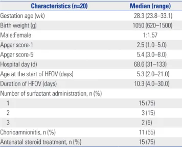
The study population consisted of 20 VLBW infants with a me- dian gestational age of 28.3 weeks and median birth weight of 1050 g. The 1-minute Apgar score was 2.5, and the 5-minute Ap- gar score was 5.4 points. Age at the start of HFOV was 5.3 days, and the duration of HFOV was 10.3 days. Surfactant replace- ment therapy was administered to all the infants, and five in- fants received multiple therapy. No patients with congenital anomalies affecting the cardiac, respiratory or central nervous system were found. Eleven infants had a maternal history of chorioamnionitis, and 15 received steroid treatment (Table 1).
The ventilator parameters were set as follows: inspiratory-to- expiratory (I/E) time ratio, 1:1; frequency, 10.5 Hz; amplitude, 23.6 cm H2O; and MAP, 11.7 cm H2O. The measured VT was 1.7 mL/kg, minute volume was 0.7 mL/kg, and DCO2 was 43.5 mL2/s. Median results of the blood gas test were as follows:
pH, 7.31; pCO2, 52.6 mm Hg; and SpO2, 90.5% (Table 2).
In a comparison of the normocapnia and hypercapnia groups, the VT, minute volume, and DCO2 were statistically sig- nificantly different. The mean VT of the normocapnia group was 2.1 mL/kg, minute volume was 0.9 mL/kg, and DCO2 was 68.4 mL2/s (Table 3). The relationship between pCO2 and DCO2 was negative. The correlation between the VT and pCO2
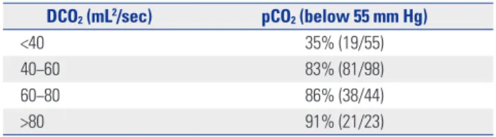
was also negative, shown in Fig. 1. When we divided infants into 4 groups according to the DCO2 value, the percentage of infants
Table 1. Patient Characteristics
Characteristics (n=20) Median (range)
Gestation age (wk) 28.3 (23.8–33.1)
Birth weight (g) 1050 (620–1500)
Male:Female 1:1.57
Apgar score-1 2.5 (1.0–5.0)
Apgar score-5 5.4 (3.0–8.0)
Hospital day (d) 68.6 (31–133)
Age at the start of HFOV (days) 5.3 (2.0–21.0)
Duration of HFOV (days) 10.3 (4.0–30.0)
Number of surfactant administration, n (%)
1 15 (75)
2 3 (15)
3 2 (5)
Chorioamnionitis, n (%) 11 (55)
Antenatal steroid treatment, n (%) 15 (75) HFOV, high-frequency oscillatory ventilation.
Table 2. Ventilator Parameters and Results of Blood Gas Analysis Parameters and gas analysis (n=217) Median (range)
I:E 1:1
Frequency (Hz) 10.5 (9.0–12.0)
Amplitude (cm H2O) 23.6 (15.0–30.0)
MAP (cm H2O) 11.7 (7.0–15.0)
FiO2 0.46 (0.30–0.80)
Tidal volume (mL/kg) 1.7 (1.2–2.3)
Minute volume (mL/kg) 0.7 (0.2–1.2)
DCO2 (mL2/sec) 43.5 (20.5–88.3)
pH 7.31 (7.08–7.51)
pCO2 (mm Hg) 52.6 (32.8–88.6)
SpO2 (%) 90.5 (82.0–100.0)
HFOV, high-frequency oscillatory ventilation; I:E, inspiratory-to-expiratory time ratio; MAP, mean arterial pressure; FiO2, fraction of inspired oxygen;
DCO2, carbon dioxide diffusion coefficient; pCO2, plasma CO2; SpO2, peripher- al capillary oxygen saturation.
Table 3. Comparisons of Parameters between Normocapnia and Hypercapnia Group
Parameters Normocapnia (n=105) Hypercapnia (n=112) p value
Frequency, mean (±SD, Hz) 11.2 (±1.2) 10.9 (±1.1) 0.035
Amplitude, mean (±SD, cm H2O) 24.6 (±6.2) 23.7 (±4.2) <0.001
MAP, mean (±SD, cm H2O) 11.9 (±5.3) 12.1 (±4.8) <0.018
FiO2, mean (±SD) 0.32 (±0.16) 0.49 (±0.14) <0.001
Tidal volume, mean (±SD, mL/kg) 2.1 (±0.5) 1.6 (±0.3) <0.001
Minute volume, mean (±SD, mL/kg) 0.9 (±0.2) 0.6 (±0.3) <0.001
DCO2, mean (±SD, mL2/sec) 68.4 (±32.7) 32.4 (±15.7) <0.001
SpO2, mean (±SD, %) 91.5 (±5.9) 90.1 (±4.9) 0.542
MAP, mean arterial pressure; FiO2, fraction of inspired oxygen; DCO2, carbon dioxide diffusion coefficient; SpO2, peripheral capillary oxygen saturation; SD, stan- dard deviation.
100 90 80 70 60 50 40 30 20 10 0
100 90 80 70 60 50 40 30 20 10 0
pCO2 (mm Hg) pCO2 (mm Hg)
0 10 20 30 40 50 60 70 80 90 100 0 0.5 1.0 1.5 2.0 2.5 3.0 3.5
DCO2 (mL2/sec) VT-Hf (mL/kg)
y=-0.6144x+81.253 R2=0.563
y=-19.503x+85.597 R2=0.5832
Fig. 1. Correlation analysis between DCO2 and pCO2 and between VT and pCO2. DCO2 values showed negative correlation with pCO2. VT showed neg- ative correlation with pCO2. DCO2, carbon dioxide diffusion coefficient; pCO2, plasma CO2; VT, tidal volume.
with normocapnia corresponding to a pCO2 <55 mm Hg was 83% for a DCO2 40−60 mL2/s in each DCO2 group (Table 4).
According to the receiver operating characteristic curves, the
area under the curve for normocapnic status was 0.833 (p=
0.041). The estimated optimal cut-off points to predict the re- maining normocapnic status was a VT of 1.75 mL/kg (sensi-
tivity 73%, specificity 80%) (Fig. 2).
DISCUSSION
HFOV is widely used as a treatment of choice and rescue ther- apy for respiratory failure in preterm infants. The use of elec- tive HFOV can result in a reduction in the risk of chronic lung disease.7 However, the respiratory mechanics in infants venti- lated with the Dräger Babylog VN500 ventilator or other HFOV devices has much individual variance, thus few studies have reported appropriate data. The present study is meaningful, as it is the first one to assess the respiratory mechanics using the Dräger Babylog VN500 ventilator and to analyse the venti- lation status of Korean VLBW infants.
Severe hypercapnia and hypocapnia may cause neonatal brain injury. These conditions can induce changes in the ce- rebral blood volume.16 The association between hypocapnia and periventricular leukomalacia in preterm infants is well documented.17,18 Severe hypercapnia is associated with organ dysfunction, including barotrauma, renal dysfunction, cardio- vascular dysfunction, and an increased motality risk in the in- tensive care unit (odds ratio 1.93).19 Furthermore, a high pCO2
value has been shown to be a risk factor for severe intraven- tricular haemorrhage.20 Therefore, it is essential to maintain
normocapnia in patients under ventilator care. However, fre- quent sampling to check the pCO2 level is limited in VLBW in- fants.
HFOV is effective for ventilation control compared to con- ventional mechanical ventilation.21 Targeting the ideal DCO2
may induce a more stable PCO2 level, which has the potential to reduce both hypercarbia and hypocarbia. However, the DCO2 has various values depending on the size of the lung and underlying conditions, and the absolute target value has not yet been confirmed.22 In a previous study, the mean values of DCO2 according to body weight were reported according to the weight ranges in extremely low birth weight infants, in those weighing between 1000 g and 2000 g, and in those weigh- ing over 2000 g.14 In the present study, we confirmed that DCO2 and pCO2 values were more useful to maintain normo- capnia with proper ventilation than DCO2 values of above 40 mL2/s in VLBW infants. In a neonatal animal model, no signifi- cant changes on CO2 elimination are observed during HFOV with I/E ratios of 1:1 and 1:2 both.23
Considering the correlation between lung pressure and lung inflation, maintenance of adequate VT between under-in- flation and over-inflation is essential to prevent barotrauma.24,25 It has been reported that the VT improved CO2 elimination, and it had a greater effect than the frequency of ventilation.23,26 HFOV is useful for maintaining the VT within the target range of normocapnia, and it is associated with a low incidence of hy- pocapnia and hypercapnia compared to conventional ventila- tion. The most important consideration is that the optimal ini- tial value of VT is not yet clearly known. VT decreases with increasing frequency, and it increases with increasing compli- ance and the tracheal tube diameter. One study used a target VT of 2 mL/kg, as the expected dead space was thought to be ap- proximately 2.2 mL/kg in healthy, awake neonates.27
To date, few studies have evaluated the optimal VT target in neonates during HFOV, because these values vary with gesta- tional or postnatal age and underlying disease. In a study of in- fants who had been ventilated by HFOV at all time during their hospital days, the median gestation age was 27.2 weeks (range, 23.3−41.0 weeks) and respiratory distress syndrome (RDS) was most commonly diagnosed (56%), followed by lung hypoplasia (14%). The median delivered normocapnic VT during HFOV was 1.67 mL/kg, and the median suggested hypocapnic and hypercapnic VTs were 1.94 mL/kg and 1.54 mL/kg, respective- ly.28 Other studies demonstrated that the median delivered normocapnic VT during HFOV was 1.74 mL/kg, median hypo- capnic VT was 1.97 mL/kg, and median hypercapnic VT was 1.54 mL/kg in preterm infants during the initial phase of RDS.12 A previous study showed that the adequate VT was 1.46 mL/kg for ELBW infants, 1.57 mL/kg for infants weighing 1000−2000 g, and 2.27 mL/kg for infants weighing over 2000 g. In the pres- ent study, the mean VT for normocapnia was 2.1 mL/kg, and the minute volume was 0.9 mL/kg in VLBW infants.14
Another study showed that an increase in VT from 2 to 3 mL/kg Table 4. Incidence of Normocapnia According to the DCO2 Value Ranges
DCO2 (mL2/sec) pCO2 (below 55 mm Hg)
<40 35% (19/55)
40−60 83% (81/98)
60−80 86% (38/44)
>80 91% (21/23)
DCO2, carbon dioxide diffusion coefficient; pCO2, plasma CO2. 1.0
0.8
0.6
0.4
0.2
0.0
Sensitivity
0.0 0.2 0.4 0.6 0.8 1.0
1-specificity
AUC 0.833
Fig. 2. ROC curve analysis. On the ROC curve analysis, setting of the tid- al volume at 1.75 mL/kg was able to maintain normocapnia with a sensi- tivity of 73%, specificity of 80%, and an AUC of 0.833. ROC, receiver op- erating characteristic curves; AUC, area under the curve.
resulted in a decrease in the pCO2 level from 60±11 mm Hg to 51±8 mm Hg with concomitant increases in the amplitude and DCO2 in a newborn piglet model, and the DCO2 value was 68.4 mL2/s.29 In this study, the correlation between the VT and pCO2 was also significantly negative.
The present study has a limitation in the exact analysis of nor- mocapnia and hypercapnia because of the small number of patients and failure to distinguish between arterial, venous, and capillary blood. The individual variances about lung patholo- gy and underlying condition were not adjusted. Because fixed frequency was not used though 10−12 Hz small ranges, the ef- fect of decreased VT with increasing frequency was ignored.
Actually, when the frequency changes from 10 Hz to 12 Hz, the dependency of oscillatory volume on frequency changes from 6.6 mL to 4.6 mL in 10 mbar MAP.30 Because of the small num- ber of patients, we couldn’t divide the result of the blood test by frequency of the HFOV. We also examined only short-term physiological and respiratory variables; thus, the potential long- term benefits are only speculative.
In conclusion, it is advisable to maintain a normocapnia state with a VT of 1.75 mL/kg in VLBW infants and a DCO2 val- ue of ≥40 mL2/s. Further studies are needed to establish this topic further.
ACKNOWLEDGEMENTS
This work was supported by a National Research Foundation of Korea grant, funded by the Korea government (Number 2011-0014207).
ORCID
Seul Mi Lee https://orcid.org/0000-0002-3687-4872 Soon Min Lee https://orcid.org/0000-0003-0174-1065
REFERENCES
1. Bouchut JC, Godard J, Claris O. High-frequency oscillatory venti- lation. Anesthesiology 2004;100:1007-12.
2. Cools F, Askie LM, Offringa M, Asselin JM, Calvert SA, Courtney SE, et al. Elective high-frequency oscillatory versus conventional ventilation in preterm infants: a systematic review and meta-analy- sis of individual patients’ data. Lancet 2010;375:2082-91.
3. Guo YX, Wang ZN, Li YT, Pan L, Yang LF, Hu Y, et al. High-frequen- cy oscillatory ventilation is an effective treatment for severe pediat- ric acute respiratory distress syndrome with refractory hypoxemia.
Ther Clin Risk Manag 2016;12:1563-71.
4. Poddutoor PK, Chirla DK, Sachane K, Shaik FA, Venkatlakshmi A.
Rescue high frequency oscillation in neonates with acute respira- tory failure. Indian Pediatr 2011;48:467-70.
5. Sun H, Cheng R, Kang W, Xiong H, Zhou C, Zhang Y, et al. High- frequency oscillatory ventilation versus synchronized intermit- tent mandatory ventilation plus pressure support in preterm infants with severe respiratory distress syndrome. Respir Care 2014;59:
159-69.
6. Weber K, Courtney SE, Pyon KH, Chang GY, Pandit PB, Habib
RH. Detecting lung overdistention in newborns treated with high- frequency oscillatory ventilation. J Appl Physiol (1985) 2000;89:
364-72.
7. Claris O, Salle BL. High frequency oscillatory ventilation and the prevention of chronic lung disease. Pediatr Pulmonol Suppl 1997;
16:33-4.
8. Lia Graciano A, Freid EB. High-frequency oscillatory ventilation in infants and children. Curr Opin Anaesthesiol 2002;15:161-6.
9. Ochiai R. [What should we know about respiratory physiology for the optimal anesthesia management?]. Masui 2016;65:442-51.
10. Boynton BR, Hammond MD, Fredberg JJ, Buckley BG, Villanueva D, Frantz ID 3rd. Gas exchange in healthy rabbits during high-fre- quency oscillatory ventilation. J Appl Physiol (1985) 1989;66:
1343-51.
11. Fredberg JJ, Glass GM, Boynton BR, Frantz ID 3rd. Factors influ- encing mechanical performance of neonatal high-frequency ven- tilators. J Appl Physiol (1985) 1987;62:2485-90.
12. Iscan B, Duman N, Tuzun F, Kumral A, Ozkan H. Impact of vol- ume guarantee on high-frequency oscillatory ventilation in pre- term infants: a randomized crossover clinical trial. Neonatology 2015;108:277-82.
13. Chang HK. Mechanisms of gas transport during ventilation by high-frequency oscillation. J Appl Physiol Respir Environ Exerc Physiol 1984;56:553-63.
14. González-Pacheco N, Sánchez-Luna M, Ramos-Navarro C, Na- varro-Patiño N, de la Blanca AR. Using very high frequencies with very low lung volumes during high-frequency oscillatory ventila- tion to protect the immature lung. A pilot study. J Perinatol 2016;
36:306-10.
15. John J, Harcourt ER, Davis PG, Tingay DG. Dräger VN500’s oscil- latory performance has a frequency-dependent threshold. J Pae- diatr Child Health 2014;50:27-31.
16. Ito H, Ibaraki M, Kanno I, Fukuda H, Miura S. Changes in the ar- terial fraction of human cerebral blood volume during hypercap- nia and hypocapnia measured by positron emission tomography.
J Cereb Blood Flow Metab 2005;25:852-7.
17. Hatzidaki E, Giahnakis E, Maraka S, Korakaki E, Manoura A, Saitakis E, et al. Risk factors for periventricular leukomalacia. Acta Obstet Gynecol Scand 2009;88:110-5.
18. High-frequency oscillatory ventilation compared with conven- tional mechanical ventilation in the treatment of respiratory fail- ure in preterm infants. The HIFI Study Group. N Engl J Med 1989;
320:88-93.
19. Nin N, Muriel A, Peñuelas O, Brochard L, Lorente JA, Ferguson ND, et al. Severe hypercapnia and outcome of mechanically venti- lated patients with moderate or severe acute respiratory distress syndrome. Intensive Care Med 2017;43:200-8.
20. Levene MI, Fawer CL, Lamont RF. Risk factors in the development of intraventricular haemorrhage in the preterm neonate. Arch Dis Child 1982;57:410-7.
21. Friesecke S, Stecher SS, Abel P. High-frequency oscillation venti- lation for hypercapnic failure of conventional ventilation in pul- monary acute respiratory distress syndrome. Crit Care 2015;19:
201.
22. Kurata T, Ohta Y, Kondo T, Kuwahira I, Hayashi Y. O2 uptake and CO2 elimination during mechanical ventilation with high frequen- cy oscillation. Tokai J Exp Clin Med 1991;16:133-43.
23. Sánchez-Luna M, González-Pacheco N, Santos M, Blanco A˚, Or- den C, Belik J, et al. Effect of the I/E ratio on CO2 removal during high-frequency oscillatory ventilation with volume guarantee in a neonatal animal model of RDS. Eur J Pediatr 2016;175:1343-51.
24. Muhlethaler V, Malcolm G. Mechanical ventilation in the new- born; a simplified approach. Part 2: High-frequency ventilation. J
Paediatr Child Health 2014;50:E10-3.
25. Dargaville PA, Tingay DG. Lung protective ventilation in extremely preterm infants. J Paediatr Child Health 2012;48:740-6.
26. Wong R, Deakers T, Hotz J, Khemani RG, Ross PA, Newth CJ. Vol- ume and pressure delivery during pediatric high-frequency oscil- latory ventilation. Pediatr Crit Care Med 2017;18:e189-94.
27. Lagneaux D, Mossay C, Geubelle F, Christiaens G. Alveolar data in healthy, awake neonates during spontaneous ventilation: a pre- liminary investigation. Pediatr Pulmonol 1988;5:225-31.
28. Zimová-Herknerová M, Plavka R. Expired tidal volumes measured by hot-wire anemometer during high-frequency oscillation in pre- term infants. Pediatr Pulmonol 2006;41:428-33.
29. Sánchez Luna M, Santos González M, Tendillo Cortijo F. High- frequency oscillatory ventilation combined with volume guaran- tee in a neonatal animal model of respiratory distress syndrome.
Crit Care Res Pract 2013;2013:593915.
30. Stachow R. High-frequency ventilation−basics and practical ap- plications. Lübeck, Germany:Drägerwerk AG;1995.