https://doi.org/10.5468/ogs.2018.61.5.626 pISSN 2287-8572 · eISSN 2287-8580
Introduction
Conventional cancer treatments include surgery, chemothera- py, or radiotherapy, and are usually applicable in patients with early-stage or even locally advanced cervical cancer. However, there is no standard treatment for patients with metastatic cervical cancer because of its heterogeneous manifestations.
The median survival time for metastatic cervical cancer is only 8 to 13 months [1]. The 5-years survival rate for metastatic cervical cancer is 16.5% compared to 91.5% for localized cervical cancer [2].
Isolated para-aortic lymph node (LN) metastasis after defini- tive initial treatment is quite rare, with radiologically detect- able lesions varying from 1.7% to 2.1% [3].
Surgical resection and additional adjuvant therapy is con- sidered the most recommended modality. However, there are controversies regarding the treatment modalities for isolated para-aortic LN metastasis in uterine cervical cancer. Salvage radiotherapy or chemo-radiotherapy without surgery are sometimes done due to the risk of incomplete surgery and anatomical difficulties in accessing and removing metastatic
lesions. It is difficult for surgeons to decide the extent of surgical resection without exact and clear anatomical infor- mation. To this effect, we tried 3-dimensional (3D) computed tomography (CT) angiography to visualize the area surround- ing the lesion. We report a case of para-aortic lymphadenec- tomy for isolated enlarged LN after visualization with 3D CT angiography.
Salvage para-aortic lymphadenectomy in recurrent cervical cancer after visualization with 3-dimensional computed tomography angiography
Tomoyasu Kato
1, Ki Ho Seol
2, Jung Soo Youn
3, Dae Gy Hong
31Department of Gynecology, National Cancer Center Hospital, Tokyo, Japan; 2Department of Radiation Oncology, Catholic University of Daegu, School of Medicine; 3Department of Obstetrics and Gynecology, Kyungpook National University Medical Center, Daegu, Korea
We report a case of salvage lymphadenectomy for an isolated metastatic lesion in the para-aortic lymph node (LN) in a 49-year old woman with a history of cervical cancer, initially treated with radical hysterectomy and adjuvant radiotherapy. Preoperative 3-dimensional (3D) computed tomography (CT) angiography clearly revealed a huge retro- crural metastatic LN with distinct demarcation. A metastatic lesion, more than 10 cm in size, was located behind the vena cava, aorta, and left kidney, encompassing the left renal and lumbar arteries. The metastatic LN was excised along with the left kidney. On histologic examination, the tumor was found to have invaded the pelvis of the left kidney. Compared with conventional imaging techniques, 3D CT angiography can more clearly visualize such lesions.
Thus, 3D CT angiography provides useful anatomical information, such as the exact size and location, and provides clear visualization and demarcation.
Keywords: Cervical cancer; Lymphadenectomy; Recurrence; Computed tomography angiography
Articles published in Obstet Gynecol Sci are open-access, distributed under the terms of the Creative Commons Attribution Non-Commercial License (http://creativecommons.
org/licenses/by-nc/3.0/) which permits unrestricted non-commercial use, distribution, and reproduction in any medium, provided the original work is properly cited.
Copyright © 2018 Korean Society of Obstetrics and Gynecology
Received: 2017.07.23. Revised: 2017.09.18. Accepted: 2017.09.20.
Corresponding author: Dae Gy Hong
Department of Obstetrics and Gynecology, Kyungpook National University Medical Center, 807 Hoguk-ro, Buk-gu, Daegu 41404, Korea
E-mail: [email protected] https://orcid.org/0000-0003-4646-9317
Case report
A 49-year-old woman visited us due to increased levels of car- cinoembryonic antigen (CEA). In December 2012, the patient had undergone type III abdominal radical hysterectomy with bilateral salpingo-oophorectomy, and pelvic lymphadenec- tomy. Postoperative histopathologic examination indicated invasive adenosquamous cell carcinoma with involvement of the resected vaginal margin, left parametrium, and left pelvic LN. The patient received adjuvant intensity-modulated radiotherapy to the vagina and pelvic draining LNs (common, internal and external iliac, and presacral LN regions). The pre-
scribed dose for the target volume was 50 Gy with 2 Gy frac- tions administered daily, 5 days per week.
Forty-five months after initial treatment, CEA levels in- creased up to 314 ng/mL (normal range <2.5 ng/mL). CT revealed a metastatic lesion more than 10 cm in size in the retroaortic and retrocaval basins. F18-fluorodeoxyglucose pos- itron emission tomography (PET)-CT showed no other distant metastasis except the para-aortic lesion.
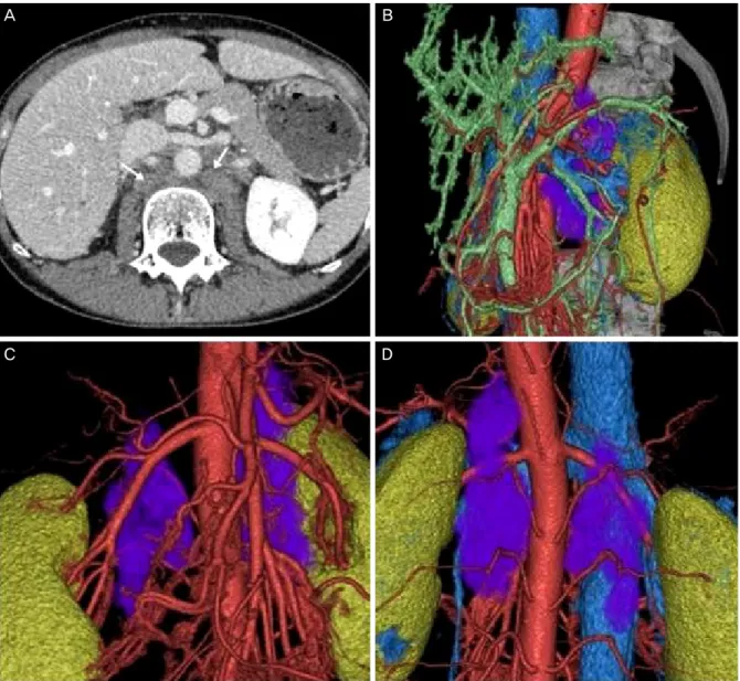
Preoperative 3D CT angiography showed a huge retro- crural metastatic LN with clear visualization and demarcation of the lesion. A metastatic lesion more than 10 cm in size was located behind the vena cava, aorta, and left kidney, encom-
A B
C D
Fig. 1. (A) Computed tomography (CT) image; arrows indicate the retro-crural metastatic lesions. (B) Three-dimensional (3D) CT angiography image, left side view; the purple color indicates the metastatic lesions. (C) 3D CT angiography image, anterior view. (D) 3D CT angiography im- age, posterior view.
passing the left renal and lumbar arteries (Fig. 1). 3D CT an- giography was performed with a 64 channel MDCT scanner (Aquilion 64; Toshiba Medical Systems, Tchigi, Japan). A total of 2.0 mL/kg of body weight of an iodine contrast medium, Iohexol (Omnipaque 300 syringe; Daiichi Sankyo, Tokyo, Japan) was administered at a rate of 3.5 mL/s with a power injector (Dual Shot GX; Nemoto Kyorindo, Tokyo, Japan), which was then flushed with 35 mL of normal saline solution, also at 3.5 mL/s. The obtained images were transferred to a computer workstation and reconstituted using 3D computer graphics software (Ziostation 2; Ziosoft, Tokyo, Japan).
Lymphadenectomy and left nephrectomy were performed as salvage surgery. The lesion was a 10×15-cm sized meta- static LN located on the left side of the vertebra. The lesion extended from the left kidney to the right renal vessels, en- compassing the left renal artery and lumbar artery behind the aorta and vena cava. There were severe adhesions be- tween the metastatic lesion and the posterior portion of the
left kidney.
There was no invasion into the psoas muscle or the verte- bra. All metastatic lesions were completely removed.
Histopathologic diagnosis revealed metastatic adenosqua- mous cell carcinoma in all the 5 resected para-aortic LNs.
The tumor had also invaded into the pelvis of the left kidney (Fig. 2). Cytological analysis of the peritoneal washing was negative for malignant cells.
Discussion
When para-aortic disease occurs, treatment is decided de- pending on the extent of the disease, primary treatment, performance status, or comorbidities present, if any. The man- agement of this case was based on the size of the lesion (more than 10 cm), anatomical location (in the retroaortic and retro- caval basin), and histology (adenosquamous cell carcinoma).
Salvage radiotherapy was not considered appropriate for this case as the enlarged metastatic lesion would require an exter- nal beam radiotherapy dose of approximately 55–60 Gy with conventional fractionation [4]. In the current case, the meta- static lesion was located in the retroaortic and retrocaval basin encompassing the lumbar and left renal artery. Such a high radiotherapy dose can lead to significant injury of adjacent organs such as duodenum, small intestine, and kidney, con- sidering the radiation tolerance [5]. Huang et al. [6] reported that adenosquamous cell carcinoma of cervix showed poor survival outcome compared with squamous cell carcinoma treated using the same radiotherapy protocol. Therefore, we decided to reduce the tumor volume to increase the response to the adjuvant therapy such as systemic chemotherapy or chemoradiotherapy.
Salvage surgery has a limited role in patients with isolated para-aortic LN metastasis considering the high associated morbidity and mortality rates (10% to 16%) [7]. Recently with the development of laparoscopy surgical resection has reduced morbidity. However, for larger metastatic lesions both laparotomic and laparoscopic approaches are not easy as the procedure needs lifting up of the aorta and the vena cava, along with meticulous dissection along the neighboring organs. Metastatic LN in the retroaortic or retrocaval basins sometimes show extracapsular invasion into neighboring or- gans, encompassing the neighboring arteries and veins [8].
Before salvage surgery for a metastatic lesion that is located
Fig. 2. (A) Postoperative photography; arrows indicate the cut endsof left renal vein and left renal artery. (B) Resected retro-crural meta- static lymph node and left kidney; arrow indicates tumor invasion into the renal parenchyme.