Intragastric gavage with denatonium benzoate acutely induces neuronal activation in the solitary tract nucleus via the vagal afferent pathway 1
6
0
0
전체 글
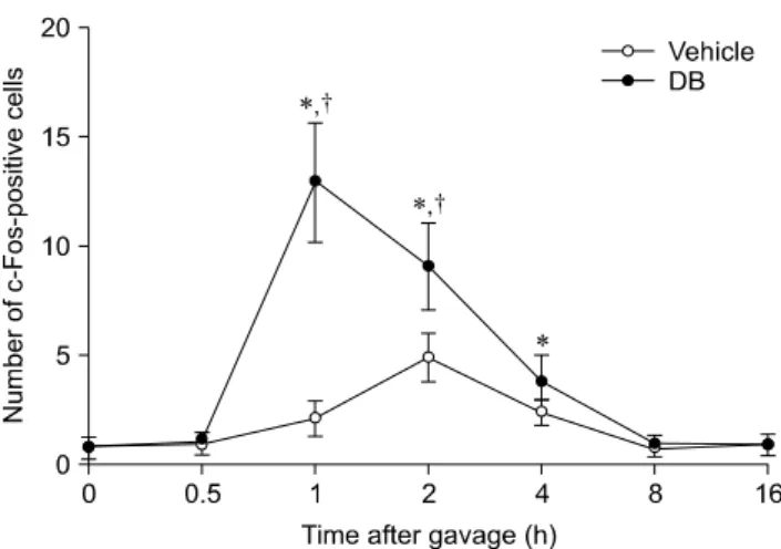
(2) 460 Hyo Young Jung et al.. Materials and Methods Experimental animals Male Sprague-Dawley rats (n = 128) were purchased from the Japan SLC (Japan). The animals were used at 6 weeks of age and housed under standard laboratory o conditions with adequate temperature (22 C) and humidity (60%) as well as a 12-h light/dark cycle. All rats had free access to solid diet (D12450Bi; Research Diets, USA) and tap water. Handling and care of the rats conformed to guidelines that comply with current international laws and policies (National Institutes of Health [NIH] Guide for the Care and Use of Laboratory Animals, NIH Publication No. 85-23, 1985, revised 1996). The study protocols were also approved by the Institutional Animal Care and Use Committee (IACUC) of Seoul National University, Korea (approval no. SNU-120103-10). All experiments were conducted to minimize both the number of animals used and their suffering due to the procedures performed. Subdiaphragmatic vagotomy The animals (n = 5 in each group) were anesthetized with a mixture of 2.5% isoflurane (Baxtor, USA) in 33% oxygen and 67% nitrous oxide. Subdiaphragmatic vagotomy was performed according to the technique described by Gschossmann et al. [5]. Briefly, a midline laparotomy incision was made to expose the peritoneal cavity. The external muscle coat of the esophagus 1 cm from the cardia distally was stripped. Since vagal branches are connected to the muscle coat, this results in a subdiaphragmatic vagotomy. To ensure completeness of the vagotomy, the left gastric artery was isolated and stripped of all connective tissues and nerves, including an accessory branch of the vagus nerve. Another group of rats underwent a sham operation. The animals were housed individually after the surgery and received a liquid diet (Ensure; Abbott Laboratories, USA) with water ad libitum. The rats were allowed to recover for 7∼10 days before the subsequent experiment. Intragastric gavage with DB The rats (n = 7 per group) were treated with either 0.5 mL of distilled water or 0.5 mL of DB (30 mg/kg; Sigma, USA) after a 12-h fast. To monitor the dose-dependent effects of DB on c-Fos expression, the animals received 0.5 mL DB (30 mg/kg, 100 mg/kg, or 300 mg/kg, n = 5 for each dosage) intragastrically after a 12-h fast. Tissue processing For histology, vehicle- or DB-treated rats were anesthetized with 30 mg/kg Zoletil 50 (Virbac, France) at 0 (immediately after gavage), 0.5 (30 min), 1, 2, 4, 8, or 16 h after gavage (n = 7 for each time point). The rats were then perfused transcardially with 0.1 M phosphate-buffered. saline (PBS, pH 7.4) followed by 4% paraformaldehyde in 0.1 M phosphate buffer (PB, pH 7.4). Brains were then removed and postfixed in 4% paraformaldehyde in 0.1 M phosphate buffer for 12 h at room temperature. The brains were cryoprotected by infusion with 30% sucrose o overnight at 4 C and cut coronally into 30-μm-thick sections using a cryostat (Leica Biosystems, Germany). The sections were transferred to six-well plates (SPL life science, Korea) containing PBS for further processing.. Immunohistochemistry for c-Fos To assess c-Fos expression, the sections were carefully processed simultaneously under identical conditions. Brain tissue sections from −12.80 mm and −13.30 mm posterior to the bregma according to the rat atlas [14] were selected for each animal. The sections were sequentially treated with 0.3% hydrogen peroxide (H2O2) in PBS for 15 min at room temperature and 5% normal goat serum (Vector Laboratories, USA) in 0.1 M PBS. They were next incubated with rabbit anti-c-Fos antibody (1 : 10,000; EMD Millipore, USA) overnight at room temperature, and subsequently exposed to biotinylated goat anti-rabbit IgG (1 : 200; Vector Laboratories) with streptavidin peroxidase complex for 2 h at room temperature (1 : 200, Vector Laboratories). Next, antibody binding was visualized by staining with 3,3’-diaminobenzidine tetrahydrochloride (Sigma). c-Fos-positive nuclei in all groups were examined using an image analysis system equipped with a computer-based Charge-Coupled Device camera (Optimas 6.5 software; CyberMetrics, USA). The number of c-Fos-positive cells was counted per section and the average was calculated. Statistical analysis All statistical analyses were carried out using GraphPad Prism 4.0 (GraphPad Software, USA). The data are presented as the mean ± standard error of the mean (SEM) for each group. Differences between the two groups were analyzed by Student’s t-test. Multiple parameters were assessed by a one-way or two-way analysis of variance (ANOVA). P-values < 0.05 were considered statistically significant.. Results Effects of vehicle on c-Fos expression In the vehicle-treated group, few c-Fos-positive nuclei were detected in the NTS 0 h after gavage (panel A in Fig. 1). The average number of c-Fos-positive nuclei was 0.89 ± 0.38 per section. The number of c-Fos-positive nuclei increased in the NTS 30 min after gavage and peaked 2 h after gavage (panels B-D in Fig. 1) although differences between groups were not significant. In the 2-h group, the number of c-Fos-positive nuclei was 4.91 ± 1.11 per.
(3) Effects of peripheral bitter taste ligand on brain stem 461. section (Fig. 2). Thereafter, the number of c-Fos-positive nuclei decreased in a time-dependent manner (panels E-G in Fig. 1, and Fig. 2). At 8 and 16 h after gavage, the number of c-Fos-positive nuclei was similar to that found 0 h after gavage (panels F and G in Fig. 1, and Fig. 2).. Effects of DB on c-Fos expression In the DB-treated group, few c-Fos-positive nuclei were observed in the NTS 0 h after gavage (panel A in Fig. 3). The number of c-Fos-positive nuclei slightly increased in the NTS 30 min after gavage compared to that observed in the vehicle-treated group (Fig. 2, and panel B in Fig. 3). The number of c-Fos-positive nuclei peaked 1 h after gavage and was significantly higher than that found in the vehicle-treated group (Fig. 2, and panel C in Fig. 3). The number of c-Fos-positive nuclei was 12.9 ± 2.75 per section (Fig. 2). Thereafter, the number of c-Fos-positive nuclei. Fig. 1. c-Fos-specific immunohistochemistry in the area postrema (AP) and nucleus tractus solitarius (NTS) of rats at 0 (A), 0.5 (B), 1 (C), 2 (D), 4 (E), 8 (F), and 16 h (G) after gavage with distilled water. Few c-Fos-positive nuclei were observed in the NTS 0 h after gavage. The number of c-Fos-positive nuclei (arrows) was increased 1∼2 h after gavage and decreased thereafter over time. Scale bar = 50 μm.. Fig. 2. The number of c-Fos-positive nuclei per section for each group at 0, 0.5, 1, 2, 4, 8, and 16 h after gavage with distilled water or denatonium benzoate†(DB) (n = 7 per group; *p < 0.05 vs. the 0 h post-gavage group; p < 0.05 vs. the vehicle-treated group). Data are presented as the mean ± SEM.. Fig. 3. c-Fos-specific immunohistochemistry in the NTS of rats at 0 (A), 0.5 (B), 1 (C), 2 (D), 4 (E), 8 (F), and 16 h (G) after gavage with DB. Few c-Fos-positive nuclei were observed in the NTS 0 h after gavage. c-Fos-positive nuclei (arrows) were abundantly detected 1 h after gavage and the number decreased thereafter over time. The distribution pattern of c-Fos-positive nuclei 8 or 16 h after gavage was similar to that found 0 h after gavage. Scale bar = 50 μm..
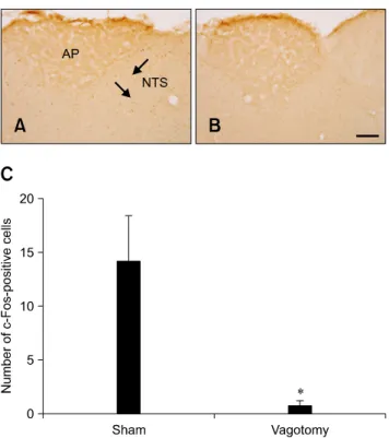
(4) 462 Hyo Young Jung et al.. Fig. 5. c-Fos expression in the NTS of rats 1 h after gavage with DB and a sham operation (A) or vagotomy (B). In the sham-operated group, c-Fos-positive nuclei (arrows) were observed in the NTS. In the vagotomy group, few c-Fos-positive nuclei were detected. (C) The number of c-Fos-positive nuclei per section for each group (n = 5 per group; *p < 0.05 vs. the sham-operated group). Data are presented as the mean ± SEM. Scale bar = 50 μm.. Fig. 4. c-Fos-specific immunohistochemistry in the NTS of rats 1 h after gavage with vehicle (A) or 30 mg/kg (B), 100 mg/kg (C), and 300 mg/kg (D) DB. In the vehicle-treated group, few c-Fos-positive nuclei were observed in the NTS. In the DB-treated group, the number of c-Fos-positive nuclei (arrows) was dose-dependently increased in the NTS. (E) The number of c-Fos-positive nuclei per section for each group (n = 5 per group; *p < 0.05 vs. the vehicle-treated group, †p < 0.05 vs. the 30 mg/kg DB-treated group, #p < 0.05 vs. the 100 mg/kg DB-treated group). Data are presented as the mean ± SEM. Scale bar = 50 μm.. decreased in a time-dependent manner although the number of these nuclei remained high in the DB-treated group compared to that in the vehicle-treated group at the 4-h time point (Fig. 2). At 8 and 16 h after gavage, the number of c-Fos-positive nuclei was similar than that found in the vehicle-treated group (Fig. 2, and panels F and G in Fig. 3).. Dose-dependent effects of DB on c-Fos expression In the vehicle-treated group, few c-Fos-positive nuclei were observed in the NTS 1 h after gavage (panel A in Fig. 4). In the DB-treated animals, c-Fos-positive nuclei were abundantly detected in the NTS 1 h after gavage (panels. B-E in Fig. 4). Interestingly, the number of c-Fos-positive nuclei increased in a dose-dependent manner. The number of nuclei in the 300 mg/kg DB-treated group was about 3.36-fold of that found in the 30 mg/kg DB-treated group.. Effects of vagotomy on DB-induced c-Fos expression In the sham group, some c-Fos-positive nuclei were detected in the NTS 1 h after DB gavage (panel A in Fig. 5) whereas only a few c-Fos-positive nuclei were observed in the NTS of the vagotomized group 1 h after DB gavage (panel B in Fig. 5). In the vagotomized group, the number of c-Fos-positive nuclei (0.78 ± 0.42 per section) was significantly lower than that in the sham group (panel C in Fig. 5).. Discussion Nociception and perception of bitter tastants are facilitated by T2R-expressing cells including brush cells in the rat gastrointestinal tract [9,13,15,17]. Detection and perception induce signal transduction by vagal afferents, and play an important role in gut-brain communication. Activation of vagal afferents causes neuron excitation in the NTS.
(5) Effects of peripheral bitter taste ligand on brain stem 463. followed by reflex activation of the vagal efferent neurons that alters gastrointestinal function [4]. In the current study, we monitored chronological changes of c-Fos-positive cells in the NTS following T2R ligand gavage at various time points that mimics neuronal taste perception [8,12,22]. The number of c-Fos-positive nuclei in the NTS increased 1 h after gavage with distilled water although this increase was not statistically significant. In contrast, c-Fos-positive nuclei numbers in the NTS significantly increased 2 h after gavage before decreasing in a time-dependent manner. These results suggest that volume distension of the stomach may enhance neuronal c-Fos expression in the NTS. In the DB-treatment group, the number of c-Fos-positive nuclei was significantly increased 1 h after DB gavage. This finding indicates that gavage with a T2R ligand further activates the neuronal expression of c-Fos in the NTS along with volume distension. It was reported that c-Fos expression does not significantly increased 2 h after gavage with DB alone although this procedure tends to increase the number of c-Fos-positive neurons in the NTS compared to control conditions [6]. In the present study, we observed a further increase of c-Fos expression in the NTS 2 h after DB gavage. We also observed a dose-dependent increase of c-Fos-positive cells in the NTS at the same time points. The discrepancy between our data and findings published in the literature may be associated with the dosages used. It has been reported that antinoceptive effects in the brain are mediated by the gastric vagal nerve [5]. Additionally, DB serotonin delivered by intragastric administration has been found to discharge in the brain via the vagus nerve and is blocked by p-chlorophenylalanine that selectively depletes brain serotonin [19]. We also performed subdiaphragmatic vagotomy to investigate the correlation between vagal afferents and increased c-Fos expression in the NTS after DB gavage. The vagotomized group showed a significant loss of c-Fos expression in the NTS after DB gavage. This result was similar to ones from a previous study indicating that increases in the number of Fos-positive NTS neurons by a T2R ligand mixture are prevented by subdiaphragmatic vagotomy [7]. Thus, DB potently activates the NTS soon after gavage via the vagal afferent pathway. In conclusion, our results demonstrated that DB, a T2R ligand, enhances neuronal c-Fos expression in the NTS in a dose-dependent manner after gavage through the vagal afferents.. Acknowledgments This research was supported by the Korea Food Research Institute (grant no. E0131203).. Conflict of Interest There is no conflict of interest.. References 1. Akabas MH, Dodd J, Al-Awqati Q. A bitter substance induces a rise in intracellular calcium in a subpopulation of rat taste cells. Science 1988, 242, 1047-1050. 2. Dragunow M, Faull R. The use of c-fos as a metabolic marker in neuronal pathway tracing. J Neurosci Methods 1989, 29, 261-265. 3. Furudono Y, Ando C, Kobashi M, Yamamoto C, Yamamoto T. The role of orexigenic neuropeptides in the ingestion of sweet-tasting substances in rats. Chem Senses 2005, 30 (Suppl 1), i186-l87. 4. Grundy D. Sensory signals from the gastrointestinal tract. J Pediatr Gastroenterol Nutr 2005, 41 (Suppl 1), S7-9. 5. Gschossmann JM, Mayer EA, Miller JC, Raybould HE. Subdiaphragmatic vagal afferent innervation in activation of an opioidergic antinociceptive system in response to colorectal distension in rats. Neurogastroenterol Motil 2002, 14, 403-408. 6. Hao S, Dulake M, Espero E, Sternini C, Raybould HE, Rinaman L. Central Fos expression and conditioned flavor avoidance in rats following intragastric administration of bitter taste receptor ligands. Am J Physiol Regul Integr Comp Physiol 2009, 296, R528-536. 7. Hao S, Sternini C, Raybould HE. Role of CCK1 and Y2 receptors in activation of hindbrain neurons induced by intragastric administration of bitter taste receptor ligands. Am J Physiol Regul Integr Comp Physiol 2008, 294, R33-38. 8. Harrer MI, Travers SP. Topographic organization of Fos-like immunoreactivity in the rostral nucleus of the solitary tract evoked by gustatory stimulation with sucrose and quinine. Brain Res 1996, 711, 125-137. 9. Höfer D, Püschel B, Drenckhahn D. Taste receptor-like cells in the rat gut identified by expression of α-gustducin. Proc Natl Acad Sci U S A 1996, 93, 6631-6634. 10. Houpt TA, Berlin R, Smith GP. Subdiaphragmatic vagotomy does not attenuate c-Fos induction in the nucleus of the solitary tract after conditioned taste aversion expression. Brain Res 1997, 747, 85-91. 11. Janssen S, Laermans J, Verhulst PJ, Thijs T, Tack J, Depoortere I. Bitter taste receptors and α-gustducin regulate the secretin of ghrelin with functional effects on food intake and gastric emptying. Proc Natl Acad Sci USA 2011, 108, 2094-2099. 12. King CT, Travers SP, Rowland NE, Garcea M, Spector AC. Glossopharyngeal nerve transection eliminates quinine-stimulated fos-like immunoreactivity in the nucleus of the solitary tract: implications for a functional topography of gustatory nerve input in rats. J Neurosci 1999, 19, 3107-3121. 13. Mueller KL, Hoon MA, Erlenbach I, Chandrashekar J, Zuker CS, Ryba NJ. The receptors and coding logic for bitter taste. Nature 2005, 434, 225-229..
(6) 464 Hyo Young Jung et al.. 14. Paxinos G, Watson C. The Rat Brain in Stereotaxic Coordinates. 6th ed. pp. 139-144. Academic Press, Amsterdam, 2007. 15. Rozengurt N, Wu SV, Chen MC, Huang C, Sternini C, Rozengurt E. Colocalization of the α-subunit of gustducin with PYY and GLP-1 in L cells of human colon. Am J Physiol Gastrointest Liver Physiol 2006, 291, G792-802. 16. Sternini C. Taste receptors in the gastrointestinal tract. IV. Functional implications of bitter taste receptors in gastrointestinal chemosensing. Am J Physiol Gastrointest Liver Physiol 2007, 292, G457-461. 17. Sutherland K, Young RL, Cooper NJ, Horowitz M, Blackshaw LA. Phenotypic characterization of taste cells of the mouse small intestine. Am J Physiol Gastrointest Liver Physiol 2007, 292, G1420-1428. 18. Travers JB, Urbanek K, Grill HJ. Fos-like immunoreactivity. 19. 20. 21.. 22.. in the brain stem following oral quinine stimulation in decerebrate rats. Am J Physiol 1999, 277, R384-394. Uneyama H, Tanaka T, Torii K. Gut nutrient sensing by the abdominal vagus. Nihon Yakurigaku Zasshi 2004, 124, 210-218. Wu SV, Chen MC, Rozengurt E. Genomic organization, expression, and function of bitter taste receptors (T2R) in mouse and rat. Physiol Genomics 2005, 22, 139-149. Wu SV, Rozengurt N, Yang M, Young SH, Sinnett-Smith J, Rozengurt E. Expression of bitter taste receptors of the T2R family in the gastrointestinal tract and enteroendocrine STC-1 cells. Proc Natl Acad Sci U S A 2002, 99, 2392-2397. Yamamoto T, Sawa K. Comparison of c-Fos-like immunoreactivity in the brainstem following intraoral and intragastric infusions of chemical solutions in rats. Brain Res 2000, 866, 144-151..
(7)
수치
관련 문서
Modern Physics for Scientists and Engineers International Edition,
작곡가의 원곡이 지휘자와 연주가에 의해 새롭게 태어나듯이, 우리가 배우고 체득한 모든 이론들과 지식, 테크닉들은 우리 자신에게서 새롭게
DFO administered db/db mice decreased iron metabolism related protein expression in GA muscle ..... In the TA muscle of db/db mice administered with DFO,
2재화 2요소 헥셔-올린 모형에서는 어느 한 경제에서 어느 한 요소의 양이 증가하면, 그 요소를 집약적으로 사용하는 산업의 생산량은 증가하고 다른
Ross: As my lawfully wedded wife, in sickness and in health, until
glen plaids 글렌 플레이드와 캐시미어 카디건, 캐리지 코트, 그리고 케이프 -> 격자무늬의 캐시미어로 된 승마용 바지, 마부용 코트, 말 그림이 수
□ Heat is defined as transferred energy dew to temperature differences through boundary.. - Conduction by molecular
다양한 번역 작품과 번역에 관한 책을 읽는 것은 단순히 다른 시대와 언어, 문화의 교류를 넘어 지구촌이 서로 이해하고 하나가
