INTRODUCTION
Vascular access (VA) events including stenosis and thrombo- sis are major causes of VA failure with hemodialysis (HD).
Surveillance of VA is currently recommended because under- dialysis due to the vascular access problem can be minimized, and the rate of thrombosis can be reduced (1-3). Vascular access blood flow (VABF) is one of the clinical predictors of steno- sis and thrombosis and has been used for surveillance of VA in most HD centers. Previous reports have shown that low VABF (access flow rate less than 600 mL/min in arteriove- nous grafts [AVGs]and less than 400 to 500 mL/min in arte- riovenous fistulas [AVFs]) is a risk factor for vascular access stenosis and thrombosis (1, 4-6). These reports also indicat- ed that a sequential decrease in VABF (more than 25% over 4 months) can predict VA stenosis and thrombosis (5, 7, 8).
In these reports, however, VABF was a short-term predictor of stenosis and thrombosis (almost within 3 months) (4, 5, 7, 9, 10).
The early detection of stenosis and thrombosis and the early correction of lesions have not been clearly demonstrated to
prolong VA survival. Tessitore et al. (6) reported that a higher baseline VABF was the only variable associated with a favor- able outcome for VA longevity. However, HD patients were enrolled with various VA ages, and the study was not designed to evaluate long-term predictors. In order to evaluate whether early VABF may predict long-term VA patency, we performed a prospective long-term observational study among patients newly started on HD.
MATERIALS AND METHODS Patient characteristics
A total of 57 patients at the Gachon University Gil Hospi- tal outpatient dialysis center were included. They started HD between January 1, 2005 and December 31, 2007, and all of the patients had de novo VAs. Patients with maturation fail- ure of VA were excluded from the study, as it was impossible to measure VABF using the ultrasound dilution technique.
This study was approved by the institutional review board
728
Hyung Soo Kim1, Jin-woong Park1, Jae Hyun Chang1, Jaeseok Yang1, Hyun Hee Lee1, Wookyung Chung1, Yeon Ho Park2, and Sejoong Kim1
Departments of Internal Medicine1and Surgery2, Gachon University of Medicine and Science, Incheon, Korea
Address for Correspondence Sejoong Kim, M.D.
Department of Internal Medicine, Gachon University Gil Hospital, 21 Namdong-daero 774 beon-gil, Namdong-gu, Incheon 405-760, Korea Tel : +82.32-460-8305; Fax : +82.32-460-3431 E-mail : imsejoong@hanmail.net
Early Vascular Access Blood Flow as a Predictor of Long-term Vascular Access Patency in Incident Hemodialysis Patients
The long-term clinical benefits of vascular access blood flow (VABF) measurements in hemodialysis (HD) patients have been controversial. We evaluated whether early VABF may predict long-term vascular access (VA) patency in incident HD patients.
We enrolled 57 patients, of whom 27 were starting HD with arteriovenous fistulas (AVFs) and 30 with arteriovenous grafts (AVGs). The patients’ VABF was measured monthly with the ultrasound dilution technique over the course of the first six months after the VA operation. During the 20.4-month observational period, a total of 40 VA events in 23 patients were documented. The new VA events included 13 cases of stenosis and 10 thrombotic events. The lowest quartile of average early VABF was related to the new VA events. After adjusting for covariates such as gender, age, hypertension, diabetes, VA type, hemoglobin levels, body mass index, parathy- roid hormone, and calcium-phosphorus product levels, the hazard ratio of VABF (defined as <853 mL/min in AVF or <830 mL/min in AVG) to incident VA was 3.077 (95% confidence interval, 1.127-8.395; P=0.028). There were no significant rela- tionships between early VABF parameters and VA thrombosis. It is concluded that early VABF may predict long-term VA patency, particularly VA stenosis.
Key Words : Renal Dialysis; Blood Flow Velocity; Vascular Patency; Indicator Dilution Techniques
Received : 12 June 2009 Accepted : 23 October 2009
ⓒ 2010 The Korean Academy of Medical Sciences.
This is an Open Access article distributed under the terms of the Creative Commons Attribution Non-Commercial License (http://creativecommons.org/licenses/by-nc/3.0) which permits unrestricted non-commercial use, distribution, and reproduction in any medium, provided the original work is properly cited.
of Gachon University Gil Hospital (#GIRBA 1780). All of the patients were dialyzed with biocompatible membranes (F5 or F6; Fresenius Medical Care AG, Bad Homberg, Ger- many) three times per week using Fresenius 4008H (Frese- nius Medical Care AG).
Vascular access characteristics
All of the permanent de novo VAs were actively used for chronic HD three times per week. The time difference between the VA operation and the first use of VA was at least six weeks in AVFs and at least four weeks in AVGs. Baseline character- istics, including the type of VA and its anatomic location, were collected for all VAs starting HD (Table 1).
Study design
This study was designed as a prospective cohort observa- tional study. VABF was measured with the ultrasound dilu- tion technique (Transonic Systems, Inc., Ithaca, NY, USA) at one month intervals until six months after the VA opera- tion. The measurement was performed within the first half- hour of the dialysis session for each patient at a blood flow rate of 250-300 mL/min (11, 12). Ultrafiltration was turned off 3 min before the start of the measurements in order to avoid the effect of hemoconcentration during the measurements. All patients were monitored for VA events such as VA stenosis and thrombosis. Patients who had physical findings of persistent swelling of the arm, prolonged bleeding after needle withdraw- al or altered characteristics of pulse or thrill in the outflow
vein also had their VABF measured, and they were referred for angiography if their VABF was less than 600 mL/min in grafts and less than 400 to 500 mL/min in fistulas (1).
Definitions
The endpoint of interest was the development of new VA events at the follow-up examination. VA events included VA stenosis, which was defined as a 50% or greater decrease in luminal diameter, and thrombotic events on fistulography or vascular operation (13). All VAs were monitored for the occurrence of VA events until the last follow-up visit.
Data on the early VABF parameters, which we call the early VABFs, were collected between the first use of VA up to six months after the VA operation. When the VA events occurred within the first six months, data were collected from the first use of the VA to the time of the VA events. The early VABF parameters consisted of the average, minimum, and initial values of early VABF rates, and these were calculated from the early VABFs. The initial value of VABF was defined as the VABF measured at the first use of the VA.
Statistical analyses
The data are presented as medians (interquartile range) for continuous variables and as proportions for categorical vari- ables. Normally distributed data were analyzed with unpaired t-tests; skewed data were analyzed with the Mann-Whitney U test. The early VABF parameters were divided into the low- est quartile and all other quartiles, according to the VA types.
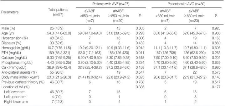
Patients with AVF (n=27) aVABF
≥853 mL/min (n=20) aVABF
<853 mL/min (n=7)
Parameters Total patients
(n=57) P
Patients with AVG (n=30) aVABF
≥830 mL/min (n=23) aVABF
<830 mL/min (n=7)
P
Male (%) 25 (43.9) 3 13 0.305 2 7 0.925
Age (yr) 54.0 (44.0-63.0) 59.0 (47.0-69.0) 51.0 (39.5-59.3) 0.293 63.0 (41.0-65.0) 52.0 (45.0-67.0) 0.980
Hypertension (%) 48 (84.2) 7 18 0.306 4 19 0.163
Diabetes (%) 30 (52.6) 4 8 0.432 4 14 0.860
Hemoglobin (g/dL) 10.7 (9.75-11.5) 10.2 (9.20-12.1) 10.9 (9.33-11.6) 0.912 11.1 (10.3-11.7) 10.7 (9.80-11.1) 0.606 PTH (mg/dL) 159 (86.2-321) 52.0 (17.2-163) 186 (126-420) 0.011 187 (126-708) 136 (62.6-290) 0.263 Calcium (mg/dL) 8.30 (7.65-9.25) 8.20 (7.40-9.50) 8.30 (7.85-9.28) 0.618 7.90 (7.00-9.10) 8.40 (7.50-9.30) 0.201 Phosphorus (mg/dL) 4.40 (3.65-5.25) 3.80 (3.10-5.30) 4.40 (3.85-4.85) 0.234 4.70 (3.80-5.50) 4.60 (3.40-5.60) 0.659 Ca×P (mg2/dL2) 36.9 (29.6-42.4) 32.8 (25.4-38.2) 37.2 (30.8-46.0) 0.158 37.1 (33.1-41.6) 37.1 (28.6-48.0) 0.980
Anti-platelet agents (%) 55 (96.5) 7 19 0.547 7 22 0.575
Body mass index (kg/m2) 23.0 (21.2-26.3) 21.4 (19.9-32.4) 22.8 (20.9-24.2) 0.825 26.6 (23.6-31.7) 22.9 (21.3-27.2) 0.148
Previous catheter history (%) 46 (80.7) 6 16 0.738 5 19 0.517
Location of VA (%) 0.385 0.177
Left lower arm 46 (80.7) 7 15 6 18
Left upper arm 4 (7.0) 0 1 0 3
Right lower arm 7 (12.3) 0 4 1 2
Table 1. Demographics and other characteristics of subjects
Values are medians (interquartile ranges).
AVF, arteriovenous fistula; AVG, arteriovenous graft; aVABF, average of early vascular access blood flow; PTH, parathyroid hormone; Ca×P, calcium and phosphorus products.
The event-free survival based on the early VABF parameters was analyzed using the Kaplan-Meier method and tested using the log-rank test. The hazard ratios between the early VABF parameters and the development of vascular events were deter- mined by univariate Cox-regression analysis. A multivariable Cox-regression analysis then was performed to evaluate the effects of the early VABF parameters, with adjustment for con- founding variables. Covariates were assessed at the baseline examination and included gender, age, hypertension, diabetes, vascular access type, hemoglobin levels, body mass index, parathyroid hormone (PTH), and calcium-phosphorus prod- uct levels.
The analyses were conducted using SPSS for Windows ver- sion 11.0 software (SPSS Inc., Chicago, IL, USA). P<0.05 was considered to be statistically significant.
RESULTS
The baseline demographics are listed in Table 1. The medi- an age of the patients was 54.0 (44.0-63.0) yr, and the propor- tion of male patients was 44%. The VA types included AVFs in 27 patients and AVGs in 30 patients. About half of the patients had diabetes mellitus, and most of the VAs were located in the left lower arms. Anti-platelet agents were pre- scribed to most of the patients.
The lowest quartiles of average early VABF rates were 853 mL/min in AVFs and 830 mL/min in AVGs (Table 2). We compared the lowest quartile of average early VABF with the other quartiles according to the VA types (Table 1). There were no significant differences between the lowest quartile group and the other quartile groups in clinical factors such as gen- der, age, hypertension, diabetes, hemoglobin levels, calcium- phosphorus product levels, anti-platelet agents, body mass index, previous catheter history, and location of VA, although the baseline PTH levels were slightly lower in the lowest quar- tile group with AVF. Two surgeons created all of the VAs in our study. Surgeon A operated from January to July 2005 and Surgeon B operated from August 2005 to December 2007, and there was no significant difference in the proportion of patients in the lowest quartile group and the other quartiles between the two surgeons (P=0.711 in AVF, P=0.847 in AVG). None of the enrolled patients had outflow tract obstruc- tion.
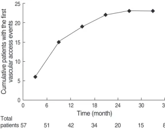
A total of 40 VA events in 23 patients were documented
throughout the total observational period (0.404 events/
patient-year). During the 20.4±10.6 month observation, six patients were discontinued from follow-up: four patients died, and two patients received renal transplantation. On 15 patients, fistulography was performed for screening VA mal- function. Eleven cases were identified based on the low level of VABF using the ultrasound dilution technique, and four cases were identified from the abnormal physical examina- tion findings. Six cases showed fistulographic stenoses that were located at the anastomic sites or juxta anastomic proxi- mal veins in AVF, and seven cases showed stenoses that were located at the venous anastomic sites or the arterial anastom- ic sites in AVG. Two cases had mild stenosis (less than 50%
in luminal diameter), and had no further intervention. Ten cases of thrombotic events showed abnormal findings such as no thrill and no pulsation of VA, and all of them received thrombectomy. In summary, the new VA events included 13 cases of stenosis, and 10 cases of thrombosis.
Within 12 months after the VA operation, the number of the new VA events was 15 out of a total of 23 events (65%).
The mean time between the VA operation and the new VA events was about 10 months in the 23 patients who experi- enced the VA events (Fig. 1).
Risk factors for the development of the new vascular access events
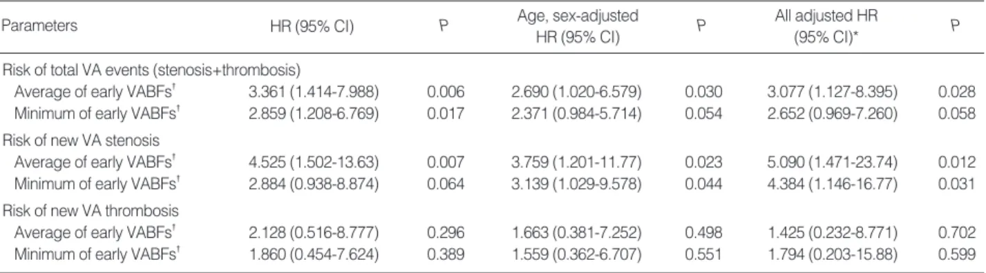
We analyzed the unadjusted and adjusted hazard ratios for
AVF, arteriovenous fistula; AVG, arteriovenous graft; VABF, vascular access blood flow.
AVF (n=27) Parameters
25 percentile 50 percentile 75 percentile
AVG (n=30)
25 percentile 50 percentile 75 percentile
Average of early VABFs (mL/min) 853 1,128 1,550 830 1,104 1,468
Minimum of early VABFs (mL/min) 740 880 1,290 798 985 1,308
Table 2. Early vascular access blood flow parameters according to vascular access types Cumulative patients with the first vascular access events
25
20
15
10
5
0
Fig. 1. Cumulative incidence of new vascular access events.
0 6 12 18 24 30 36
Time (month)
57 51 42 34 20 15 8
Total patients
early VABF parameters associated with the development of the new VA events (Table 3). The lowest quartile of average early VABF (<853 mL/min in AVF, <830 mL/min in AVG) was associated with the development of total VA events after adjustment for gender, age, hypertension, diabetes, vascular access type, hemoglobin levels, body mass index, parathyroid hormone, and calcium-phosphorus product levels (hazard ratio [HR], 3.077; 95% confidence interval [CI], 1.127-8.395;
P=0.028). HR for the lowest quartile of minimum early VABF for the risk of VA events was 2.652, but this was not statis- tically significant. The initial value of VABF was not related to the new VA events. The lowest quartiles of the average and minimum values obtained from the early VABFs were signifi- cantly associated with the development of the new VA steno- sis after adjustment of covariates. However, the unadjusted and adjusted hazard ratios for all of the parameters associat- ed with the development of the new thrombosis were not statistically significant.
VA patency until the new vascular access events
After the stratification of groups according to VA types, patients with the lowest quartile of average early VABF had worse total VA event-free survival rates than those in the other quartiles (P=0.002). Patients with the lowest quartile of aver- age values had also lower stenosis-free survival probabilities than those in other quartiles (P=0.003) (Fig. 2). However, there was no significant relationship between thrombosis- free survival and the lowest quartile of average early VABF (data not shown).
Total event-free survival and stenosis-free survival were also significantly worse in patients with the lowest quartiles of minimum early VABF (P=0.013 and P=0.011, respectively);
this was similar to the results obtained for patients in the lowest quartiles of average early VABF. However, thrombo- sis-free survival did not show a significant difference between the lowest quartile and other quartiles.
Gender, age, hypertension, diabetes, vascular access type, hemoglobin levels, body mass index, parathyroid hormone levels and Ca×P values were included as *covariates; �Parameters were measured during the first 6 month period. Hazard ratios (HR) are calculated for being in the lowest quartile compared with not being in the lowest quartile for the parameters indicated above.
CI, confidence interval; AVF, arteriovenous fistula; AVG, arteriovenous graft; BMI, body mass index; VA, vascular access; VABF, vascular access blood flow.
Parameters P Age, sex-adjusted P P
HR (95% CI)
All adjusted HR (95% CI)*
HR (95% CI)
Risk of total VA events (stenosis+thrombosis)
Average of early VABFs� 3.361 (1.414-7.988) 0.006 2.690 (1.020-6.579) 0.030 3.077 (1.127-8.395) 0.028 Minimum of early VABFs� 2.859 (1.208-6.769) 0.017 2.371 (0.984-5.714) 0.054 2.652 (0.969-7.260) 0.058 Risk of new VA stenosis
Average of early VABFs� 4.525 (1.502-13.63) 0.007 3.759 (1.201-11.77) 0.023 5.090 (1.471-23.74) 0.012 Minimum of early VABFs� 2.884 (0.938-8.874) 0.064 3.139 (1.029-9.578) 0.044 4.384 (1.146-16.77) 0.031 Risk of new VA thrombosis
Average of early VABFs� 2.128 (0.516-8.777) 0.296 1.663 (0.381-7.252) 0.498 1.425 (0.232-8.771) 0.702 Minimum of early VABFs� 1.860 (0.454-7.624) 0.389 1.559 (0.362-6.707) 0.551 1.794 (0.203-15.88) 0.599 Table 3. Association between early VABF and the new VA events
Stenosis-free survival
1.0
0.8
0.6
0.4
0.2
0.0
Fig. 2. Stenosis-free survival according to the average vascular access blood flow (VABF): (A) Stenosis-free survival in patients with arteri- ovenous fistulae (AVF) and (B) in patients with arteriovenous grafts (AVG). The dotted lines represent the patients in the lowest quartile of average early VABF (<853 mL/min in AVF and <830 mL/min in AVG), and the solid lines represent the patients in the other quartiles (P=0.003).
0 6 12 18 24 30 36 42 48
Time (month)
>853 mL/min
<853 mL/min
A
Stenosis-free survival
1.0
0.8
0.6
0.4
0.2
0.0
0 6 12 18 24 30 36 42
Time (month)
>830 mL/min
<830 mL/min
B
Subgroup analysis in patients with all of the vascular events
Ten patients (4 in AVFs and 6 in AVGs) had 2 or more events, and 4 patients had 3 or more events during the obser- vational period. Six patients had new VA events within 6 months of the VA operation. Vascular access survival could not be analyzed because only 2 patients had vascular access failures.
DISCUSSION
The aim of the study was to evaluate whether early VABF can predict long-term VA patency in incident HD patients.
We found that low levels of average and minimum early VABF may increase the risk of the development of total VA events, especially with regard to the new VA stenosis, and can pre- dict poor event-free survival or stenosis-free survival proba- bilities. Thus, patients with low average or minimum levels of VABF within six months after the VA operation should be monitored more carefully and frequently.
Several reports have shown that a low VABF or a sequen- tial decrease in VABF increases the risk of VA stenosis and thrombosis within a short-term period (1, 4, 5, 7, 9, 10). Pre- vious trials studying the association between VABF and VA longevity enrolled HD patients with a mean VA age of more than 18 months (6). In contrast, our current study enrolled incident HD patients with de novo VAs and evaluated the clinical usefulness of early VABF parameters as long-term pre- dictors of VA stenosis and thrombosis.
VABF measured up to six months after the VA operation was defined as early VABF. Practically, the maturation peri- od is four to six weeks after the operation for AVFs and two or more weeks for AVGs. By our definition, early VABF in- cluded three to four values of VABF from every patient because we measured VABF on a monthly basis. These serial data could be more representative of VA patency than a single measure- ment of VABF. In addition, according to the natural history of VA, the VA patency rates rapidly decrease during the early period, and loss of VA patency occurs at a rate of 25% within 9 months (14). Moist et al. (15) reported that a longer time between graft creation and study enrollment is associated with a decreased risk of graft thrombosis. Thus, VA surveil- lance should be started right after the first use of the VA, and these early VABFs could have a more important role than VABF values from the late period. This is another reason that we enrolled only patients with de novo VAs.
The average and minimum early VABF values were signif- icant predictors of event-free survival of VAs in our study.
The initial VABF parameters displayed a wide range of val- ues and were not useful in predicting the VA events, as the early values of VABF could change after the first use of the VA.
The current data demonstrated that early VABF parameters were not significantly associated with VA thrombotic events.
In contrast, other reports have shown that a low VABF has also been associated with an increased risk for VA thrombosis (5, 9, 16-18). There are several possible explanations for this dis- crepancy. First, most of the thrombotic events may follow VA stenoses, and some of the patients with a VA stenosis in our study might have been treated before thrombosis occurred.
In addition, anti-platelet agents were prescribed for most of the enrolled patients; this could have reduced the development of thrombosis (15, 19).
There were some limitations to our study. First, the sam- ple size was not large enough to stratify the subgroups. How- ever, we followed up patients for durations that were longer than used in any of the previous clinical trials (4, 7, 10, 17) in order to overcome the effects of a relatively small number of patients. In addition, we enrolled incident HD patients with de novo VAs in order to avoid a lead-time bias. Second, overall VA patency could not be analyzed because there were few VA failures in the enrolled patients. We considered pri- mary patency as the end point, which could be viewed as a surrogate indicator of overall VA patency. A large-scale trial is needed to clarify this potential relationship between early VABF parameters and overall VA survival. Third, several reports revealed that VABF measurement using the ultrasound dilu- tion technique allows non-invasive continuous monitoring of VA and has good accuracy and reproducibility (1, 11, 12, 20-22). However, the ultrasound dilution technique method cannot be used until the patient is using VA for HD; thus we could not evaluate information on VA maturation.
Some reports showed that the early detection of stenosis and thrombosis and the early correction of lesions cannot pro- long VA overall survival (1, 2, 23). In contrast, other reports demonstrated that well-programmed regular surveillance not only fosters the ability to detect VA events, but also prevents the morbidity and mortality associated with the failure to detect VA dysfunction (1, 3). We showed that low values of early VABF parameters may predict VA events, especially stenosis in incident hemodialysis patients. This suggests that we should focus on the high risk group with low values of early VABF parameters for effective surveillance of VA patency.
In summary, we found that low values of early VABF param- eters were associated with the development of VA events, especially VA stenosis, and poor event-free survival or steno- sis-free survival probability. This suggests that some of the VABF parameters in the early period of VA creation may pre- dict long-term VA patency in incident HD patients. These results indicate that some groups of patients with low values of early VABF parameters should be monitored more care- fully to prevent VA events.
ACKNOWLEDGEMENTS
We thank all of the medical and nursing staff of the dial- ysis center at Gachon University Gil Hospital for their assis-
tance. We also appreciate the work done by the research-nurs- ing staff (Yun Young Choi and Sung Mi Lee) with regard to the collection of clinical data. Some parts of this study were presented at the 2009 World Congress of Nephrology.
REFERENCES
1. Vascular Access 2006 Work Group. Clinical practice guidelines for vascular access. Am J Kidney Dis 2006; 48 (Suppl 1): S176-247.
2. Tonelli M, James M, Wiebe N, Jindal K, Hemmelgarn B. Ultrasound monitoring to detect access stenosis in hemodialysis patients: a sys- tematic review. Am J Kidney Dis 2008; 51: 630-40.
3. Hakim RM, Breyer J, Ismail N, Schulman G. Effects of dose of dial- ysis on morbidity and mortality. Am J Kidney Dis 1994; 23: 661-9.
4. Bosman PJ, Boereboom FT, Eikelboom BC, Koomans HA, Blan- kestijn PJ. Graft flow as a predictor of thrombosis in hemodialysis grafts. Kidney Int 1998; 54: 1726-30.
5. Tessitore N, Bedogna V, Gammaro L, Lipari G, Poli A, Baggio E, Firpo M, Morana G, Mansueto G, Maschio G. Diagnostic accuracy of ultrasound dilution access blood flow measurement in detecting stenosis and predicting thrombosis in native forearm arteriovenous fistulae for hemodialysis. Am J Kidney Dis 2003; 42: 331-41.
6. Tessitore N, Lipari G, Poli A, Bedogna V, Baggio E, Loschiavo C, Mansueto G, Lupo A. Can blood flow surveillance and pre-emptive repair of subclinical stenosis prolong the useful life of arteriovenous fistulae? A randomized controlled study. Nephrol Dial Transplant 2004; 19: 2325-33.
7. Neyra NR, Ikizler TA, May RE, Himmelfarb J, Schulman G, Shyr Y, Hakim RM. Change in access blood flow over time predicts vas- cular access thrombosis. Kidney Int 1998; 54: 1714-9.
8. Schwab SJ, Oliver MJ, Suhocki P, McCann R. Hemodialysis arteri- ovenous access: detection of stenosis and response to treatment by vascular access blood flow. Kidney Int 2001; 59: 358-62.
9. Wang E, Schneditz D, Nepomuceno C, Lavarias V, Martin K, Mor- ris AT, Levin NW. Predictive value of access blood flow in detect- ing access thrombosis. ASAIO J 1998; 44: M555-8.
10. May RE, Himmelfarb J, Yenicesu M, Knights S, Ikizler TA, Schul- man G, Hernanz-Schulman M, Shyr Y, Hakim RM. Predictive mea- sures of vascular access thrombosis: a prospective study. Kidney Int 1997; 52: 1656-62.
11. Bosman PJ, Boereboom FT, Bakker CJ, Mali WP, Eikelboom BC, Blankestijn PJ, Koomans HA. Access flow measurements in hemodial- ysis patients: in vivo validation of an ultrasound dilution technique.
J Am Soc Nephrol 1996; 7: 966-9.
12. Krivitski NM. Theory and validation of access flow measurement by dilution technique during hemodialysis. Kidney Int 1995; 48: 244-50.
13. Aruny JE, Lewis CA, Cardella JF, Cole PE, Davis A, Drooz AT, Grassi CJ, Gray RJ, Husted JW, Jones MT, McCowan TC, Meranze SG, Van Moore A, Neithamer CD, Oglevie SB, Omary RA, Patel NH, Rholl KS, Roberts AC, Sacks D, Sanchez O, Silverstein MI, Singh H, Swan TL, Towbin RB, Trerotola SO, Bakal CW. Quality improvement guidelines for percutaneous management of the throm- bosed or dysfunctional dialysis access. J Vasc Interv Radiol 2003;
14: S247-53.
14. Papanikolaou V, Papagiannis A, Vrochides D, Imvrios G, Gakis D, Fouzas I, Antoniadis N, Takoudas D. The natural history of vascu- lar access for hemodialysis: a single center study of 2,422 patients.
Surgery 2009; 145: 272-9.
15. Moist LM, Churchill DN, House AA, Millward SF, Elliott JE, Kribs SW, DeYoung WJ, Blythe L, Stitt LW, Lindsay RM. Regular mon- itoring of access flow compared with monitoring of venous pressure fails to improve graft survival. J Am Soc Nephrol 2003; 14: 2645-53.
16. Lindsay RM, Blake PG, Malek P, Posen G, Martin B, Bradfield E.
Hemodialysis access blood flow rates can be measured by a differ- ential conductivity technique and are predictive of access clotting.
Am J Kidney Dis 1997; 30: 475-82.
17. Kim YO, Yang CW, Yoon SA, Chun KA, Kim NI, Park JS, Kim BS, Kim YS, Chang YS, Bang BK. Access blood flow as a predictor of early failures of native arteriovenous fistulas in hemodialysis patients.
Am J Nephrol 2001; 21: 221-5.
18. Hoeben H, Abu-Alfa AK, Reilly RF, Aruny JE, Bouman K, Perazel- la MA. Vascular access surveillance: evaluation of combining dynam- ic venous pressure and vascular access blood flow measurements. Am J Nephrol 2003; 23: 403-8.
19. Saran R, Dykstra DM, Wolfe RA, Gillespie B, Held PJ, Young EW.
Association between vascular access failure and the use of specific drugs: the Dialysis Outcomes and Practice Patterns Study (DOPPS).
Am J Kidney Dis 2002; 40: 1255-63.
20. Schwarz C, Mitterbauer C, Boczula M, Maca T, Funovics M, Heinze G, Lorenz M, Kovarik J, Oberbauer R. Flow monitoring: performance characteristics of ultrasound dilution versus color Doppler ultrasound compared with fistulography. Am J Kidney Dis 2003; 42: 539-45.
21. Ha SJ, Lee YJ, Cho BH, Jung KH, Moon JY, Lee SH, Lee TW, Ihm CG. Glucose pump technique is as good as ultrasound dilution tech- nique for vascular access surveillance in hemodialysis patients. Kore- an J Nephrol 2007; 26: 448-54.
22. Lee KH, Park JY, Choi SJ, Kim JK, Hwang SD, Joh JH. Clinical utility of access blood flow measurement by ultrasound dilution in hemodialysis patients. Korean J Nephrol 2005; 24: 265-73.
23. Shahin H, Reddy G, Sharafuddin M, Katz D, Franzwa BS, Dixon BS. Monthly access flow monitoring with increased prophylactic angioplasty did not improve fistula patency. Kidney Int 2005; 68:
2352-61.