https://doi.org/10.9721/KJFST.2019.51.3.278
278
©The Korean Society of Food Science and Technology
Rhamnogalacturonan I-rich fractions from cherry tomatoes
stimulate phagocytosis in RAW 264.7 macrophages
Dahyun Hwang1,2, Young-Hee Lim3,4,Kwang-Soon Shin5, and Jong-Ho Koh6,*
1Department of Biomedical Laboratory Science, College of Life and Health Sciences, Hoseo University 2The Research Institute for Basic Sciences, Hoseo University
3Department of Public Health Science (BK21PLUS Program), Graduate School, Korea University 4Department of Laboratory Medicine, Korea University Guro Hospital
5Department of Food Science and Biotechnology, Kyonggi University 6Department of Bio-Food Analysis, Bio-campus, Korea Polytechnics College
Abstract Tomato (Lycopersicon esculentum) is widely known for its beneficial effects on human health. To investigate the beneficial effects of polysaccharides from cherry tomato, cherry tomato polysaccharides (CTP) were prepared, the component sugars were analyzed, and the immunomodulatory activities in RAW 264.7 macrophages were assessed. CTP mainly contained arabinose (Ara) and galactose (Gal), suggesting that CTP might be enriched with an arabinogalactan (AG) moiety. The Ara and Gal present in CTP are likely components of AG-II (35.4%), namely arabino-β-(3,6)-galactan. To investigate the immunomodulatory activity of CTP, cytokine levels and iNOS2, COX-2, and NF-κB protein levels were analyzed, and NF-κB nuclear translocation and phagocytosis were observed by immunofluorescence. CTP significantly increased the levels of TNF-α, MCP-1, and IL-6. CTP also increased iNOS2 and COX-2 expression as well as NF-κB nuclear translocation in RAW 264.7 cells. CTP significantly stimulated phagocytosis activity. These results showed that CTP stimulates macrophage activity, which can boost the innate immune response. CTP with high AG-II content could be used as a prebiotic to strengthen immunity.
Keywords: cherry tomato, polysaccharide, rhamnogalacturonan I, phagocytosis, immunostimulating activity
Introduction
Plant polysaccharides activate macrophages and also increase phagocytosis in vitro (Schepetkin and Quinn, 2006). Recent studies have shown that a variety of polysaccharides can act as immuno-modulators, helping to stimulate the immune system so that it can eliminate tumors and foreign invaders more effectively (Razali et al., 2014). In plant polysaccharides (starch, cellulose, hemicellulose, and pectin), pectins are the major component of plant cell wall and known as the most complex heteropolysaccharide. Pectins are consisted of fragments of linear and branched regions of polysaccharide such as homogalacturonan (HG), rhamnogalacturonan (RG)-I, RG-II, xylogalacturonan (XG), and apiogalacturonan (AG) (Popov and Ovodov, 2013). The composition and structural features of pectin determine pharmacological activities in humans and animals. Recently, anti-cancer and immunostimulatory activities have been reported in pectic polysaccharide, and the activities were contributed from RG-I and RG-II regions rather than the HG region (Kim et al., 2016).
Macrophages play an important role in regulating both innate
and adaptive immune responses and they defend against infection and cancer (Yoon et al., 2003). Macrophages secrete cytokines such as tumor necrosis factor α (TNF-α), interleukin-6 (IL-6), and monocyte chemoattractant protein-1 (MCP-1), and inflammatory mediators such as NO, hydrogen peroxide (H2O2), and prostaglandins (PGs), against pathogens. IL-6 and TNF-α activate surrounding cells to produce chemokines or adhesion molecules, thereby recruiting various inflammatory cells to the infection sites (Lee et al., 2010). Cyclooxygenase-2 (COX-2) and iNOS are two major inflammatory mediators. NO and PGs are produced as a result of iNOS and COX-2 stimulation, respectively (Kim et al., 2005; Marletta, 1993). Another major immune-stimulating factor is nuclear factor kappa-light-chain-enhancer of activated B cells (NF-κB). NF-κB, which is primarily composed of the protein p50 and p65, exists in a latent form in the cytosol as a dimer complex bound to the inhibitor protein, IκB. Upon exposure to internal stimuli, IκB degrades, which allows NF-κB release and translocation to the nucleus to regulate the expression of multiple target genes (Kim et al., 2007; Li et al., 2014). Another macrophage stimulation activity is phagocytosis. Phagocytosis in leukocytes plays a role as the first-line host defense against potential pathogens; thus, macrophage activation means an increase in innate immune response (Kim et al., 2007).
Tomato (Lycopersicon esculentum) is a popular fruit consumed in many countries. Tomatoes contain fiber, oligosaccharides and polysaccharides, which act as prebiotic compounds in the gut environment (Bornet et al., 2002; Napolitano et al., 2009). *Corresponding author: Jong-Ho Koh, Department of Bio-Food
Analysis, Bio-campus, Korea Polytechnics College, Nonsan, Chung-nam 32943, Korea
Tel: +82-41-746-7354 E-mail: [email protected] Received May 24, 2019; accepted May 28, 2019
However, little information is available on the relationship between polysaccharides from cherry tomatoes and immune activity. In this study, we analyzed the sugar composition of polysaccharides prepared from cherry tomatoes and evaluated the potential immune-stimulating activity of the polysaccharides on the RAW 264.7 murine macrophage cell line.
Materials and Methods
Chemicals
Dulbecco’s Modified Eagle Medium (DMEM), fetal bovine serum (FBS), and penicillin/streptomycin for the cultivation of cells were obtained from HyClone (Logan, UT, USA). 2-(2-Methoxy-4-nitrophenyl)-3-(4-nitrophenyl)-5-(2,4-disulfophenyl)-2H-tetrazolium, monosodium salt (WST-1) reagent was purchased from Roche (Indianapolis, IN, USA). Mouse inflammation cytometric bead array kit (CBA), and mouse TNF-α (Mono/Mono), MCP-1, and IL-6 enzyme-linked immunosorbent assay (ELISA) were purchased from BD Biosciences (San Diego, CA, USA). Lipopolysaccharide (LPS) and gum arabic were purchased from Sigma (St. Louis, MO, USA). All other chemicals and reagents were obtained from Sigma.
Crude polysaccharide preparation
The cherry tomato “Yoyo”, harvested from Chungcheongnamdo, South Korea. After processing with a pulper (AG-5500, Angel Juicer Co, Pusan, South Korea), cherry tomato juice was obtained, followed by freeze drying. The resultant crude powders were mixed with sterilized distilled water, stirred at room temperature for 10 min and centrifuged at 15,000×g for 30 min at 4oC. The supernatant was filtered through a 40-μm pore mesh (BD Biosciences). The filtrate was mixed with 1% Viscozyme L (Novozymes Inc., Copenhagen, Denmark; 54:1, v/v) and reacted in a shaking water bath at 50oC for 4 h at 200 rpm, then centrifuged at 1,000×g for 10 min to remove sediments. In this study, Viscozyme L was selected among several enzymes (Viscozyme L, Pectinex BE 3L, Pectinex AFP L4, and Citrozym Ultra L obtained from Novozymes) based on their yields (Koh et al., 2015). The supernatant was precipitated overnight with a final concentration of 80% ethanol (v/v) at 4oC and centrifuged at 7,500×g for 20 min. The supernatant was removed and the resulting precipitate was resolved with distilled water. The resulting precipitate was then dialyzed against water for 4 days at 4oC using a cellulose membrane tube (D-9402, 76 mm wide, >12,000 MW, Sigma). After insoluble materials were removed by centrifugation, the non-dialyzable portion was lyophilized to obtain CTP. CTP used in all experiments were dissolved in distilled water.
Chemical component analysis
Neutral sugar and uronic acid contents were determined using the phenol-sulfuric acid method (DuBois et al., 1956) and the m-hydroxybiphenyl method (Blumenkrantz and Asboe-Hansen, 1973), using D-galactose and D-galacturonic acid as standards, respectively. The aldose component of the polysaccharide was analyzed as
alditol acetate prepared by NaBH4 reduction and acetic anhydride acetylation after hydrolysis with 2 M trifluoroacetic acid for 1.5 h at 121oC (Jones and Albersheim, 1972). For uronic acid determination, the reduced hydrolysates were loaded onto the Dowex-1 resin (Sigma) and the bonded aldonic acids were eluted using 1.0 N HCl. The eluted fraction was evaporated to dryness at 40oC in a stream of filtered air and the resulting lactones were reduced under neutral conditions (Jones and Albersheim, 1972).After acetylation, the resulting carboxyl-reduced alditol acetates were analyzed using gas liquid chromatography (GLC) (Zhao et al., 1991). GLC was performed using an HL-6890 Series II gas chromatograph (Hewlett-Packard, Palo Alto, CA, USA) equipped with a flame ionization detector (FID) using a Supelco SP-2380 capillary column (30 m× 0.25 mm i.d., 0.2μm film, Bellefonte, PA, USA).
Determination of arabino-β-3,6-galactan
The amounts of arabino-β-3,6-galactan (AG-II) in the polysaccharide were measured by β-D-glucosyl Yariv reagent (Biosupplies, Rarkville, Australia) using a single radial gel diffusion according to the procedure of Holst and Clarke (van Holst and Clarke, 1985). The 10μL of CTP samples and a standard gum arabic (Sigma) were applied to a gel plate containing 0.004% (w/v) β-D-glucosyl Yariv, 75 mM NaCl, 0.01% (w/v) sodium azide, and 1% (w/v) agarose. The contents of AG-II in CTP were determined by calibration curve based on the square value of the halo formed using gum arabic as a standard.
Cell culture and cell viability
RAW 264.7 mouse macrophage cells were purchased from the Korea Cell Line Bank (KCLB, Seoul, Korea). The RAW 264.7 was maintained in DMEM supplemented with 10% FBS, 100 units/mL penicillin, and 100μg/mLstreptomycin at 37oC in an atmosphere of 5% CO2 −95% air. When 70-80% confluence was reached, the cells were seeded in a 96-well plate at a density of 1×10⁶ cells/mL and incubated overnight. After overnight culture, cells were treated with various CTP concentrations (1, 10, 50, and 100μg/mL) for 24 h. WST reagents (10 μL) were added and the viability was measured using a SpectraMax microplate reader (Molecular Devices, Sunnyvale, CA, USA) at 450 nm. The relative cell viability (%) was expressed as a relative percentage to the untreated control cells.
NO and cytokine measurement
RAW 264.7 macrophages were cultured overnight in a 96-well plate at a density of 1×10⁶ cells/mL in DMEM-FBS, and the cells were treated with various CTP concentrations (1, 10, 50, and 100 μg/mL) for 24 h. LPS was used as a positive control. The culture medium was collected for NO and cytokine (IL-6, TNF-α, and MCP-1) analysis. NO secretion was measured using a microplate assay method as previously described (Kang et al., 2011). The nitrite concentration was determined by measuring the absorbance at 540 nm. Sodium nitrite (NaNO2) was used as a standard. The TNF-α, MCP-1, and IL-6 was measured using an ELISA kit (BD Biosciences), according to the manufacturer’s instructions.
Quantitative real time polymerase chain reaction (qPCR) RAW 264.7 cells (1×10⁶ cells/mL) were cultured with various concentrations of CTP (1, 10, 50, and 100μg/mL) in 6-well plates. After 24 h, cells were treated with 1 mL of TRIzol reagent (Invitrogen, Carlsbad, CA, USA) and total RNA was extracted according to the manufacturer’s protocol. Total RNA concentration was quantified with a NanoDrop ND-1000 Spectrophotometer (Thermo Scientific, Wilmington, DE, USA). cDNA was then prepared using a Revert Aid First Strand cDNA kit (Fermentas, Burlington, Ontario, Canada), and qPCR was performed using a StepOnePlusTM Real-Time PCR System (Applied Biosystems, Foster City, CA, USA). PCR primers were purchased from Bioneer (Seoul, Korea) as follows: glyceraldehyde-3-phosphate dehydrogenase (GAPDH) (sense: 5'-TGT GAA CGG ATT TGG CCG TA-3', antisense: 5'-ACT GTG CCG TTG AAT TTG CC-3'); COX-2 (sense: CCC AGA GCT CCT TTT CAA CC-3', antisense: 5'-ATT TGG CAC 5'-ATT TCT TCC CC-3'); iNOS2 (sense: 5'-CAC CTT GGA GTT CAC CCA GT-3', antisense: 5'-ACC ACT CGT ACT TGG GAT GC-3'); TNF-α (sense: 5'-AGC ACA GAA AGC ATG ATC CG-3', antisense: 5'-GTT TGC TAC GAC GTG GGC TA-3'); and IL-6 (sense: 5'-CGA TGA TGC ACT TGC AGA AA-3', antisense: 5'-TGG AAA TTG GGG TAG GAA GG-3'). The reaction was preheated to 95oC for 10 min followed by 40 cycles at 95oC for 15 s, 60oC for 15 s, and 72oC for 30 s. qPCR data was quantified as previously described (Livak and Schmittgen, 2001). Relative gene expression was quantified based on equal amounts of RNA (1μg). GAPDH was used as an internal control reference gene.
Western blot analysis
RAW 264.7 cells (1×10⁶ cells/mL) were cultured with various concentrations of CTP (1, 10, 50, and 100μg/mL) in 60-mm dishes. After 24 h, the cells were collected and lysed for protein extraction. iNOS2 and COX-2 analysis was performed on whole-cell extracts using Pro-Prep protein extraction solution (Intron, Seoul, Korea). NF-κB analysis was performed on the nuclear protein fraction using NE-PER Nuclear and Cytoplasmic Extraction Reagents (Thermo, Rockford, IL, USA). Equal amount (30 µg) of protein from each sample was separated by 10% sodium dodecyl sulfate-polyacrylamide gel electrophoresis (SDS-PAGE). The separated proteins were transferred to a polyvinylidene difluoride (PVDF) membrane (Millipore, Bed-ford, MA, USA) using a Trans-Blot semi-dry transfer cell (Bio-Rad, Hercules, CA, USA). The membrane was blocked with 5% non-fat skimmed milk in phosphate buffered saline (PBS) with 0.05% Tween 20 (PBS-T) overnight at 4oC. The membrane was washed three times with PBS-T, then incubated with a 1:500 dilution of anti-iNOS2 (Santa Cruz Biotechnology, Dallas, TX, USA) antibody, 1:2,000 dilution of an anti-COX-2 antibody (Novus Bio, Littleton, CO, USA), and 1:2,000 dilution of an anti-NF-κB antibody (Abcam, Cambridge, UK) for 1 h at room temperature. GAPDH, used as an endogenous control, was detected with a 1:5,000 dilution of anti-GAPDH antibody (Thermo Scientific, Waltham, MA, USA). The membrane was washed three times with PBS-T, and incubated further with a secondary antibody. The 1:10,000 dilution of goat anti-rabbit IgG (H+L) horseradish
peroxidase conjugated antibody (Thermo Scientific) was used as a secondary antibody for anti-iNOS, anti-COX-2, and anti-NF-κB. The 1:50,000 dilution of goat anti-mouse IgG (H+L) horseradish peroxidase conjugated antibody (Thermo Scientific) was used as
Fig. 1. Single radial gel diffusion (A) and reactivity (B) between β-glucosyl Yariv reagent and CTP. β-Glucosyl Yariv reagent specifically binds and precipitates AG-II. Gum arabic was used as a standard. The arabino-β-3,6-galactan (%) in CTP was calculated to relative percentage against the standard at a same dose.
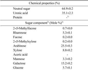
Table 1. Chemical composition of CTP Chemical properties (%)
Neutral sugar 64.9±0.2
Uronic acid 35.1±2.3
Protein -1)
Sugar component2) (Mole %)3)
2-O-Methylfucose 0.7±0.0 Rhamnose 5.3±0.1 Fucose 0.2±0.0 2-O-Methylxylose 0.2±0.0 Arabinose 25.5±0.30 Xylose 8.8±0.2 Aceric acid -Mannose 3.3±0.2 Galactose 15.2±0.20 Glucose 5.7±0.1 1)Not detected
2)Monosaccharides were analyzed using alditol acetates. 3)Mole % was calculated from the detected total carbohydrate.
the secondary antibody for anti-GAPDH. Bound antibodies were detected using WesternBright Sirius western blotting detection kit (Advansta, Menlo Park, CA, USA). Blot images were obtained using the imaging system FluorChem E (Proteinsimple, CA, USA) and band intensity was measured using analysis tools.
NF-κB immunofluorescence
Cells were grown on coverslips and fixed in 4% paraform-aldehyde/PBS overnight at 4oC, then permeabilized with 0.01% Triton X-100 in PBS for 5 min. The cells were washed with PBS and blocked with 10% normal donkey serum/PBS for 30 min at room temperature. Cells were then incubated overnight with a 1:1,000 dilution of an anti-NF-κB antibody at 4oC. Cells were
washed three times with PBS and then incubated for 60 min at room temperature with a 1:1,000 dilution of goat anti-rabbit IgG, DyLight 650 (Thermo Scientific). After further washing with PBS, nuclei were counterstained with a 1:10,000 dilution of a 4,6-diamidino-2-phenylindole (DAPI, Sigma) for 10 min at room temperature. Excess dye was removed by washing three times with PBS, and the coverslip was placed onto a slide and mounted using VECTASHIELD® (Vector Laboratories, Burlingame, CA, USA). Images were acquired digitally and processed using the operation
software EZ-C1 for the Nikon C1 plus confocal laser scanning microscope (Nikon, Tokyo, Japan).
Phagocytosis
To measure phagocytosis activity, RAW 264.7 cells (1×10⁶ cells/ mL) were seeded in a 6-well (for FACS analysis) or a 24-well plate (for immunofluorescence), then incubated for 24 h. The next day, cells were cultured with various CTP concentrations (1, 10, 50, and 100μg/mL) and incubated for 24 h. To quantify the phagocytosis activity, a phagocytosis assay kit IgG FITC (Cayman, MI, USA) was used. The cells were analyzed using a confocal microscopy (Nikon C1 plus confocal laser scanning microscope, Nikon).
Statistical analysis
All statistical analyses were performed using SPSS version 12.0 for Windows (SPSS, Chicago, IL, USA). The results of experiments were expressed as the mean±standard deviation (SD). The statistical significance of the difference was determined using the Student’s t-test. A p-value of <0.05 was considered the threshold for statistical significance. Statistical differences among groups were evaluated using an analysis of variance (ANOVA), followed by Duncan’s multiple range tests.
Fig. 2. Nitric oxide (NO) and cytokine production in CTP-treated RAW 264.7 cells. (A) NO, (B) TNF-α, (C) IL-6, and (D) MCP-1. Data are expressed as the mean±SD of three independent experiments performed in triplicate. *p<0.05, **p<0.01, and ***p<0.001, Student’s t-test, compared with the negative control (treated with distilled water).
Results and Discussion
CTP chemical composition analysis
Lycopene contents and nutritional components of tomato pomace and juice were previously reported (Koh et al., 2015). The CTP chemical composition from cherry tomatoes is shown in Table 1. CTP consisted of 65 and 35% of neutral sugars and uronic acid, respectively. CTP consisted of mainly Ara and Gal, over 40%, which suggests that CTP might consist mainly of AG moiety of RG-I, a type of pectic polysaccharide in plant cell wall. In fact, it is well-known that RG-I is composed by a backbone with repeating units of rhamnose (Rha) and galacturonic acid (GalA) and various side chains, such as arbinans, galactans, type I and/or type II AG (Yapo, 2011). Having determined of Ara and Gal in CTP, a single radial gel diffusion assay using β-glucosyl Yariv reagent (βGlcY) was performed. βGlcY specifically reacts and precipitates with the structure of AG-II, characterizing by a backbone of (1,3)-D-galactan with branch points of 6-linked β-D-Gal of one, two, or three residues in length, and changed color to red (Caffall and Mohnen, 2009; Paulsen et al., 2014). AG-II
amounts in CTP were determined using a gum Arabic standard curve (Fig. 1A). Ara and Gal (total 40.7%) existed in CTP consist mostly of AG-II (35.4%), namely arabino-β-(3,6)-galactan (Fig. 1B). AG-II-containing polysaccharide has been proved its immuno-modulatory properties such as the induction of macrophage iNOS expression and NO production and enhanced cytokines production for human and murine macrophage (Schepetkin et al., 2005; Schepetkin and Quinn, 2006). In addition, RG-I content, calculated as the sum of Ara, Gal, and Rha contents, accounted for 46.0% of CTP, which explains that CTP are enriched mainly of pectic polysaccharide and AG-II is a major moiety. A large amount of uronic acid (35.1%) suggested that CTP is likely to contain quite a lot of homogalacturonan (HG), another region of pectic polysaccharide. The bioavailability of tomatoes might be affected by their processing method, such as enzyme treatment. The use of multi-enzymatic complexes, such as Viscozyme-L, ruptures the cell walls and favors the extraction of useful compounds (protein and sugars) from the vegetable tissues (Dueñas et al., 2007). The proportions among HG, XG, RG-I, and RG-II are variable in various pectic polysaccharides from plant cell wall; however, Fig. 3. mRNA and protein expression levels of iNOS2 and COX-2. Gene expression levels of iNOS2 (A) and COX-2 (B) in CTP-treated RAW 264.7 cells. Protein expression levels determined by western blot (C) and quantified (D) in CTP-treated RAW 264.7 cells. NC (negative control) represents distilled water-treated control group and GAPDH was used as an endogenous control. *p<0.05, **p<0.01, and ***p<0.001, Student’s t-test, compared with the negative control (treated with distilled water).
typically HG is the most abundant (about 65%), RG-I accounts for 20 to 35%, and XG and RG-II account for under 10% (Harholt et al., 2010).
Effect of CTP on RAW 264.7 cell viability
CTP had no cytotoxic effect on the cells up to 100μg/mL. CTP significantly (p<0.05) increased RAW 264.7 cell viability compared with the negative control treated with distilled water. The cell viabilities were 111.2±1.86, 122.9±1.12, 136.3±8.19, and 130.5±2.20 in cells treated with 1, 10, 50, and 100μg/mL of CTP, respectively, compared with the negative control (100%).
Effect of CTP on NO and cytokines production
The inflammatory mediator NO produced by phagocytes and participates in the regulation of various immune responses (Wang et al., 2009). CTP significantly (p<0.05) increased NO production in RAW 264.7 cells in a dose-dependent manner (Fig. 2A). NO levels in cells treated with 1, 10, 50, and 100μg/mL of CTP increased 1.0-, 2.3-, 16.4-, and 19.4-fold, respectively, compared with the negative control. LPS (1 μg/mL), a component of the gram-negative bacterial cell wall, was used as a positive control. LPS stimulates NO, COX-2, IL-6, and TNF-α production, which
are activated through nuclear factor-κB (NF-κB) activation in the inflammatory pathway (Karin, 2009; Karin and Greten, 2005).
Cytokine production is a major indicator of macrophage activation. CTP treatment significantly increased TNF-α (Fig. 2B), IL-6 (Fig. 2C), and MCP-1 (Fig. 2D) levels in a dose-dependent manner. The type of cytokines produced will determine the macrophage effector function. IL-6 and TNF-α are important cytokines that are involved in innate immune responses against pathogens. They can enhance various functional responses and contribute to the systemic inflammatory effects (Liu et al., 2013). MCP-1 in macrophages acts as one of the most important chemokines, which participates in the direct migration of white blood cells to infected or damaged tissue. Cell motility is a macrophage characteristic toward antigens, and it contributes to the inflammation defense process (Tajima et al., 2008). The TNF-α, IL-6, and MCP-1 concentrations in 100μg/mL CTP-treated cells increased 227.8-, 187.5-, and 38.0-fold, respectively, compared with the negative control. Among the three cytokines, TNF-α level in the 50 and 100 μg/mL CTP-treated cells were higher than that in LPS-treated cells. Cytokine generation in this study suggests that CTP can stimulate innate immunity.
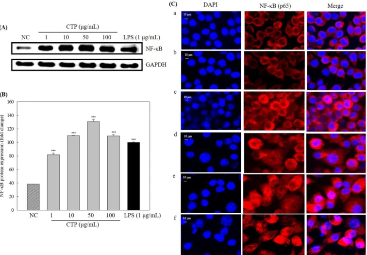
Fig. 4. Western blot analysis of NF-κB (A) and quantified (B) in CTP-treated RAW 264.7 cells and immunofluorescence of the nuclear translocation of NF-κB (p65) by confocal (C). (a) Negative control, (b) CTP 1 μg/mL, (c) CTP 10 μg/mL, (d) CTP 50 μg/mL, (e) CTP 100 μg/ mL, (f) LPS 1 μg/mL. *p<0.05, **p<0.01, and ***p<0.001, Student’s t-test, compared with the negative control (treated with distilled water).
Effect of CTP on iNOS2 and COX-2 expression
iNOS2 and COX-2 are important enzymes that mediate inflammatory processes. NF-κB is known to play a pivotal role in immune and inflammatory responses through the regulation of genes encoding pro-inflammatory cytokines, adhesion molecules, chemokines, growth factors, and inducible enzymes such as COX-2 and iNOS2 (Lawrence et al., 2001). Increase of NO, TNF-α, and IL-6 is also dependent on activation of the transcription factor NF-κB (Lawrence and Fong, 2010). NF-κB translocation to the nucleus results in upregulation of inflammatory cytokines and iNOS2 and COX-2 mRNA and protein expression. CTP increased expression levels of iNOS2 (Fig. 3A) and COX-2 (Fig. 3B) genes in a dose dependent manner compared with the negative control. iNOS2 and COX-2 protein expression in whole lysate increased in CTP-treated RAW 264.7 cells (Fig. 3C). iNOS2 expression increased 1.2-, 1.8-, 3.3-, and 4.0-fold in the cells treated with 1, 10, 50, and 100 μg/mL concentration of CTP, respectively, compared with the negative control (Fig. 3D). COX-2 expression also increased 1.7-, 11.6-, 48.0-, and 69.9-fold in the cells treated with 1, 10, 50, and 100 μg/mL concentration of CTP, respectively, compared with the negative control (Fig. 3D).
Effect of CTP on NF-κB expression and translocation to the nucleus
NF-κB protein levels in nuclear lysate considerably increased in CTP-treated RAW 264.7 cells compared with the negative control (Fig. 4A). NF-κB levels increased 2.1-, 2.9-, 3.4-, and 2.9-fold in the cells treated with 1, 10, 50, and 100μg/mL concentration of CTP, respectively, compared with the negative control (Fig. 4B). To confirm NF-κB (p65) nuclear translocation, an immunofluorescence assay was performed and the images were observed with confocal microscopy. NF-κB (p65) was mainly translocated to the nucleus in CTP- or LPS-treated RAW 264.7 (Fig. 4C). These results demonstrated that CTP induced NF-κB (p65) nuclear translocation. Effect of CTP on macrophage phagocytosis
Phagocytosis is one of the major processes of the innate immune response against foreign pathogens and a major mechanism to remove cellular debris. The ability of the activated RAW 264.7 cells to phagocytose foreign particles was observed by a confocal microscope using fluorescent IgG-coated latex beads (Fig. 5A). The mean of fluorescence value in macrophages is directly proportionally to phagocytic index. The fluorescence intensity Fig. 5. Phagocytic activity in CTP-treated RAW 264.7 cells was analyzed by confocal microscopy (A) and the activity was quantified using EZ-C1 software (B). Cells were treated with or without CTP; (a) Negative control, (b) CTP 1 μg/mL, (c) CTP 10 μg/mL, (d) CTP 50 μg/ mL, (e) CTP 100 μg/mL, (f) LPS 1 μg/mL. **p<0.01 and ***p<0.001, Student’s t-test, compared with the negative control (treated with distilled water).
increased proportional to the CTP concentration, which suggests that CTP stimulated phagocytosis in a dose-dependent manner (Fig. 5B). The phagocytic activities increased 0.96-, 1.08-, 2.76-, and 2.80-fold in the cells treated with 1, 10, 50, and 100μg/mL CTP, respectively, compared with the negative control.
Conclusion
Cherry tomato polysaccharides (CTP) are enriched mainly of pectic polysaccharide, and AG-II in RG-I is a major moiety of CTP. CTP increased production of NO and cytokines, TNF-α, MCP-1, and IL-6. CTP promoted iNOS2 and COX2 secretion, caused nuclear translocation of NF-κB (p65), and increased phagocytic activity, resulting in the increase of immunostimulating activity. Therefore, CTP could be used as prebiotics to strengthen immunity.
References
Blumenkrantz N, Asboe-Hansen G. New method for quantitative determination of uronic acids. Anal. Biochem. 54: 484-489 (1973)
Bornet F, Brouns F, Tashiro Y, Duvillier V. Nutritional aspects of short-chain fructooligosaccharides: natural occurrence, chemistry, physiology and health implications. Dig. Liver Dis. 34: S111-S120 (2002)
Caffall KH, Mohnen D. The structure, function, and biosynthesis of plant cell wall pectic polysaccharides. Carbohydr. Res. 344: 1879-1900 (2009)
DuBois M, Gilles KA, Hamilton JK, Rebers P, Smith F. Colorimetric method for determination of sugars and related substances. Anal. Chem. 28: 350-356 (1956)
Dueñas M, Hernández T, Estrella I. Changes in the content of bioac-tive polyphenolic compounds of lentils by the action of exoge-nous enzymes. Effect on their antioxidant activity. Food Chem. 101: 90-97 (2007)
Harholt J, Suttangkakul A, Scheller HV. Biosynthesis of pectin. Plant Physiol. 153: 384-395 (2010)
Jones TM, Albersheim P. A gas chromatographic method for the determination of aldose and uronic acid constituents of plant cell wall polysaccharides. Plant Physiol. 49: 926-936 (1972)
Kang SR, Park KI, Park HS, Lee DH, Kim JA, Nagappan A, Kim EH, Lee WS, Shin SC, Park MK, Han DY, Kim GS. Anti-inflammatory effect of flavonoids isolated from Korea Citrus aurantium L. on lipopolysaccharide-induced mouse macrophage RAW 264.7 cells by blocking of nuclear factor-kappa B (NF-κB) and mitogen-activated protein kinase (MAPK) signalling path-ways. Food Chem. 129: 1721-1728 (2011)
Karin M. NF-κB as a critical link between inflammation and cancer. Cold Spring Harb. Perspect. Biol. 1: 1-15 (2009)
Karin M, Greten FR. NF-κB: linking inflammation and immunity to cancer development and progression. Nat. Rev. Immunol. 5: 749-759 (2005)
Kim H, Hong HD, Suh HJ, Shin KS. Structural and immunological feature of rhamnogalacturonan I-rich polysaccharide from Korean persimmon vinegar. Int. J. Biol. Macromol. 89: 319-327 (2016) Kim SF, Huri DA, Snyder SH. Inducible nitric oxide synthase binds,
S-nitrosylates, and activates cyclooxygenase-2. Science 310: 1966-1970 (2005)
Kim AJ, Kim YO, Shim JS, Hwang JK. Immunostimulating activity of crude polysaccharide extract isolated from Curcuma xanthor-rhiza Roxb. Biosci. Biotechnol. Biochem. 71: 1428-1438 (2007)
Koh JH, Hong JH, Oh JH. Characterization of tomato pomace and reaction conditions of industrial pectinase to treat tomato pomace. Food. Eng. Prog. 19: 279-284 (2015)
Lawrence T, Fong C. The resolution of inflammation: anti-inflamma-tory roles for NF-κB. Int. J. Biochem. Cell Biol. 42: 519-523 (2010)
Lawrence T, Gilroy DW, Colville-Nash PR, Willoughby DA. Possible new role for NF-κB in the resolution of inflammation. Nat. Med. 7: 1291-1297 (2001)
Lee JB, Ohta Y, Hayashi K, Hayashi T. Immunostimulating effects of a sulfated galactan from Codium fragile. Carbohydr. Res. 345: 1452-1454 (2010)
Li L, Wang L, Wu Z, Yao L, Wu Y, Huang L, Liu K, Zhou X, Gou D. Anthocyanin-rich fractions from red raspberries attenuate inflammation in both RAW264. 7 macrophages and a mouse model of colitis. Sci. Rep. 4: 6234 (2014)
Liu W, Xu N, Yuan H, Li S, Liu L, Pu Z, Wan J, Wang H, Chang Y, Li R. Immunomodulatory activity of recombinant ricin toxin binding subunit B (RTB). Int. J. Mol. Sci. 14: 12401-12410 (2013)
Livak KJ, Schmittgen TD. Analysis of relative gene expression data using real-time quantitative PCR and the 2 ΔΔCT method. Meth-ods 25: 402-408 (2001)
Marletta MA. Nitric oxide synthase structure and mechanism. J. Biol. Chem. 268: 12231-12234 (1993)
Napolitano A, Costabile A, Martin-Pelaez S, Vitaqlione P, Klinder A, Gibson GR, Foqliano V. Potential prebiotic activity of oligosac-charides obtained by enzymatic conversion of durum wheat insol-uble dietary fibre into solinsol-uble dietary fibre. Nutr. Metab. Cardiovasc. Dis. 19: 283-290 (2009)
Paulsen B, Craik D, Dunstan D, Stone B, Bacic A. The Yariv reagent: Behaviour in different solvents and interaction with a gum arabic arabinogalactanprotein. Carbohydr. Polym. 106: 460-468 (2014)
Popov S, Ovodov YS. Polypotency of the immunomodulatory effect of pectins. Biochem. 78: 823-835 (2013)
Razali FN, Ismail A, Abidin NZ, Shuib AS. Stimulatory effects of polysaccharide fraction from Solanum nigrum on RAW 264.7 murine macrophage cells. PLoS ONE 9: e108988 (2014)
Schepetkin IA, Faulkner CL, Nelson-Overton LK, Wiley JA, Quinn MT. Macrophage immunomodulatory activity of polysaccharides isolated from Juniperus scopolorum. Int. Immunopharmacol. 5: 1783-1799 (2005)
Schepetkin IA, Quinn MT. Botanical polysaccharides: macrophage immunomodulation and therapeutic potential. . Int. Immunophar-macol. 6: 317-333 (2006)
Tajima T, Murata T, Aritake K, Urade Y, Hirai H, Nakamura M, Ozaki H, Hori M. Lipopolysaccharide induces macrophage migra-tion via prostaglandin D2 and prostaglandin E2. J. Pharmacol. Exp. Ther. 326: 493-501 (2008)
van Holst G-J, Clarke AE. Quantification of arabinogalactan-protein in plant extracts by single radial gel diffusion. Anal. Biochem. 148: 446-450 (1985)
Wang Y, Cui X, Tai G, Ge J, Li N, Chen F, Yu F, Liu Z. A critical role of activin A in maturation of mouse peritoneal macrophages in vitro and in vivo. Cell Mol. Immunol. 6: 387-392 (2009) Yapo BM. Pectic substances: From simple pectic polysaccharides to
complex pectins-A new hypothetical model. Carbohydr. Polym. 86: 373-385 (2011)
Yoon YD, Han SB, Kang JS, Lee CW, Park SK, Lee HS, Kang JS, Kim HM. Toll-like receptor 4-dependent activation of macroph-ages by polysaccharide isolated from the radix of Platycodon grandiflorum. Int. Immunopharmacol. 3: 1873-1882 (2003) Zhao JF, Kiyohara H, Yamada H, Takemoto N, Kawamura H.
Heter-ogeneity and characterisation of mitogenic and anti-complemen-tary pectic polysaccharides from the roots of Glycyrrhiza uralensis Fisch et DC. Carbohydr. Res. 219: 149-172 (1991)