https://doi.org/10.5624/isd.2018.48.2.121
Introduction
Enchondral calcification and ossification of the hya- line laryngeal cartilages begins when skeletal maturity is attained and progresses thereafter as a physiological process. Calcified triticeous and thyroid cartilages are the laryngeal cartilages that are most frequently visualized on panoramic radiographs.1
Calcified laryngeal cartilages(CLC) are an incidental radiographic finding with no clinical importance. How- ever, the detection of laryngeal cartilages(triticeous and thyroid cartilages) on panoramic radiographs is important
because they may be confused with carotid artery calcifi- cations(CAC) in the carotid bifurcation, which are a risk factor for stroke.2,3
On panoramic images, calcified triticeous cartilages are located in the soft tissues of the pharynx inferior to the greater horn of the hyoid bone and adjacent to the supe- rior border of C4. Triticeous cartilages are mostly oval or circular.4 The periphery of calcified triticeous cartilage is well-defined and smooth. Ossification of thyroid cartilage was found to have radiographic manifestations ranging from linear shadows to dense laminar calcifications in patients in their third decade and beyond.5 Usually, the superior horn of a calcified thyroid cartilage appears me- dial to C4 and is superimposed on the prevertebral soft tissue.1 Generally, only the top 2 to 3mm of this calcified cartilage is visible at the lower edge of the panoramic ra- diograph. If the cassette is positioned too far inferiorly, a
Is the diagnosis of calcified laryngeal cartilages on panoramic radiographs possible?
Leyla Berna Çağırankaya1,*, Nursel Akkaya1, Gökçen Akçiçek1, Hatice Boyacıoğlu Doğru1
1Department of Dentomaxillofacial Radiology, Faculty of Dentistry, Hacettepe University, Ankara, Turkey
AbstrAct
Purpose: Detecting laryngeal cartilages(triticeous and thyroid cartilages) on panoramic radiographs is important because they may be confused with carotid artery calcifications in the bifurcation region, which are a risk factor for stroke. This study assessed the efficiency of panoramic radiography in the diagnosis of calcified laryngeal cartilages using cone-beam computed tomography(CBCT) as the reference standard.
Materials and Methods: A total of 312 regions(142 bilateral, 10 left, 18 right) in 170 patients(140 males, 30 females) were examined. Panoramic radiographs were examined by an oral and maxillofacial radiologist with 11 years of experience. CBCT scans were reviewed by 2 other oral and maxillofacial radiologists. The kappa coefficient(κ) was calculated to determine the level of intra-observer agreement and to determine the level of agreement between the 2 methods. Diagnostic indicators(sensitivity, specificity, accuracy, and false positive and false negative rates) were also calculated. P values <.05 were considered to indicate statistical significance.
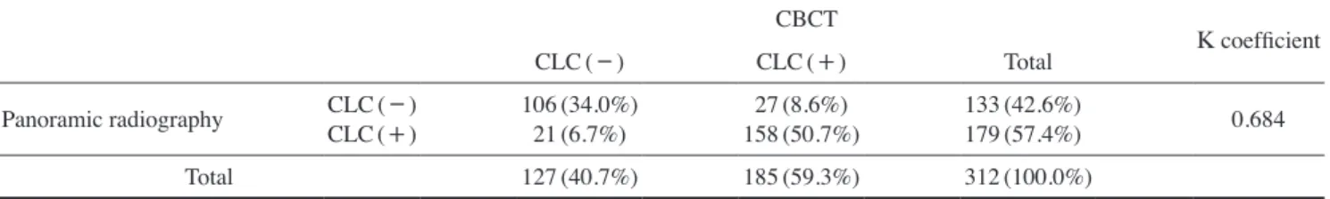
results: Eighty-two images were re-examined to determine the intra-observer agreement level, and the kappa coefficient was calculated as 0.709(P<.05). Statistically significant and acceptable agreement was found between the panoramic and CBCT images(κ=0.684 and P<.05). The sensitivity, specificity, diagnostic accuracy rate, the false positive rate, and the false negative rate of the panoramic radiographs were 85.4%, 83.5%, 84.6%, 16.5%, and 14.6%, respectively.
conclusion: In most cases, calcified laryngeal cartilages could be diagnosed on panoramic radiographs. However, due to variation in the calcifications, diagnosis may be difficult.(Imaging Sci Dent 2018; 48: 121-5)
Key words: Laryngeal Cartilages; Diagnostic Imaging; Radiography, Panoramic; Cone-Beam Computed Tomography
Copyright ⓒ 2018 by Korean Academy of Oral and Maxillofacial Radiology
This is an Open Access article distributed under the terms of the Creative Commons Attribution Non-Commercial License(http://creativecommons.org/licenses/by-nc/3.0) which permits unrestricted non-commercial use, distribution, and reproduction in any medium, provided the original work is properly cited.
Imaging Science in Dentistry·pISSN 2233-7822 eISSN 2233-7830 Received February 19, 2018; Revised April 17, 2018; Accepted May 18, 2018
*Correspondence to : Prof. Leyla Berna Çağırankaya
Department of Dentomaxillofacial Radiology, Faculty of Dentistry, Hacettepe University, Ankara 06100, Turkey
Tel) 90-312-305-2205, Fax) 90-312-305-4221, E-mail) lbartvinli@yahoo.com
greater length of this cartilage will be visualized.3 CLC may show different degrees of calcification. They gener- ally demonstrate a homogeneous radiopacity, but may oc- casionally have an outer cortex.1
Interestingly, it is not well known how accurately CLC can be diagnosed on panoramic radiographs.
Although it has been suggested that on panoramic ra- diographs, the shape, outline, and location of triticeous cartilage can help differentiate it from CAC;4 the diagnos- tic indicators(sensitivity, specificity, accuracy, and false positive and false negative rates) of panoramic radiogra- phy for the diagnosis of CLC have not been evaluated.
Thus, this study assessed the efficiency of panoramic ra- diography in the diagnosis of CLC using cone-beam com- puted tomography(CBCT) as the reference standard.
Materials and Methods
This retrospective study was based on 170 pairs of pan- oramic radiographs and CBCT scans covering the rele- vant region(140 males, 30 females; mean age, 41.4 years;
range, 8-80 years). Initially, the CBCT scans were select- ed and then coupled with available panoramic images.
CBCT scans obtained between December 2014 and De- cember 2015 were chosen from the records of the Depart- ment of Dentomaxillofacial Radiology, Faculty of Den- tistry, Hacettepe University(n=2177). The study proto- col was reviewed and approved by the Non-Interventional Clinical Research Ethics Board of Hacettepe University.
For this type of study, formal consent is not required. All procedures performed in studies involving human partici- pants were in accordance with the ethical standards of the institutional and/or national research committee and with the 1964 Helsinki declaration and its later amendments or comparable ethical standards.
Panoramic radiographs
Digital panoramic images were obtained using Ortho- phos XG 5(Sirona Dental Company, Bensheim, Germa- ny; 60-90kVp, 3-16mA, 14s) or Veraview IC5(Morita Corporation, Kyoto, Japan; 60-70kVp, 1-7.5mA, 5.5-10 s) equipment. Exposure parameters were selected accord- ing to the patient’s size. Radiographs on which the hyoid bone was too low in the neck to be visualized were ex- cluded. The images were anonymized before the analysis, and were numbered and saved.
Panoramic radiographs were examined to detect thyroid and triticeous cartilage calcifications according to the im- aging features presented in an oral radiology textbook1 by
an oral and maxillofacial radiologist(GA) with 11 years of experience. A calibration study was carried out for the observer before the examination of the panoramic images.
Both types of calcifications were designated as laryngeal cartilage calcifications.
CBCT scans
The CBCT scans were acquired with an i-CAT Next Generation(Imaging Sciences International, Hatfield, PA, USA) unit. A tube voltage of 120kVp was used for all ac- quisitions, with variation in the field of view, acquisition voxel size, tube current(3-7mA), and exposure time. The images were viewed with i-CAT Vision software(ver- sion 1.9.3.14, Imaging Science International, Hatfield, PA, USA). The CBCT scans were reviewed by 2 oral and maxillofacial radiologists.
For optimal viewing, a task-specific image display for soft-tissue calcifications of the neck was used. The rec- ommended protocol used a slice thickness of 30mm and maximum intensity projection images.6 Therefore, in the present study, each scan was initially viewed with this protocol. Once a calcification was identified, thinner slic- es were obtained if more detail was needed.
Statistical analysis
The kappa coefficient(κ) was calculated to determine the level of intra-observer agreement for the panoram- ic radiography evaluations and to determine the level of agreement between the 2 methods.
The discrimination of radiographs with and without la- ryngeal cartilage calcification and diagnostic indicators (sensitivity, specificity, and accuracy) for the panoramic radiography evaluations were also calculated. Data anal- ysis was performed using SPSS Statistics version 17.0 (SPSS Inc., Chicago IL, USA.). P values <.05 were con- sidered to indicate statistical significance.
results
A total of 312 regions(142 bilateral, 10 left, 18 right) were evaluated in 170 patients(140 males, 30 females).
The observer re-evaluated 82 panoramic images to assess the level of intra-observer agreement. The kappa coeffi- cient was calculated as 0.709, which was statistically sig- nificant(P<.05).
There was a statistically significant and acceptable rela- tionship between the CBCT and panoramic image evalua- tions(κ=0.684, P<.05).
The sensitivity, specificity, and diagnostic accuracy
rate of panoramic radiography for the discrimination of positive-CLC and negative-CLC images according to the CBCT gold standard were 85.4%(158 of 185), 83.5%(106 of 127), and 84.6%(264 of 312), respectively. In 127 CBCT images, CLC was not detected, while in 21 pan- oramic images it was mistakenly detected(false positive rate, 16.5%). Furthermore, in the CBCT images, 185 cas- es of CLC were detected, while in 27 panoramic images it was mistakenly not detected(false negative rate, 14.6%) (Table 1).
discussion
In the dental literature, laryngeal calcifications have been described in relation to their appearance on pan- oramic radiographs, with a great emphasis on differen- tiating triticeous ossification from CAC. To the best of our knowledge, the present study is the first to assess the efficiency of panoramic radiography for the detection of CLC.Recently, a study in a large population(age range, 18- 97 years; mean age, 65 years) utilizing computed to- mography images revealed that the presence of triticeous cartilage as a part of the laryngeal skeleton was quite common(53.1%).7 In contrast, the frequency of calcified triticeous cartilage on panoramic radiographs was report- ed to be 5% in men and 12% in women among 40 years and over.4 This disparity in the prevalence is partially due to the technique of panoramic radiography. The hyoid bone is the reference point for the diagnosis of laryngeal cartilages on panoramic radiography. However, in some cases, the hyoid bone may be too low to be visualized on panoramic radiographs.
A complicating factor in relation to the differential di- agnosis is that the age range of the greatest prevalence of CAC coincides with the age range associated with the oc- currence of patterns of mineralization and/or calcification that have been reported to be responsible for erroneous diagnoses, such as ossification of the triticeous, cricoid,
and thyroid cartilages.2 Furthermore; Alqahtani et al.,7 in a study of computed tomography, reported a wide range of variation in the morphology, shape, ossification, and position of the triticeous cartilage within the thyrohyoid ligament. Due to this variation, symmetric contralateral comparison is not appropriate for the diagnosis of CLC.
Initial decisions regarding soft-tissue calcification in the soft tissues of the lateral neck of panoramic imag- es involve determining the location of the calcification relative to the hyoid bone and further characterization relative to other structures or appearances. In addition to CLC, calcified lymph nodes in the carotid sheath and CAC can be found below the hyoid bone. Calcified trit- iceous cartilages have been described as single, small, round radiopacities below the greater horn of the hyoid.8 Kamikawa et al.2 positioned radiopaque spheres in the anatomic structures of the cervical region that can be sites of calcification in cadaver specimens, and found that 75%
of the examiners correctly indicated the reference point at the bifurcation of the carotid artery and 79.2% report- ed a triticeous cartilage as CAC. This study showed that the localization of radiopacities in the cervical region of panoramic radiographs alone is not enough for the diag- nosis of calcified laryngeal structures. In contrast, Ahmad et al.4 showed that the shape, outline, and location of the triticeous cartilage can help differentiate it from CAC.
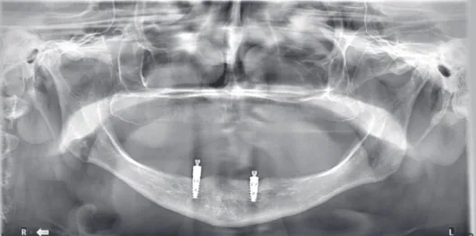
The current study showed that the diagnosis of CLC is possible with panoramic radiography in most cases. How- ever, when small, ill-defined opacities are present below the greater horn of the hyoid(Figs. 1-4), supplemental radiographs are needed because even small areas of CAC could be associated with an elevated risk of stroke or oth- er cardiovascular events.9
In the present study, both types of calcifications were designated as laryngeal cartilage calcifications, and the exclusive efficiency of panoramic radiography for the di- agnosis of calcified triticeous cartilage was not estimated.
This retrospective study was based on CBCT scans that were obtained at our dental faculty from patients with dis-
Table 1. Frequency distributions of calcified laryngeal cartilages(CLC) according to cone-beam computed tomography(CBCT) and pan- oramic evaluations
CBCT Κ coefficient
CLC(-) CLC(+) Total
Panoramic radiography CLCCLC(-)(+) 10621(34.0%)(6.7%) 15827(50.7%)(8.6%) 133179(57.4%)(42.6%) 0.684
Total 127(40.7%) 185(59.3%) 312(100.0%)
Fig. 1. A panoramic radiograph reveals small, ill-defined opacities below the greater horn of the hyoid on the right side, while calcifica- tion is demonstrated in the carotid region on cone-beam computed tomographic images.
Fig. 2. A panoramic radiograph reveals small, ill-defined opacities below the greater horn of the hyoid on the right side, while calcified laryngeal cartilages are demonstrat- ed on cone-beam computed tomo- graphic images.
Fig. 3. Axial maximum intensity projection of the patient in Figure 1 shows the location of the opacity. This image demonstrates cal- cification in the carotid region.
Fig. 4. Axial maximum intensity projection of the patient in Fig- ure 2 shows the location of the opacity. This image demonstrates calcified laryngeal cartilages. Calcified laryngeal cartilages can be differentiated from calcification in the carotid region because they are medial to the greater horn of the hyoid bone.
eases in the oral and maxillofacial region. Therefore, in most cases, the field of view did not allow triticeous carti- lages to be distinguished from thyroid cartilages.
In conclusion, the present study showed that CLC could be diagnosed on panoramic radiographs in most cases.
However, diagnosis may be difficult due to variation in the calcifications.
references
1. Carter LC. Soft tissue calcifications and ossifications. In: White SC, Pharoah MJ. Oral radiology: principles and interpretation.
7th ed. St. Louis: Elsevier; 2014. p. 524-41.
2. Kamikawa RS, Pereira MF, Fernandes A, Meurer MI. Study of the localization of radiopacities similar to calcified carotid atheroma by means of panoramic radiography. Oral Surg Oral Med Oral Pathol Oral Radiol Endod 2006; 101: 374-8.
3. Carter LC. Discrimination between calcified triticeous cartilage and calcified carotid atheroma on panoramic radiography. Oral Surg Oral Med Oral Pathol Oral Radiol Endod 2000; 90: 108- 10.
4. Ahmad M, Madden R, Perez L. Triticeous cartilage: prevalence on panoramic radiographs and diagnostic criteria. Oral Surg Oral Med Oral Pathol Oral Radiol Endod 2005; 99: 225-30.
5. Mupparapu M, Vuppalapati A. Ossification of laryngeal carti- lages on lateral cephalometric radiographs. Angle Orthod 2005;
75: 196-201.
6. Scarfe WC, Li Z, Aboelmaaty W, Scott SA, Farman AG. Max- illofacial cone beam computed tomography: essence, elements and steps to interpretation. Aust Dent J 2012; 57 Suppl 1: 46- 7. Alqahtani E, Marrero DE, Champion WL, Alawaji A, Kousou-60.
bris PD, Small JE. Triticeous cartilage CT imaging character- istics, prevalence, extent, and distribution of ossification. Otol- aryngol Head Neck Surg 2016; 154: 131-7.
8. MacDonald D, Chan A, Harris A, Vertinsky T, Farman AG, Scarfe WC. Diagnosis and management of calcified carotid artery artheroma: dental perspectives. Oral Surg Oral Med Oral Pathol Oral Radiol 2012; 114: 533-47.
9. Garoff M, Ahlqvist J, Levring Jäghagen E, Johansson E, Wester P. Carotid calcification in panoramic radiographs: radiographic appearance and the degree of carotid stenosis. Dentomaxillofac Radiol(in press).