Endocrinol Metab 2015;30:110-115
http://dx.doi.org/10.3803/EnM.2015.30.1.110 pISSN 2093-596X · eISSN 2093-5978
Case Report
Multiple Myeloma in a Patient with Acromegaly
Yu Mi Kang1,*, Jong Han Choi1,*, Min Jung Lee1, Ari Ahn2, Chan-Jeoung Park2, Kiju Chang3, Seyoung Seo3, Sun In Hong3, Min-Seon Kim1
1Division of Endocrinology and Metabolism, Department of Internal Medicine, Departments of 2Laboratory Medicine, 3Internal Medicine, Asan Medical Center, University of Ulsan College of Medicine, Seoul, Korea
Acromegaly is a slowly progressing condition resulting from excess growth hormone (GH), generally caused by a GH-secreting pituitary adenoma. Cancer is the third most common cause of mortality in patients with acromegaly, and insulin-like growth factor 1 (IGF-1) is known to influence tumor formation by increasing cell proliferation and inhibiting apoptosis. Multiple myeloma (MM) is a plasma cell neoplasm, and previous studies have suggested the possible role of IGF-1 in its development of MM. However, no cases of acromegaly accompanied with MM have been reported in Asia to date. We here report the case of a 58-year-old woman with acromegaly accompanied with MM who presented with longstanding acromegalic manifestations resulting from a GH-secret- ing pituitary adenoma and also exhibited anemia, a reversed albumin/globulin ratio, and plasmacytosis on bone marrow examina- tion. Because IGF-1 has been suggested to play an important role in the development and progression of MM, the patient promptly underwent surgical removal of the pituitary adenoma via a transsphenoidal approach. Since there is currently no consensus on ther- apeutic guidelines and suggested prognosis for MM with acromegaly, long-term follow-up of such cases is needed.
Keywords: Acromegaly; Multiple myeloma; Insulin-like growth factor I
INTRODUCTION
Acromegaly is a systemic, chronic, and slowly progressing con- dition resulting from excess growth hormone (GH) and insulin- like growth factor 1 (IGF-1) [1]. It is most commonly caused by a GH-secreting pituitary adenoma [2]. Patients with acro- megaly have a 1.6- to 3.3-fold increase in age-related mortality and a 10-year reduction in life expectancy [3], and cancer is the third most common cause of mortality [4]. The role of the GH–
IGF-1 axis in the development of malignancy has been contin- uously investigated, and IGF-1 is thought to enhance cell pro- liferation and to inhibit apoptosis [4]. Breast cancer is epidemi-
ologically linked to IGF-1 levels in premenopausal women [5], and an increased IGF-1 level may increase the relative risk of developing colon cancer [6].
Multiple myeloma (MM) is a malignant proliferative disease of monoclonal plasma cells that produce monoclonal immuno- globulins [7]. Diagnosis of MM is made based on the clonal plasmacytosis of at least 15% on bone marrow examination or biopsy-proven clonal plasmacytoma, the presence of M protein in serum or urine, and any evidence of end-organ damage (i.e., hypercalcemia, renal insufficiency, anemia, and bone disease) attributable to myeloma involvement [7].
Although acromegaly is associated with malignancy, its as-
Received: 21 May 2014, Revised: 3 July 2014 Accepted: 30 July 2014
Corresponding author: Min-Seon Kim
Division of Endocrinology and Metabolism, Department of Internal Medicine, Asan Medical Center, University of Ulsan College of Medicine, 88 Olympic-ro 43-gil, Songpa-gu, Seoul 138-736, Korea
Tel: +82-2-3010-3245, Fax: +82-2-3010-6962, E-mail: mskim@amc.seoul.kr
*These authors contributed equally to this work.
Copyright © 2015 Korean Endocrine Society
This is an Open Access article distributed under the terms of the Creative Com- mons Attribution Non-Commercial License (http://creativecommons.org/
licenses/by-nc/3.0/) which permits unrestricted non-commercial use, distribu- tion, and reproduction in any medium, provided the original work is properly cited.
sociation with MM has not been thoroughly investigated. Ear- lier reports demonstrated the role of GH and IGF-1 in B lym- phocyte activation [8] and the universal expression of IGF-1 receptors in MM cells in vitro [9]. Also, in several cases, acro- megaly was associated with the malignant transformation of monoclonal gammopathy of undetermined significance (MGUS) into overt MM [10,11]. However, only a few actual cases of MM associated with acromegaly have been reported [12,13]. Here we describe the first Asian patient, who was di- agnosed with acromegaly and MM, simultaneously.
CASE REPORT
A 58-year-old woman was admitted to our department for diag- nostic workup of acromegalic features. She had noticed the en- largement of her face, hands, and feet 8 years prior to her visit,
and these were associated with hyperhidrosis, a thicker and deeper voice, and decreased libido. At that time, she had also undergone uvulectomy because of obstructive sleep apnea that had caused severe snoring. The snoring continued after surgery, albeit much more infrequently.
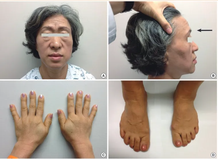
On admission, the patient’s blood pressure was 120/80 mm Hg. She had a heart rate of 86 per minute, a respiratory rate of 20 per minute, and a body temperature of 36.5°C. During physical examination, the facial enlargement with frontal boss- ing was noticed, as was the disproportionate enlargement of the tongue, nose, lips, hands, and feet (Fig. 1). Hypertrichosis was seen, especially in the lower extremities. Soft tissue swelling was noted, and the skin folds were thickened.
A complete blood count revealed a plasma hemoglobin level of 10.0 g/dL. The serum protein level was 10.5 g/dL (reference range, 6 to 8) and the serum albumin level was 2.5 g/dL (refer-
Fig. 1. (A) Physical examination of the patient revealed frontal bossing, thickened lips, and an enlarged nose. (B) Exaggerated frontal bossing (arrow) observed from the side. Disproportionately enlarged (acromegalic) hands (C) and feet (D) were also seen.
A
C
B
D
ence range, 3.5 to 5.2), reflecting a reversed albumin/globulin (A/G) ratio. Serum creatinine and calcium levels were 0.63 mg/
dL (reference range, 0.70 to 1.40) and 8.6 mg/dL (reference range, 8.6 to 10.2), respectively. Thus, the serum calcium level corrected for hypoalbuminemia was 9.2 mg/dL. The fasting plasma glucose level was 109 mg/dL and the 2-hour postprandi- al plasma glucose level was 280 mg/dL. Hemoglobin A1c was 7.8% and the C-peptide level was 4.8 ng/mL. The thyroid-stim- ulating hormone level was 1.5 µU/mL (reference range, 0.4 to 5.0) and the free thyroxine level was 1.3 ng/dL. Basal levels of adrenocorticotropic hormone, prolactin, luteinizing hormone, follicle-stimulating hormone, and estradiol were 23.0 pg/mL, 6.8 ng/mL, 17.1 mIU/mL, 44.4 mIU/mL, and 10.0 pg/mL, re- spectively, which were all within normal ranges. Serum levels of IGF-1 and human growth hormone (hGH) were elevated to 898 ng/mL (age-adjusted reference range, 71 to 284) and 42 ng/
mL (reference range, <16.0), respectively (Tables 1, 2).
Because these laboratory findings strongly suggested acro- megaly and associated diabetes mellitus, we performed a 75g
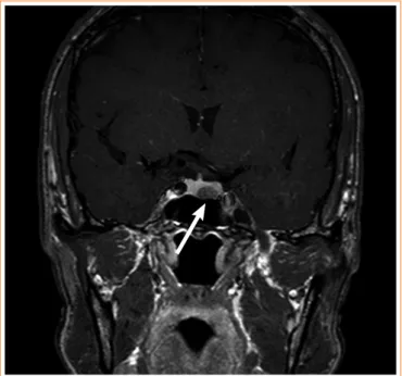
oral glucose tolerance test (OGTT) to confirm GH hypersecre- tion (Table 2). To confirm the presence of a GH-secreting pi- tuitary adenoma, we performed magnetic resonance imaging of the sella turcica. It revealed a 1.0×0.6-cm pituitary adeno- ma on the left side of the pituitary gland (Fig. 2).
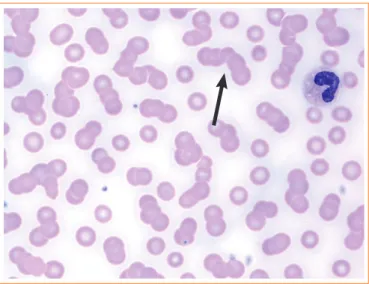
The markedly reversed A/G ratio and anemia led us to sus- pect a monoclonal gammopathy such as MM. A peripheral blood smear revealed a red blood cell rouleux formation (Fig.
3). Protein electrophoresis (PEP) and immunofixation electro- phoresis (IFE) of serum samples revealed a monoclonal gam- mopathy (M peak 4.9 g/dL) of immunoglobulin G (IgG)-λ type;
PEP and IFE of urine samples were inadequate because of low urinary protein. The patient’s serum IgG level was elevated to 5,860.0 mg/dL, and the κ and lambda free light chain ratios were 7.16 and 34.50 mg/L, respectively. To confirm the diag- nosis of MM, we performed bone marrow aspiration and biop- sy with plasma cell phenotyping. Neoplastic plasma cells were observed on bone marrow aspirate and touchprint slides (Fig.
4A, B), and packed bone marrow with neoplastic plasma cells (35.6% of the clonal plasma cells) was observed in the bone marrow biopsy specimen (Fig. 4C-F). In an attempt to find my- eloma-related organ damage, we took simple X-rays of the en- tire body, and they did not reveal any osteolytic lesions. Ac- cording to the diagnostic criteria for symptomatic MM, one should exhibit more than one of the parameters reflecting my- Table 1. Comparison of Preoperative and Postoperative Serum
Insulin-Like Growth Factor 1
Variable Measured level Age-adjusted normal reference range
Preoperative, ng/mL 898 71–284
Postoperative, ng/mL 223 71–284
Table 2. Comparison of Preoperative and Postoperative 75 g OGTT results
Variable Serum glucose, mg/dL Serum human GH, ng/mL Baseline, min
0 109 9.4
30 179 11.0
60 -a 10.8
90 268 21.1
120 280 15.6
3 Days after surgery, time, min
0 101 2.2
30 160 2.0
60 197 4.1
90 245 3.3
120 246 2.7
OGTT, oral glucose tolerance test; GH, growth hormone.
aSerum glucose level was not measured at 60 minutes.
Fig. 2. A T1-weighted coronal magnetic resonance imaging of the sella turcica indicated a 10×6-mm hypointense, nonenhanced pi- tuitary adenoma on the left side of the pituitary gland (arrow).
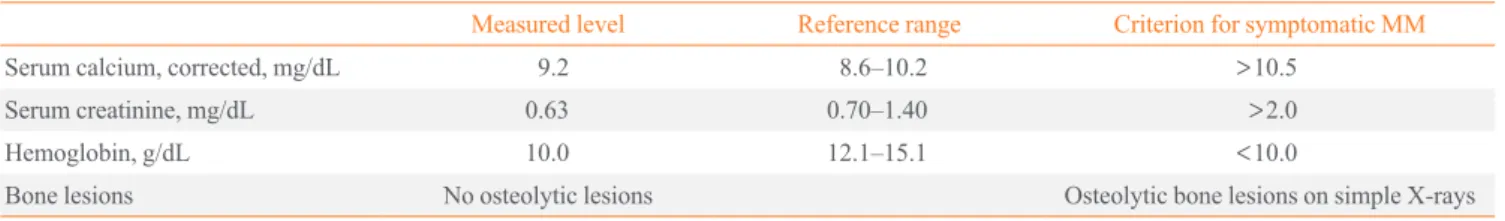
eloma-related organ dysfunction (Table 3). Because the M peak was >3 g/dL and the proportion of monoclonal plasma cells in the bone marrow was >10% without myeloma-related orgam damages, the patient was diagnosed with asymptomatic (smol-
dering) MM. However, it is of note that her serum hemoglobin level was at the cutoff value for diagnosis of symptomatic MM and that her serum calcium level was slightly below the cutoff (Table 3). In accordance with the treatment guidelines for as- ymptomatic MM, we planned close follow-up, with deferral of chemotherapy until the disease became symptomatic.
To treat the GH-secreting pituitary adenoma, the patient un- derwent tumor resection via a transsphenoidal approach. There were no immediate postoperative complications. Three days after surgery, the serum IGF-1 level was measured and the 75 g OGTT was repeated. Although the serum hGH level was low- er than it had been before surgery, the results indicated incom- plete remission immediately after surgery (Table 2). However, after the surgery, the patient exhibited clinical improvements in acromegalic features, such as decreased skin thickness and reduced size of the extremities. She also claimed that the fre- quency of snoring and daytime drowsiness had decreased dra- matically. Therefore, we decided to closely monitor the clini- cal and biochemical responses to the removal of the GH-se- creting pituitary adenoma and to continuously determine the need for further treatment.
Fig. 3. Peripheral blood smear results revealed a red blood cell rouleaux formation (arrow; Wright stain, ×1,000).
Fig. 4. (A) Neoplastic plasma cells in the bone marrow aspirate specimen (Wright stain, ×1,000). (B) Numerous clustered neoplastic plasma cells on a bone marrow touchprint slide. (C) Packed neoplastic plasma cells were prominent in a bone marrow biopsy specimen stained with H&E (×400). (D) CD138-positive neoplastic plasma cells in an immunohistochemically stained bone marrow biopsy speci- men (×400). In an immunohistochemically stained bone marrow biopsy specimen, neoplastic cells were positive for λ light chain (E, × 400) and negative for κ light chain (F, ×400).
A
D
B
E
C
F
DISCUSSION
Acromegaly is an uncommon endocrinologic disorder caused by overproduction of GH, most commonly by a GH-secreting pituitary adenomas [3]. Earlier epidemiological studies have in- dicated a possible increase in malignancy risk in patients with acromegaly because GH and IGF-I may promote proliferation and inhibit apoptosis of neoplastic cells [4]. Nabarro [1] found a markedly increased incidence of breast cancer in patients with acromegaly. Also, a large retrospective cohort study conducted by Orme et al. [3] to determine mortality and cancer incidence in 1,362 subjects with acromegaly reported a significant in- crease in the colon cancer mortality rate and a nonsignificant in- crease in mortality due to breast cancer. The authors also found that mortality due to all malignant disease and colon cancer in- creased with higher posttreatment GH levels, which suggests that GH hypersecretion may have modified the progression of coexisting malignancies, particularly colonic carcinomas.
There have been sporadic case reports of hematologic ma- lignancies such as lymphoma and leukemia associated with acromegaly [12,13]. However, the causal relationship between acromegaly and MM has not been clearly proven in clinical settings. Despite the complexity of the interactions between the bone marrow microenvironment and surrounding stromal cells, attempts have been made to determine the exact mecha- nisms of GH and IGF-1 in the development and proliferation of myeloma [5,6,14]. Murphy et al. [8] demonstrated that treatment with hGH had stimulatory effects on B-cell prolifer- ation. Georgii-Hemming et al. [9] demonstrated the universal expression of IGF-1 receptors in MM cells and postulated that IGF-1 might act like a growth factor in human MM. Also, Ge and Rudikoff [14] demonstrated a stimulatory effect of IGF-1 on six of eight myeloma cell lines, one of two B lymphoma cell lines, and neither of two T leukemia cell lines, where all cell lines expressed similar levels of IGF-1 receptors. The au- thors suggested that the IGF-1 pathway is biologically relevant
in the later stages of B-cell development.
There is neither an established therapeutic guideline nor a prognosis specific for acromegaly with MM. However, several cases illustrate that treatment of acromegaly improves the pro- gression of MGUS or MM. Kanazawa et al. [10] reported a case of MGUS in a patient with acromegaly in which the se- rum IgG level, as well as the GH and IGF-1 levels, decreased after the removal of the GH-secreting pituitary adenoma.
Moreover, Tucci et al. [11] described a patient with acromega- ly in whom a longstanding MGUS had transformed into overt MM. In this patient, serum and urine M components indicated that MM activity was correlated with serum IGF-1 levels.
These findings are solid evidence supporting the hypothesis that elevated GH and IGF-1 levels promote the proliferation of malignant plasma cells in MM and that maintaining low levels of GH and IGF-1 through intensive treatment might be a key therapeutic approach in treating coexistent MM.
In our case, the patient’s serum hemoglobin level (10.0 g/
dL) was at the cutoff level for symptomatic MM and her se- rum calcium level (9.2 mg/dL) was slightly below the cutoff, suggesting that the risk of transformation into symptomatic MM would have been very high had the acromegaly been left untreated. Follow-up measurement of serum and urine M pro- teins and simultaneous measurement of serum GH levels would be necessary to determine the long-term effect of the removal of the GH-secreting tumor on MM. Although these measurements were unavailable in our study because of the recent diagnosis, we expect a favorable prognosis because of the prompt surgical treatment of the acromegaly before the MM became symptomatic. Likewise, the lack of long-term follow-up data from previously reported cases makes it diffi- cult to predict the prognosis of acromegaly with MM. Further long-term observational studies with a larger data set will al- low us to specifically suggest appropriate therapeutic guide- lines and to extrapolate the prognosis in patients with acro- megaly and MM.
Table 3. Findings for Myeloma-Related Organ Damage
Measured level Reference range Criterion for symptomatic MM
Serum calcium, corrected, mg/dL 9.2 8.6–10.2 >10.5
Serum creatinine, mg/dL 0.63 0.70–1.40 >2.0
Hemoglobin, g/dL 10.0 12.1–15.1 <10.0
Bone lesions No osteolytic lesions Osteolytic bone lesions on simple X-rays
MM, multiple myeloma.
In summary, this is the first reported case of acromegaly ac- companied by MM from Asia. Our case strengthens the hy- pothesis of prior studies that acromegaly plays a significant role in the development of MM. Considering previous studies that have demonstrated the role of IGF-1 in plasma cell prolif- eration and the correlation between serum IGF-1 levels and the progression of monoclonal gammopathy, prompt efforts to lower circulating GH and IGF-1 levels are an important thera- peutic approach. Further research with a larger set of longer term data is needed to develop treatment guidelines specific for MM accompanying acromegaly and to predict prognosis.
CONFLICTS OF INTEREST
No potential conflict of interest relevant to this article was re- ported.
REFERENCES
1. Nabarro JD. Acromegaly. Clin Endocrinol (Oxf) 1987;26:
481-512.
2. Freda PU, Post KD. Differential diagnosis of sellar masses.
Endocrinol Metab Clin North Am 1999;28:81-117.
3. Orme SM, McNally RJ, Cartwright RA, Belchetz PE. Mor- tality and cancer incidence in acromegaly: a retrospective cohort study. United Kingdom Acromegaly Study Group. J Clin Endocrinol Metab 1998;83:2730-4.
4. Melmed S. Acromegaly and cancer: not a problem? J Clin Endocrinol Metab 2001;86:2929-34.
5. Hankinson SE, Willett WC, Colditz GA, Hunter DJ, Mi- chaud DS, Deroo B, Rosner B, Speizer FE, Pollak M. Cir- culating concentrations of insulin-like growth factor-I and
risk of breast cancer. Lancet 1998;351:1393-6.
6. Ma J, Pollak MN, Giovannucci E, Chan JM, Tao Y, Hen- nekens CH, Stampfer MJ. Prospective study of colorectal cancer risk in men and plasma levels of insulin-like growth factor (IGF)-I and IGF-binding protein-3. J Natl Cancer Inst 1999;91:620-5.
7. Riedel DA, Pottern LM. The epidemiology of multiple myeloma. Hematol Oncol Clin North Am 1992;6:225-47.
8. Murphy WJ, Rui H, Longo DL. Effects of growth hormone and prolactin immune development and function. Life Sci 1995;57:1-14.
9. Georgii-Hemming P, Wiklund HJ, Ljunggren O, Nilsson K.
Insulin-like growth factor I is a growth and survival factor in human multiple myeloma cell lines. Blood 1996;88:2250-8.
10. Kanazawa I, Yamaguchi T, Yamane Y, Murakami N, Kato Y, Sugimoto T. Acromegaly associated with monoclonal gam- mopathy of undetermined significance (MGUS). Endocr J 2006;53:687-91.
11. Tucci A, Bonadonna S, Cattaneo C, Ungari M, Giustina A, Guiseppe R. Transformation of a MGUS to overt multiple myeloma: the possible role of a pituitary macroadenoma secreting high levels of insulin-like growth factor 1 (IGF- 1). Leuk Lymphoma 2003;44:543-5.
12. Barbosa FR, Vieira Neto L, Lima GA, Wildemberg LE, Portugal R, Gadelha MR. Hematologic neoplasias and ac- romegaly. Pituitary 2011;14:377-81.
13. Hagg E, Asplund K, Holm J. Acromegaly and multiple my- eloma. Ann Intern Med 1988;109:437-8.
14. Ge NL, Rudikoff S. Insulin-like growth factor I is a dual ef- fector of multiple myeloma cell growth. Blood 2000;96:
2856-61.