Endocrinol Metab 2013;28:138-143
http://dx.doi.org/10.3803/EnM.2013.28.2.138 pISSN 2093-596X · eISSN 2093-5978
Case Report
Riedel Thyroiditis in a Patient with Graves Disease
Doo Young Lee1, Jung Sik Moon1, Ga-Eon Kim2, Hee Kyung Kim3, Ho-Cheol Kang3
1Department of Internal Medicine, Chonnam National University Hospital, Chonnam National University Medical School, Gwangju; 2Department of Pathology, Seonam University College of Medicine, Namwon; 3Department of Internal Medicine, Chonnam National University Hwasun Hospital, Chonnam National University Medical School, Hwasun, Korea
Riedel’s thyroiditis is a rare form of infiltrative and inflammatory disease of the thyroid gland and can be associated with system- ic fibrotic processes, Hashimoto thyroiditis and Graves disease. Riedel thyroiditis in combination with Graves disease however, is very rare. A 57-year-old woman with a past medical history significant for Graves disease diagnosed 30 years ago presented with an enlarging neck mass and voice changes. Due to suspicion of malignancy, thyroidectomy was performed. Histopathologic examination revealed Riedel thyroiditis. To our knowledge, the association of Riedel thyroiditis with Graves disease has not yet been reported in our country. Here we report a patient with Riedel thyroiditis evolved from antecedent Graves disease.
Keywords: Graves disease; Riedel thyroiditis
INTRODUCTION
Riedel thyroiditis (RT) is a rare form of infiltrative and fibrotic disease of the thyroid. It is characterized by breathing difficul- ties and dysphagia resulting from pressure from a rapidly en- larging thyroid. In rare cases, it can cause voice hoarseness if the enlarged thyroid damages the recurrent laryngeal nerve.
Riedel [1] first recognized the disease in 1896. The cause of RT is not yet clear, but an immune disorder is considered the most likely cause [2,3]. Recent research has shown that immu- noglobulin G4-related systemic disease (IgG4-RSD) is likely involved [4-9]. In 1985, Hay et al. [10] reported that RT was identified in 30 of 5,700 cases of thyroidectomy during the pe- riod from 1920 through 1984, according to histopathologic examination at the Mayo Clinic. The incidence rate was esti- mated at 0.06 to 1.06 per 100,000 people. In their study, the number of female patients with RT was three times higher than the number of men, and the median age of female patients was
51 years. Of 37 patients with RT, 12 experienced fibrosclero- sis of the cervix, mediastinum, or retroperitoneum within 10 years after thyroidectomy [10].
Pi et al. [11] reported a case study in which a thyroidectomy was recommended for a patient with RT that was difficult to differentiate from thyroid cancer. However, no case of concur- rent RT and Graves disease has been reported in the local liter- ature. The authors diagnosed RT in a 57-year-old woman with Graves disease who presented with paralyzed vocal cords.
Herein, we present a case of RT cured with surgical treatment, and a review of the literature.
CASE REPORT
Patient: Mrs. Kim, age 57.
Chief complaint: Recently enlarging neck mass and hoarse- ness that started 5 days before admission to the hospital.
Present medical condition: A 57-year-old woman visited the
Received: 18 June 2012, Accepted: 4 September 2012 Corresponding author: Ho-Cheol Kang
Department of Internal Medicine, Chonnam National University Hwasun Hospital, Chonnam National University Medical School, 322 Seoyang-ro, Hwasun 519-763, Korea
Tel: +82-61-379-7620, Fax: +82-61-379-7628, E-mail: drkang@chonnam.ac.kr
Copyright © 2013 Korean Endocrine Society
This is an Open Access article distributed under the terms of the Creative Com- mons Attribution Non-Commercial License (http://creativecommons.org/
licenses/by-nc/3.0/) which permits unrestricted non-commercial use, distribu- tion, and reproduction in any medium, provided the original work is properly cited.
outpatient clinic with a very hard, fixed lump that had been present on the right side of her neck for months and hoarse- ness, which started 5 days previous. The patient had previous- ly taken antithyroid agents prescribed by a private hospital but did not undergo additional tests or follow-up related to her hy- perthyroidism. She did not show any symptoms associated with thyrotoxicosis on admission.
Past medical history: She underwent intermittent treatment for hyperthyroidism over the last three decades. She has had no other symptoms.
Family medical history: No medical problems.
Physical examination: The patient was conscious and in no acute distress. She was medically stable with a blood pressure
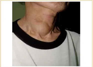
of 130/90 mm Hg, pulse of 76 beats per minute, respiration rate of 20 breaths per minute, and a temperature of 36.5ºC. Globe protrusion, scleral icterus, and conjunctival anemia were not observed. Enlarged cervical lymph nodes and jugular vein en- gorgement were also not present. A hard and fixed thyroid nod- ule that measured approximately 5×4 cm was palpated, but no neck tenderness was appreciated (Fig. 1). On auscultation, nor- mal heart and breath sounds were observed. Physical exami- nation revealed no pitting edema in her extremities.
Lab results: Complete blood count revealed a white blood cell count of 6,460/mm3 (neutrophil 57.4%, lymphocyte 36.6%, monocyte 7.7%, eosinophil 0.6%), hemoglobin of 11.6 g/dL, hematocrit of 35.2%, and platelet count of 295×103/mm3. Urine test was normal. Serum biochemical examination revealed a blood urea nitrogen/creatinine of 25/0.6 mg/dL, total protein/
albumin of 7.4/4.3 g/dL, aspartate aminotransferase/alanine aminotransferase of 22/20 IU/L, Na/K/Cl of 142/5.1/105 mEq/
L, Ca of 4.6 mEq/L (normal range, 4.5 to 5.2), erythrocyte sedimentation rate of 19 mm/hr, and C-reactive protein <0.2 mg/day. Thyroid function tests revealed thyroid stimulation hormone of 0.07 μIU/mL (normal range, 0.4 to 4.5), free T4 of 3.86 ng/dL (normal range, 0.7 to 2), TBII of 51% (normal range, 0 to 15), thyroglobulin of 493 ng/dL (normal range, 2 to 60), antithyroid microsomal antibody of 3.1 U/mL (normal range, 0 to 8), and negative thyroglobulin antibody of 22 U/
mL (normal range, 0 to 55), consistent with Graves disease.
Electrocardiography: Normal sinus rhythm.
Chest radiography: Signs of emphysema and right to left shift of the trachea as a consequence of the thyroid nodule.
Fig. 1. Gross appearance of the neck. A very hard, fixed nodule is seen in the right side of the neck.
Fig. 2. Panoramic ultrasonographic view of the thyroid. A large,
heterogeneously echoic mass is seen in the right lobe of the thyroid. Fig. 3. A hypodense mass with scattered internal dense calcifica- tions is evident in a postcontrast study.
Thyroid ultrasound: A well-defined irregular and hypoecho- ic nodule measuring 4 cm in diameter, along with large scat- tered dense calcifications, was observed in the right thyroid lobe (Fig. 2). However, enlarged cervical lymph nodes and in- vasion into the parathyroid glands were not present.
Thyroid fine needle aspiration (FNA) cytology: Since re- peated nonaspiration technique failed to obtain a specimen, we employed an aspiration technique which yielded a very small amount of cytologic specimen. In cytopathologic examination, no malignant cells were detected, but adenomatous goiter was suspected. Although thyroid cancer was not diagnosed by FNA, the large, solid mass and paralyzed vocal cords on the right side suggested the possibility of thyroid cancer. Therefore, a surgical approach was determined for histological diagnosis and treatment.
Computed tomography (CT) scan of the neck: There was a
4.3×4.0 cm nodular lesion occupying most of the right lobe of the thyroid. A peripheral calcified lesion was also identified.
Very severe marginal calcification was detected in the inferior portion of the nodule, which was barely observable in ultraso- nography (Fig. 3). Unilateral fixed vocal cords were revealed in a paramedian position. Vocal cord paralysis was suspected on the right side (Fig. 4).
Surgery: A total thyroidectomy was performed. A 4.5-cm sol- id mass of the right thyroid lobe was closely adhered to the surrounding airway and recurrent laryngeal nerve. It was im- possible to completely dissect the mass from the nerve.
Histological examination: The resected mass was covered by a thin capsule, and its cut surface showed mixed strips of white, yellowish-brown, and pink colors. Peripheral dense calcifica- tion was also seen. Microscopic examination revealed that thyroid follicles were replaced by extensive inflammatory and fibrous tissue. Inflammation was also noticed in the vein, which was encased in fibrous tissue, but no malignant cells were found.
Based on these pathologic characteristics, RT was diagnosed (Fig. 5).
Outcome: The patient recovered from RT, and there were no signs of complications. She is currently taking 100 μg levo- thyroxine while undergoing postsurgical outpatient follow-up.
She has not yet recovered from her vocal cord paralysis.
DISCUSSION
RT is a very unusual form of thyroid disease, characterized by extensive fibrosis of the thyroid tissue [1]. RT was first recog- nized as one type of inflammatory thyroiditis, but it is increas- ingly seen as a distinct disease associated with autoimmune disorders and systemic multifocal fibrosclerosis. The hypothe- Fig. 4. Neck computed tomography showed abnormal configura-
tion of the right vocal cord, suggestive of vocal cord paralysis.
Fig. 5. (A) Enlarged right lobe of the thyroid with calcification. (B) Extensively hyalinized fibrous tissue replaced the thyroid gland. Fi- brosis and inflammation extended beyond the thyroid capsule (H&E stain, ×100). (C) Medium-sized veins encased by fibrosis indicat- ing inflammation of their walls. The inflammation is of the monocuclear type, with a predominance of lymphocytes (H&E stain, ×400).
A B C
sis of an association between RT and an autoimmune mecha- nism has several supportive findings [4]; for example, thyroid- specific autoantibodies have been detected in many patients with RT; RT occurs in conjunction with other autoimmune dis- eases such as Hashimoto thyroiditis, Graves disease, and per- nicious anemia; infiltration with lymphocytes, plasma cells, and histiocytes is often evident in RT; local vasculitis is com- monly found on pathology; and lastly, RT responds well to systemic steroids. On the other hand, RT was not considered an autoimmune disease in some studies for the following rea- sons: the lymphocyte subpopulation of white blood cells and serum complement levels are usually normal, and the increase in thyroid antibody may be a response elicited by antigens in thyroid tissue.
Systemic multifocal fibrosclerosis is a syndrome character- ized by fibrosis involving various organs, including those of the retroperitoneum and mediastinum, the orbit, lung, bile duct, lacrimal gland, and parotid gland. In several studies, RT was regarded as one clinical symptom of multifocal fibrosclerosis [4-6]. Particularly, Fatourechi et al. [7] reported that 38% of patients with RT showed a fibrotic process that extended be- yond the neck. Recent studies have claimed that RT is more likely associated with multifocal fibrosclerosis and IgG4-RSD.
IgG4-RSD is a disease in which a mass may be formed by in- flammatory activation of IgG4-positive plasma cells that are related to fibrosclerosis and obliterative phlebitis [8]. RT is con- sidered a type of multifocal fibrosclerosis seen in IgG4-RSD due to its immunohistochemical profile [9]. Heufelder and Hay [12] reported the incidence of RT in patients with Graves dis- ease, and McIver et al. [13] discussed Graves disease in patients with unilateral RT. In the latter study, Graves disease occurred within 3 years after RT stabilization, indicating the important role of an autoimmune mechanism in causing RT. This case study also suggests the involvement of an autoimmune mecha- nism in RT because the patient had medical history of hyper- thyroidism over the last three decades and Graves disease on admission.
Most patients with RT are admitted to the hospital because of a rapidly enlarging goiter and its pressure on surrounding tissue. Thyroid nodules extend to both lobes in most cases, are solid like a stone, and are fixed as a result of invasion and ad- hesion to surrounding tissue. The pressure that the enlarged thyroid puts on surrounding structures causes shortness of breath, dysphagia, suffocation, and hoarseness. The differen- tial diagnoses include thyroid cancer, thyroid lymphoma, and Hashimoto thyroiditis. However, fibrotic proliferation to sur-
rounding tissue is not common in these diseases. Lab results in RT are usually nonspecific; most patients with these diseases maintain normal thyroid function. According to Cleveland Clinic’s report, 64% of total patients had normal thyroid func- tion, 32% had hypothyroidism, and 4% had hyperthyroidism [2]. Nearly 45% to 67% of patients were positive for antithy- roid antibody, but the titer of antibody was lower than that in patients with Hashimoto thyroiditis.
RT is a very rare disease, and its clinical characteristics are not easily differentiated from those of thyroid carcinoma, pri- mary thyroid lymphoma, and undifferentiated carcinoma. The final diagnosis therefore depends on histological findings. As Harigopal et al. [14] suggested, RT can be diagnosed based on triplicated findings of FNA performed with an adequate amount of specimen, physical examination, antibody test, and radio- logical test. However, obtaining adequate amount of specimen necessary for testing is not easy in FNA and core needle biop- sy. Also, suspicious cells may be found in collected tissues.
Therefore, surgical biopsy is usually performed instead of these methods. In this study, thyroid cancer was suspected based on radiological imaging, while FNA revealed only adenomatous goiter. A thyroidectomy was performed for histological diag- nosis and treatment. In thyroidectomy, the dissection process is often very difficult as the fibrotic mass can extend to the bronchus, cervical muscle, esophagus, and nerve plexus. The thyroid frequently is hard like cartilage and does not have blood vessels, so it often does not bleed during dissection. In this study, the dissection was difficult as the mass was adhered to the airway and recurrent laryngeal nerve.
It is important to differentiate characteristics of RT from those of Hashimoto thyroiditis. Along with fibrosis of thyroid tissue, fibrosis of surrounding structures and obstructive phle- bitis differentiates RT from characteristics of Hashimoto thy- roiditis. In terms of immunohistochemical profile, overall fi- brosis and infiltration with granulocytes, monocytes, and eo- sinophils are more severe in RT, while fibrosis with histiocytes and CD8+ T&B lymphocytes is less severe compared to Hashi- moto [15]. Harach and Williams [16] presented differences between RT and Hashimoto’s using immunohistochemical staining. According to their study results, RT contained a great- er number of λ-IgA in histiocytes, while Hashimoto showed a greater number of κ-IgG [16]. Although differences between RT and Hashimoto are identifiable through clinical, histologi- cal, and immunological tests, caution is needed to obtain the proper diagnosis.
Radiological evaluation of RT is nonspecific. Although im-
age scans show decreased thyroid uptake and an inhomoge- neous pattern in RT, other kinds of thyroiditis can also present this way. RT is seen on ultrasound as an irregular and hypoecho- ic lesion due to extensive fibrosis. A CT scan is the best test to confirm the invasion of the mass to surrounding organs. In CT imaging, a lesion appears hypodense, but the lesion’s density is reinforced after the administration of contrast agent. A le- sion appears low signal intensity in T1 and T2-weighted mag- netic resonance imaging [17].
Most RT cases have good outcome. Mortality rarely occurs as a result of complications such as pneumonia and breathing difficulties caused by the pressure of the enlarged mass on the trachea.
Small-scale studies and case reports have been conducted to address the effects of drug treatment on RT, reporting good outcomes with the use of corticosteroids [12,18]. This medi- cine proved to be effective as early treatment in life-threatening extending fibrosis. Treatment with corticosteroids often takes a long-term course as symptoms often get worse after the drug is stopped. The antiestrogen tamoxifen is also effective in re- straining the expansion of connective tissue. A study achieved good outcomes after using tamoxifen in a patient with progres- sive RT, although the patient took corticosteroids after surgery [19,20]. The mechanism of tamoxifen is unknown, but it ap- pears to be related to transforming growth factor-β stimulation, which suppresses immature fibroblast and epithelial cells.
A surgical treatment is required when tissue sample is nec- essary for diagnosis, drug therapy fails, and pressure symptom is severe. A wedge resection in isthmic portion of the thyroid enables the surgeon to obtain tissue while separating the left and right lobes and easing pressure. Since the dissection of a mass is not easy due to adhesion to surrounding tissue, any ex- tensive surgery such as subtotal or total thyroidectomy is not recommended.
This study presents a case of RT diagnosed in a woman who had treatment for hyperthyroidism over the last three decades and Graves disease on admission. The solid and fixed mass accompanied by hoarseness did not rule out the risk of thyroid cancer; so, a thyroidectomy was performed and RT was diag- nosed. Concurrent RT and Graves disease is rare and herein reported for the first time in South Korea. In conclusion, we report important effects of an autoimmune mechanism on the onset of RT with clinical, radiological, and histological find- ings in the patient with concurrent RT and Graves disease.
In summary, we present a case of a 57-year-old woman who was admitted to our hospital with a solid thyroid mass accom-
panied by paralysis of the recurrent laryngeal nerve on the right side. She had received intermittent treatment for Graves dis- ease for three decades. Since her clinical characteristics sug- gested a possible risk of thyroid cancer, a thyroidectomy was performed, and RT was diagnosed. We report her clinical char- acteristics and a review of the literature and emphasize the need for including RT in the differential diagnosis of autoim- mune thyroid diseases presenting with a hard nodule.
CONFLICTS OF INTEREST
No potential conflict of interest relevant to this article was re- ported.
REFERENCES
1. Riedel BM. Die chronische, zur bildung eisenharter tumor- en fuehrende entzundung der schildduese. Verh Dtsch Ges Chir 1896;25:101-5.
2. Schwaegerle SM, Bauer TW, Esselstyn CB Jr. Riedel’s thyroiditis. Am J Clin Pathol 1988;90:715-22.
3. Wan SK, Chan JK, Tang SK. Paucicellular variant of ana- plastic thyroid carcinoma. A mimic of Reidel’s thyroiditis.
Am J Clin Pathol 1996;105:388-93.
4. Papi G, LiVolsi VA. Current concepts on Riedel thyroid- itis. Am J Clin Pathol 2004;121 Suppl:S50-63.
5. Tutuncu NB, Erbas T, Bayraktar M, Gedik O. Multifocal idiopathic fibrosclerosis manifesting with Riedel’s thyroid- itis. Endocr Pract 2000;6:447-9.
6. Zimmermann-Belsing T, Feldt-Rasmussen U. Riedel’s thy- roiditis: an autoimmune or primary fibrotic disease? J In- tern Med 1994;235:271-4.
7. Fatourechi MM, Hay ID, McIver B, Sebo TJ, Fatourechi V.
Invasive fibrous thyroiditis (Riedel thyroiditis): the Mayo Clinic experience, 1976-2008. Thyroid 2011;21:765-72.
8. Divatia M, Kim SA, Ro JY. IgG4-related sclerosing dis- ease, an emerging entity: a review of a multi-system dis- ease. Yonsei Med J 2012;53:15-34.
9. Dahlgren M, Khosroshahi A, Nielsen GP, Deshpande V, Stone JH. Riedel’s thyroiditis and multifocal fibrosclerosis are part of the IgG4-related systemic disease spectrum. Ar- thritis Care Res (Hoboken) 2010;62:1312-8.
10. Hay ID. Thyroiditis: a clinical update. Mayo Clin Proc 1985;60:836-43.
11. Pi GY, Lee YS, Hong SW, Chang HS, Park CS. A case of Riedel’s thyroiditis. J Korean Surg Soc 2012;82:317-20.
12. Heufelder AE, Hay ID. Evidence for autoimmune mecha- nisms in the evolution of invasive fibrous thyroiditis (Rie- del’s struma). Clin Investig 1994;72:788-93.
13. McIver B, Fatourechi MM, Hay ID, Fatourechi V. Graves disease after unilateral Riedel’s thyroiditis. J Clin Endocri- nol Metab 2010;95:2525-6.
14. Harigopal M, Sahoo S, Recant WM, DeMay RM. Fine-nee- dle aspiration of Riedel’s disease: report of a case and re- view of the literature. Diagn Cytopathol 2004;30:193-7.
15. Papi G, Corrado S, Carapezzi C, De Gaetani C, Carani C.
Riedel’s thyroiditis and fibrous variant of Hashimoto’s thyroiditis: a clinicopathological and immunohistochemi- cal study. J Endocrinol Invest 2003;26:444-9.
16. Harach HR, Williams ED. Fibrous thyroiditis: an immuno- pathological study. Histopathology 1983;7:739-51.
17. Perez Fontan FJ, Cordido Carballido F, Pombo Felipe F, Mosquera Oses J, Villalba Martin C. Riedel thyroiditis: US, CT, and MR evaluation. J Comput Assist Tomogr 1993;17:
324-5.
18. Yasmeen T, Khan S, Patel SG, Reeves WA, Gonsch FA, de Bustros A, Kaplan EL. Clinical case seminar: Riedel’s thy- roiditis: report of a case complicated by spontaneous hypo- parathyroidism, recurrent laryngeal nerve injury, and Horn- er’s syndrome. J Clin Endocrinol Metab 2002;87:3543-7.
19. Few J, Thompson NW, Angelos P, Simeone D, Giordano T, Reeve T. Riedel’s thyroiditis: treatment with tamoxifen.
Surgery 1996;120:993-8.
20. Jung YJ, Schaub CR, Rhodes R, Rich FA, Muehlenbein SJ. A case of Riedel’s thyroiditis treated with tamoxifen:
another successful outcome. Endocr Pract 2004;10:483-6.